Violence Exposure, Affective Style, and Stress-Induced Changes in Resting State Functional Connectivity
Heather E. Dark
Nathaniel G Harnett
Adam M. Goodman
Muriah D. Wheelock
Sylvie Mrug
SimpleOriginal

Summary

Among 233 participants, childhood violence exposure and affective style influenced stress-related brain connectivity changes, revealing neural pathways linked to emotion regulation and stress responses.

2020

Violence Exposure, Affective Style, and Stress-Induced Changes in Resting State Functional Connectivity

Keywords Childhood violence exposure; functional brain connectivity; stress response; amygdala; hippocampus; prefrontal cortex; affective style; internalizing symptoms; emotion regulation; adolescent development

Abstract

Chronic childhood stress is linked to greater susceptibility to internalizing disorders in adulthood. Specifically, chronic stress leads to changes in brain connectivity patterns, and, in turn, affects psychological functioning. Violence exposure, a chronic stressor, increases stress reactivity and disrupts emotion regulation processes. However, it is unclear to what extent violence exposure affects the neural circuitry underlying emotion regulation. Individual differences in affective style also moderate the impact of stress on psychological function and can thus alter the relationship between violence exposure and brain function. Resting-state functional connectivity (rsFC) is an index of intrinsic brain activity. Stress-induced changes in rsFC between the amygdala, hippocampus, and prefrontal cortex (PFC) are associated with emotion dysregulation and may elucidate how affective style modulates the relationship between violence exposure and brain connectivity. Therefore, the present study examined the impact of violence exposure and affective style on stress-induced changes in rsFC. Participants (n = 233) completed two 6-minute resting-state functional magnetic resonance imaging scans, one before (pre-stress) and one after (post-stress) a psychosocial stress task. The bilateral amygdala, hippocampus, and ventromedial prefrontal cortex (vmPFC) were used as seed regions for rsFC analyses. Significant stress-induced changes in the prefrontal, fronto-limbic, and parieto-limbic rsFC were observed. Further, pre-stress to post-stress differences in rsFC varied with violence exposure and affective style. These findings suggest that prefrontal, fronto-limbic, and parieto-limbic connectivity is associated with the emotional response to stress and provide new insight into the neural mechanisms through which affective style moderates the impact violence exposure has on the brain.

Childhood violence exposure can have deleterious effects on adolescent and adult psychological functioning (Hanson et al., 2008; Hart & Rubia, 2012; Mead, Beauchaine, & Shannon, 2010; Moffitt, 2013; Mrug, Loosier, & Windle, 2008; Mrug & Windle, 2010). The adverse impact violence exposure has on psychological functioning may be mediated, in part, by stress-induced changes in brain function (Mead et al., 2010; Moffitt, 2013; Thomason & Marusak, 2017; Thomason et al., 2015; Weissman et al., 2019; Weissman et al., 2020). Specifically, the interconnections among regions like the amygdala, hippocampus, and ventromedial, dorsomedial, and dorsolateral prefrontal cortex (PFC) mediate emotion expression and regulation processes that may be disrupted in adulthood by stress-related processes associated with maltreatment and/or violence exposure during childhood (Hart & Rubia, 2012; Mead et al., 2010; Moffitt, 2013; Thomason & Marusak, 2017). Disruptions between the interconnections among these brain regions may disrupt otherwise healthy stress responses (Arnsten, 2009; Lupien, McEwen, Gunnar, & Heim, 2009; Thomason & Marusak, 2017), which may ultimately lead to greater internalizing (e.g., depression and anxiety) symptomology (Burghy et al., 2012; Herringa et al., 2013; Thomason et al., 2015). Therefore, determining the impact childhood exposure to violence has on stress-induced changes in the functional connectivity of the amygdala, hippocampus, and PFC in adulthood may offer novel insight into neural processes that promote emotional dysfunction.

More than 50% of children in the United States are exposed to violence (i.e., as victims and witnesses) before 17 years of age (Finkelhor, Turner Shattuck & Hamby, 2015; Mrug et al., 2008), and prior work from cross-sectional studies indicate that this violence rarely occurs in isolation (Finkelhor, Ormrod, & Turner, 2007, 2009). Instead, violence is often experienced in multiple contexts (e.g., family, school, community) of a child’s life (Finkelhor et al., 2007, 2009; Mrug et al., 2008; Mrug & Windle, 2010; Turner, Shattuck, Finkelhor, & Hamby, 2016) and includes witnessing and victimization in the form of threats and direct physical violence (Finkelhor et al., 2015; Mrug et al., 2008; Mrug & Windle, 2010). Thus, violence exposure typically reflects intentional, interpersonal violence rather than unintentional acts (e.g., car accident, natural disaster; Forbes et al., 2012; Moffitt, 2013). Prior work suggests intentional acts of violence are stronger predictors of negative mental health outcomes (Forbes et al., 2012; Jiang, Webster, Robinson, Kassam-Adams, & Richmond, 2018). In addition, children exposed to a violent incident are more likely to be repeatedly exposed to violence throughout their childhood and adolescence (Finkelhor et al., 2007, 2009; Turner, Finkelhor, & Ormrod, 2010). Findings from cross-sectional and longitudinal studies suggest that repeated exposure to violence across multiple contexts deprives children of a safe environment in which to cope with the violence they experience (Finkelhor et al., 2007; Hooven, Nurius, Logan-Greene, & Thompson, 2012). Thus, violence often becomes a persistent condition throughout childhood and adolescence (Finkelhor et al., 2007; Hooven et al., 2012; Mrug et al., 2008; Schwab-Stone et al., 1995), rather than an isolated traumatic event (i.e., car accident, natural disaster). The intentional and interpersonal nature of childhood violence exposure, which often occurs in multiple contexts, may enhance the detrimental outcomes of violence, compared with other types of childhood trauma. Thus, childhood violence exposure appears to be a chronic environmental stressor, which may have unique implications for adolescents as they emerge into adulthood (Finkelhor et al., 2007; Hooven et al., 2012).

Children and adolescents exposed to maltreatment and violence demonstrate greater emotional arousal and hypervigilance, altering their emotional response to acute stressors (De Bellis et al., 1999; Margolin & Gordis, 2004; Saltzman, Holden, & Holahan, 2005). Further, repeated violence exposure during childhood and adolescence has been linked to higher rates of internalizing disorders (e.g., depression and anxiety) during childhood, adolescence, and adulthood (Hanson et al., 2008; Hooven et al., 2012; Mrug & Windle, 2010). Thus, violence exposure during these important developmental periods appears to disrupt the neural systems that underlie the stress response (Admon et al., 2009; Dunn et al., 2017; Mead et al., 2010).

Prospective studies demonstrate that childhood and adolescent maltreatment (e.g., violence exposure) is linked to both structural and functional changes in the brain regions (e.g., PFC, amygdala, hippocampus) that underlie stress reactivity and internalizing symptomology (Butler, Yang, Laube, Kühn, & Immordino-Yang, 2018; Lambert et al., 2017; Saxbe et al., 2018; Weissman et al., 2020). More specifically, high levels of childhood and adolescent maltreatment and violence exposure are associated with decreased PFC, amygdala, and hippocampal volumes in both adolescence and young adulthood (Dannlowski et al., 2012; Hart & Rubia, 2012; Lambert et al., 2017; Saxbe et al., 2018). In turn, cross-sectional research has found smaller amygdala and hippocampal volumes in adolescents and adults with internalizing disorders (e.g., depression, posttraumatic stress disorder [PTSD], anxiety; Gilbertson et al., 2002; Morey et al., 2012; Mueller et al., 2013; Redlich et al., 2018), suggesting that changes in these brain regions are associated with internalizing symptoms. Several studies also link childhood maltreatment, violence exposure, and adversity to changes in the brain function of adolescents and adults (Harnett et al., 2019; Herringa et al., 2013; Lambert et al., 2017; Thomason & Marusak, 2017; van der Werff et al., 2013; Weissman et al., 2019; Yamamoto et al., 2017). For instance, both prospective and retrospective studies demonstrate that adolescents and adults that experienced high levels of childhood maltreatment showed greater amygdala activity to threatening stimuli compared to those with lower levels of childhood maltreatment (Dannlowski et al., 2012; Gerin et al., 2019; Redlich et al., 2018; van Harmelen et al., 2013). Interestingly, amygdala activity appears to mediate the relationship between childhood maltreatment and internalizing symptomology in adolescents and adults (Bremner et al., 2005; Burghy et al., 2012; Gerin et al., 2019; Redlich et al., 2018; van den Bulk et al., 2016). This line of research suggests that childhood maltreatment (e.g., violence exposure) may alter amygdala function and modify the future expression of psychiatric symptoms (Gerin et al., 2019; van den Bulk et al., 2016). Finally, threat-elicited amygdala activity varies with PTSD symptom severity in adults (Bremner et al., 2005; Protopopescu et al., 2005; Shin, Rauch, & Pitman, 2006; White, Costanzo, Blair, & Roy, 2015), suggesting the amygdala may be hyperresponsive to emotionally salient stimuli in adults with PTSD (Bremner et al., 2005; Protopopescu et al., 2005; Shin et al., 2006; White et al., 2015). Taken together, prior research suggests that early life stress (e.g., violence exposure, maltreatment, adversity) alters brain function and may impact the expression of psychiatric symptoms in adulthood.

Both early life stress (e.g., violence exposure) and acute stress can influence functional brain connectivity in adolescents and adults (Herringa et al., 2013; Neumeister et al., 2018; Saxbe et al., 2018; Thomason & Marusak, 2017; Thomason et al., 2015; van der Werff et al., 2013; Veer, Oei, Spinhoven, van Buchem, Elzinga, & Rombouts, 2011; Wang et al., 2014). For instance, both prospective and retrospective studies demonstrate that childhood maltreatment is associated with decreased amygdala–ventromedial PFC (vmPFC) resting state functional connectivity (rsFC) in adolescence (Burghy et al., 2012; Herringa et al., 2013; van der Werff et al., 2013). These types of changes in amygdala–vmPFC connectivity may interrupt inhibitory control of the PFC over the amygdala and interfere with the healthy regulation of the emotional response to stress (Burghy et al., 2012; Herringa et al., 2013; van der Werff et al., 2013). The amygdala responds to salient emotional information, and projects to the vmPFC, where the vmPFC appears to evaluate the information and regulate amygdala activity (Delgado, Nearing, LeDoux, & Phelps, 2008; Johnstone, van Reekum, Urry, Kalin, & Davidson, 2007; Motzkin, Philippi, Wolf, Baskaya, & Koenigs, 2015; Phelps, Delgado, Nearing, & LeDoux, 2004; Quirk & Beer, 2006; Rauch, Delgado, Nearing, & LeDoux, 2006). In turn, the amygdala controls the peripheral expression of emotion (Cheng, Knight, Smith, & Helmstetter, 2006; Cheng, Knight, Smith, Stein, & Helmstetter, 2003; Knight, Nguyen, & Bandettini, 2005; Wood, Ver Hoef, & Knight, 2014). The vmPFC also receives projections from the dorsomedial prefrontal cortex (dmPFC) and dorsolateral prefrontal cortex (dlPFC), which direct attentional resources toward stressors (Delgado et al., 2008; Hare, Camerer, & Rangel, 2009; Morawetz, Bode, Baudewig, & Heekeren, 2017; Ochsner, Silvers, & Buhle, 2012; Roy, Shohamy, & Wager, 2012). Childhood maltreatment has also been linked to decreased amygdala–insula rsFC during adulthood (van der Werff et al., 2013). The connectivity of the amygdala and insula appears to support the identification of salient emotion-related information (Fan et al., 2015; Menon, 2015) important for responding to environmental stressors. Taken together, this prior work suggests that childhood violence exposure may disrupt the function of these brain regions in adolescence and adulthood (Herringa et al., 2013; Thomason et al., 2015; van der Werff et al., 2013). The dysfunction of these brain regions may, in turn, disrupt acute stress-related emotional processes. However, prior work has not prospectively assessed childhood violence exposure to determine its impact on acute stress-induced changes in rsFC.

The amygdala, hippocampus, and PFC process salient emotional stimuli and regulate emotional expression via projections to the autonomic nervous system and hypothalamic–pituitary–adrenal (HPA) axis (Arnsten, 2009; Diorio, Viau, & Meaney, 1993; Hakamata et al., 2017; Lupien et al., 2009; Vyas, Mitra, Rao, & Chattarji, 2002). Accordingly, the function of these brain regions underlies important emotion processes that may influence internalizing symptomology (Burghy et al., 2012; Davidson, 2003; Gerin et al., 2019; Johnstone et al., 2007; Yamamoto et al., 2017). Acute stress appears to alter the function and connectivity of these brain regions. For example, prior work has demonstrated changes in brain function and connectivity to acute laboratory stress (Dedovic et al., 2014; Dedovic et al., 2009; Gilam et al., 2017; Maron-Katz, Vaisvaser, Lin, Hendler, & Shamir, 2016; Pruessner et al., 2008; Quaedflieg et al., 2015; Wheelock et al., 2016; Wheelock et al., 2018). Acute laboratory stress alters both task-based and resting-state functional connectivity among the dlPFC, dmPFC, vmPFC, ventrolateral PFC (vlPFC), cingulate cortex, insula, inferior parietal lobule (IPL), and the hippocampus in adults (Dedovic et al., 2014; Dedovic et al., 2009; Fan et al., 2015; Gilam et al., 2017; Gold, Morey, & McCarthy, 2015; Paret et al., 2016; Quaedflieg et al., 2015; Veer et al., 2011; Wheelock et al., 2016; Wheelock et al., 2018). Specifically, stress-elicited activation of the dlPFC, dmPFC, insula, mid cingulate cortex, posterior cingulate cortex (PCC), and parietal lobe, and deactivation of the hippocampus and anterior cingulate cortex (ACC) has been observed during task-based functional magnetic resonance imaging (fMRI) studies (Dedovic et al., 2014; Dedovic et al., 2009; Wheelock et al., 2016). Additionally, amygdala connectivity with the PFC, ACC, PCC, insula, and parietal lobe increases as a function of stress (Fan et al., 2015; Gilam et al., 2017; Gold et al., 2015; Veer et al., 2011). Alterations in the rsFC among these brain regions may in turn enhance stress reactivity by impeding successful communication about stressors. For example, stress-induced changes in rsFC may reflect shifts in the allocation of emotional resources during acute stress (Maron-Katz et al., 2016). As discussed above, childhood violence exposure appears to alter the rsFC of many of these same brain regions (Saxbe et al., 2018; Thomason et al., 2015). Taken together, this prior work suggests that childhood violence exposure may alter the connectivity of brain regions that support important emotion processes (Saxbe et al., 2018). In turn, the altered connectivity of these brain regions may result in the long-term disruption of emotion processes, and ultimately lead to prolonged stress responses in adulthood (Saxbe et al., 2018).

Individual differences in affect (or dispositional mood) can be a protective or risk factor for the negative consequences of stress (Davidson, 2000, 2002, 2003; Hankin & Abramson, 2001; Karatsoreos & McEwen, 2011; Meulders, Meulders, & Vlaeyen, 2014). Affective style reflects individual differences in emotion processes (e.g., emotion reactivity and regulation) that influence emotional experiences (Davidson, 2000; Gross, 1998). For example, positive affect appears to alter the interpretation of stressful events by reframing negative impressions into positive interpretations and is therefore associated with fewer mood disorder symptoms (Harding & Mezulis, 2017). Hence, a positive affective style may serve as a protective factor, reducing stress reactivity and the later development of internalizing disorders (Davidson, 2000; Karatsoreos & McEwen, 2011; Meulders et al., 2014). In contrast, high levels of stress are linked to greater negative affect and the tendency to attribute negative events to oneself (Hankin & Abramson, 2001). These findings suggest that negative affect may function as a vulnerability factor through which childhood violence exposure promotes subsequent internalizing symptomology (Davidson, 2000; Karatsoreos & McEwen, 2011; Meulders et al., 2014). Despite converging evidence, few studies have examined the combined effects of positive and negative affect on the relationship between prior life violence exposure and the acute stress response.

The present study examined the relationship between childhood violence exposure and acute stress-induced changes in rsFC, and whether this relationship is moderated by affective style (i.e., positive and negative affect). We hypothesized that (1) acute changes in amygdala, hippocampus, and vmPFC rsFC, following stress induction, would vary as a function of prior life violence exposure, such that those with greater violence exposure would exhibit greater amygdala and hippocampus rsFC with the PFC, insula, and IPL post-stress, and (2) positive and negative affect would moderate the effect of prior life violence exposure on these stress-induced changes in rsFC. For instance, the relationship between violence exposure and pre-stress versus post-stress changes in rsFC may be blunted by high positive affect and enhanced by high negative affect. This study aims to provide novel insight into the mechanisms through which childhood exposure to violence may influence acute stress-induced changes in functional brain connectivity, and how this relationship is moderated by affective style. Understanding the relationship between childhood violence exposure and acute stress-induced changes in rsFC may provide new knowledge about how childhood violence exposure influences stress reactivity in adulthood.

Method

Participants

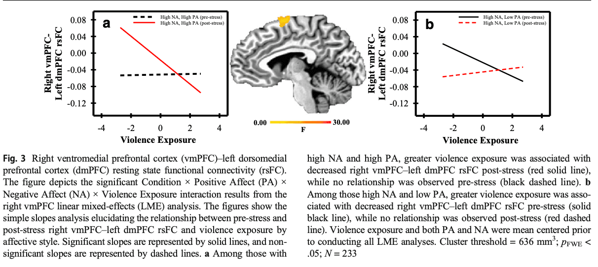
Two hundred eighty-two participants volunteered for the present study. Forty-nine participants were excluded due to excessive motion, poor data quality, or incomplete data (e.g., not completing both resting state scans); therefore, data for two hundred thirty-three emerging adults from the Birmingham site of the Healthy Passages Study were included in the present data analyses (see Table 1). The Healthy Passages Study was a longitudinal, multisite project designed to identify risk and protective factors for adolescent health (Schuster et al., 2012; Windle et al., 2004) and originally included 1,594 children at the Birmingham site. Participants in the Healthy Passages study were recruited from the 5th-grade classrooms of public schools. Data were collected at four time points between 2003 and 2017 (see Supplemental Fig. S1). The average age (mean ± SD) of the present sample at each time point was 11.24 ± 0.52 years at Time 1; 13.07 ± 0.51 years at Time 2; 16.22 ± 0.54 years at Time 3; and 19.10 ± 1.14 years at Time 4. Magnetic resonance imaging (MRI) data were collected (average age: 19.61 ± 1.20 years) after the fourth Healthy Passages time point was completed (see Supplemental Fig. S1). There was no difference in the proportion of Black American (BA) and White American (WA) participants, χ2(1) = 2.08, p = ns, in the current sample (BA = 149, WA = 84) compared with the Healthy Passages sample from Birmingham that did not participate in the present study (BA = 747, WA = 521). However, there was a difference in sex , χ2(1) = 4.73, p = .032, with a greater proportion of males in the current sample (female = 101, male = 132) compared with the Birmingham sample that did not participate in the present study (female = 648, male = 620). There was no difference in violence exposure, t(1483) = 1.17, p = ns, between the current sample and the Healthy Passages sample that did not participate in the present study. Exclusion criteria for the present study included standard MRI contraindications (e.g., metallic devices, pacemaker, metallic foreign body), left-handedness, previous head injury, loss of consciousness, spinal cord abnormalities, pregnancy, and history of claustrophobia, seizures, psychotic symptoms, and blood or circulation disorders (e.g., sickle cell, anemia, diabetes).

Table 1 Participant demographics and descriptive statistics

Procedure

Upon arrival to the laboratory, participants provided written informed consent as approved by the University of Alabama at Birmingham Institutional Review Board. The original Healthy Passages study, from which the participants in the present study were recruited, was approved by the Centers for Disease Control and Prevention and the original study site institutions. Participants completed questionnaires, and two 6-minute resting state-fMRI scans, during which they were instructed to remain still with their eyes open and not think about anything in particular. Resting state scans were completed prior to (pre-stress) and after (post-stress) a modified version of the Montreal Imaging Stress Task (MIST; Dedovic et al., 2005). The MIST is a psychosocial stress protocol designed for functional brain imaging settings and consists of computerized mental arithmetic challenges and social evaluative threat. Participants completed two MIST scans (i.e., a Control scan followed by a Stress scan). The MIST conditions were presented in a fixed order to best address the questions of interest in this project by reducing variability related to counterbalancing conditions and carryover effects that develop when the Stress condition precedes the Control condition (Wheelock et al., 2016). The version of the MIST used for the present study has been described in prior work (Goodman et al., 2016; Wheelock et al., 2016).

Measures

Violence exposure

Violence exposure was assessed using the Healthy Passages Violence Exposure measure (Eaton et al., 2006; Mrug et al., 2008; Windle et al., 2004) at each of the four time points described above. Participants reported whether they witnessed (1) a threat of physical violence, (2) actual physical violence, and (3) a threat or actual violence involving a weapon; and whether they were a victim of (1) a threat of physical violence, (2) actual physical violence, (3) a threat or actual violence involving a weapon, and (4) physical violence inflicting an injury that required medical care in the past 12 months. Participants responded to each item using a 4-point scale ranging from 0 (never) to 3 (many times). Internal consistency for the Healthy Passages Violence Exposure measure (Eaton et al., 2006; Mrug et al., 2008; Windle et al., 2004) at each wave was: Wave 1 = .748, Wave 2 = .646, Wave 3 = .705, Wave 4 = .705. Responses to each item on the scale were averaged, and the scale was then summed across all time points to create a composite index of violence exposure (Mrug et al., 2008). Violence exposure was mean centered prior to all analyses. A latent class analysis was also completed using Mplus statistical software to outline patterns of violence exposure across all four time points. The results yielded a three-class solution (see Supplemental Fig. S2). The three-class solution was then used in voxel-wise analyses (described below) to determine the effects of violence exposure trajectories on functional brain connectivity.

Positive and Negative Affect Schedule (PANAS)

The PANAS (Watson, Clark, & Tellegen, 1988) is a self-report measure that assesses trait-positive and trait-negative affect. Participants rated to what extent they felt each of 10 positive and 10 negative emotions in general using a 5-point Likert scale ranging from 1 (very slightly or not at all) to 5 (extremely) to reflect positive affect (PA) and negative affect (NA). Positive and negative emotions were independently summed to reflect PA and NA, respectively. Cronbach’s alpha was .864 for positive affect and .798 for negative affect. Both PA and NA were mean centered prior to all analyses. Participants completed the PANAS at the MRI session, prior to the MRI scan.

Self-reported stress

Self-reported stress was assessed retrospectively, outside the scanner following the completion of the post-stress resting-state fMRI scan. The self-reported stress measure included eight statements for both the Control and Stress conditions of the MIST (see the Supplemental material). Participants rated each item on a 5-point scale ranging from 1 (not at all) to 5 (extremely). The scale included four items that were positively worded (e.g., I felt I had control) and four that were negatively worded (e.g., I felt overwhelmed). Participants’ responses were summed separately for Stress and Control conditions, with total possible scores ranging from 8 to 40 for each condition (Wheelock et al., 2016; Wheelock et al., 2018). Cronbach’s alpha for the self-reported stress measure was .844 (Control MIST) and .852 (Stress MIST). Self-reported stress data for 12 participants were not collected.

Skin conductance level (SCL)

SCL data were collected using MR compatible physiological monitoring equipment (Biopac Systems; Goleta, CA). SCL data were sampled at 10 kHz using two disposable radio translucent electrodes, attached to the thenar and hypothenar eminence of the nondominant hand. Data were filtered using a 1-Hz Infinite Impulse Response (IIR) low-pass filter, resampled to 250 Hz, and transformed based on the individual participant resistance level using Acqknowledge 4.1.0 (Bach, Flandin, Friston, & Dolan, 2009). Separate averages of SCL amplitude were acquired for the pre-stress and post-stress resting-state scans. Data acquisition methods were similar to prior work (Knight & Wood, 2011; Wheelock et al., 2016). Data from 22 participants were not analyzed due to equipment malfunction or low/unmeasurable skin conductance values.

Functional MRI (fMRI)

Image acquisition

MRI data were obtained using a 3T Siemens Allegra scanner. Standard high-resolution T1 weighted structural magnetization-prepared rapid gradient-echo (MPRAGE) images were collected (TR = 2,300 ms, TE = 3.9 ms, flip angle = 12°, FOV = 25.6 cm, matrix = 256 × 256, slice thickness = 1 mm, gap = 0.5 mm) prior to the first resting-state scan to serve as an anatomical reference for the fMRI data. Resting-state blood oxygen level-dependent (BOLD) fMRI was measured with a gradient-echo echoplanar pulse sequence in an oblique axial orientation (TR = 2,000 ms, TE = 30 ms, flip angle = 70°, FOV = 24 cm, matrix = 64 × 64, voxel size = 3.75 × 3.75 × 4.0 mm, slice thickness = 4 mm, no gap).

Preprocessing

Images were preprocessed using the Analysis of Functional NeuroImages (AFNI; Cox, 1996) software package, the FMRIB Software Library (FSL; Smith et al., 2004), and MRIcron (Rorden & Brett, 2000). Echoplanar data for pre-stress and post-stress scans were reconstructed (using the Dicom to Nifti option in MRIcron) and reregistered to minimize movement artifact and generate motion correction parameters for use as covariates in subsequent analyses (using 3dvolreg in AFNI). Images were then corrected for slice timing offset with a Fourier transformation (using 3dTshift in AFNI) and spatially smoothed using a 4-mm full-width-at-half-maximum Gaussian filter (using 3dmerge in AFNI). Time-course data for tissue-based regressors, including cerebrospinal fluid (CSF) and white matter (WM), were extracted from the functional data set prior to spatial smoothing (using 3dSeg in AFNI).

Data analyses

SCL

A paired-samples t test was conducted to determine whether SCL differed pre-stress to post-stress. A linear mixed-effects (LME) model analysis was also conducted to determine whether SCL differed pre-stress to post-stress by violence exposure, PA, and NA; all mean centered. Condition was entered as a within-subjects factor (1 = pre-stress and 2 = post-stress), and violence exposure, PA, and NA were entered as continuous factors. Race/ethnicity and sex were entered as covariates. All two-way, three-way, and four-way interactions among violence exposure, PA, NA, and Condition were tested. Statistical analyses were completed using SPSS Statistics software.

Self-reported stress

A paired-samples t test was conducted to determine whether self-reported stress differed between the Control and Stress conditions of the MIST. An LME model analysis was also conducted to determine whether self-reported stress differed between the Control and Stress conditions of the MIST by violence exposure, PA, and NA (all mean centered). Condition was entered as a within-subjects factor (1 = Control MIST and 2 = Stress MIST), and violence exposure, PA, and NA were entered as continuous factors. Race/ethnicity and sex were entered as covariates. All two-way, three-way, and four-way interactions among violence exposure, PA, NA, and Condition were tested. Statistical analyses were completed using SPSS Statistics software.

FMRI

First-level analyses

Individual subject-level analyses were completed using multiple linear regression (3dDeconvolve in AFNI) to account for variables of no interest, including (1) mean CSF time course, (2) mean WM time course, (3) six motion parameters, (4) six motion derivatives, and (5) 111 band-pass time courses (band-pass filter: 0.01< f > 0.1 Hz). These variables were regressed out of the gray matter (GM) time course for each participant. Time points where >3% of voxels were greater than 5 times the median absolute deviation (e.g., outliers) of the time series were excluded from the individual subject analyses similar to prior work (Wood et al., 2015). Excluded volumes were ignored in subsequent statistical analyses. The mean number of included volumes was 177 for pre-stress scans and 175 volumes for post-stress scans (out of a total of 178 possible volumes). Thus, 1–3 volumes were excluded (~1%), on average, from each scan. Participants with less than 80% useable TRs were excluded from further analyses (n = 1). The functional dataset was then normalized to the Talairach and Tournoux stereotaxic coordinate system (Talairach & Tournoux, 1988). For each participant, a seed (6-mm sphere) was placed in six regions of interest (ROIs) based on coordinates obtained from the Talairach atlas in AFNI—the amygdala: right (x: 23 y: -5 z: -15), left (x: -23 y: -5 z: -15); hippocampus: right (x: 30 y: −24 z: −9), left (x: −30 y: −24 z: −9); and vmPFC: right (x: 12 y: 49 z: 4), left (x: −12 y: 49 z: 4)—resulting in one average time course for each of the six ROI. Six (pre-stress and post-stress) voxel-wise Pearson correlation analyses were conducted to correlate the time series of each ROI with the time series of all other voxels throughout the whole brain. The Pearson correlation analysis resulted in one pre-stress and one post-stress ROI–whole brain correlation map for each ROI (i.e., bilateral amygdala, hippocampus, and vmPFC). Each Pearson correlation value was then converted to a Fisher’s Z value to normalize the distribution for each participant, and each map was resampled to 1-mm isotropic voxels.

Group-level analyses

(1) Six paired-sample t tests (left and right = 2) were conducted in AFNI using 3dttest++ for the bilateral amygdala–whole brain, hippocampus–whole brain, and vmPFC–whole brain analyses to examine the difference between pre-stress and post-stress rsFC. To reduce familywise error (FWE), a Monte Carlo simulation was conducted (3dClustSim in AFNI), using an uncorrected significance threshold of p < .005, to determine the cluster corrected significance threshold. Smoothness was estimated based on the spherical autocorrelation function parameter (3dFWHMx in AFNI) by averaging participants’ residual time series from the first level analysis, resulting in a voxel-wise cluster threshold of 636 mm3 (pcorrected < .05). (2) An LME model analysis was conducted using 3dLME (Chen, Saad Britton, Pine, & Cox, 2013) in AFNI, to determine whether bilateral amygdala–whole brain rsFC, hippocampus–whole brain rsFC, and vmPFC–whole brain rsFC differed between pre-stress and post-stress as a function of violence exposure and as a function of the interaction between violence exposure and both PA and NA. Both PA and NA were included in each seed–whole brain analysis. A full factorial model was conducted examining all main effects and two-way, three-way, and four-way interactions. The ROI–whole brain Fisher’s Z maps were used as the dependent variable for each separate LME analysis. Pre-stress and post-stress scans were coded and entered into the model as a repeated, within-subjects factor: Condition (1 = pre-stress and 2 = post-stress). Race and sex were included as covariates in both analyses.

Follow-up analyses

After completion of the LME analysis for each ROI, follow-up analyses were conducted to further examine significant interactions. First, for each interaction term, the average Fisher’s Z values were obtained for each significant volume of activity for both pre-stress and post-stress scans. If the significant interaction included Condition (e.g., pre-stress to post-stress difference), two separate follow-up analyses were completed, one using pre-stress rsFC, and the other using post-stress rsFC as the dependent variable to determine how the interaction of violence exposure, PA, and NA varied with rsFC pre-stress versus post-stress. For main effects and significant interaction terms that did not include Condition, pre-stress rsFC and post-stress rsFC data were averaged to reflect an overall rsFC value and used as the dependent variable for follow-up analyses. Using PROCESS (Hayes, 2012; Hayes & Preacher, 2013), a multiple regression analysis was conducted to compute simple slopes for each significant interaction. Each simple slopes analysis examined the conditional effects of violence exposure on rsFC at different levels of the moderators (e.g., PA, NA): one standard deviation below the mean (low) and one standard deviation above the mean (high) (Hayes, 2012). In addition, partial correlation was used as a follow-up analysis of a significant Condition × Violence Exposure interaction. The partial correlation analysis compared violence exposure with both pre-stress and post-stress rsFC, while controlling for PA, NA, race, and sex to determine the relationship between violence exposure and rsFC during pre-stress and post-stress scans.

Violence exposure class and rsFC

Differences in pre-stress to post-stress rsFC among the three violence exposure classes were assessed using AFNI’s 3dttest++ with a covariate for violence exposure class (dependent measures: left/right amygdala–whole brain, left/right hippocampus–whole brain, and left/right vmPFC–whole brain). A voxel-wise cluster threshold of 636 mm3 (pcorrected < .05) was also applied to this analysis.

Violence exposure class and SCL

A repeated-measures ANOVA (dependent measure: SCL) was conducted using SPSS Statistics software to determine whether there were pre-stress to post-stress differences among the three violence exposure classes.

Violence exposure class and self-reported stress

A repeated-measures ANOVA (dependent measure: self-reported stress) was conducted using SPSS Statistics software to determine whether there were differences in self-reported stress among the three violence exposure classes.

Results

Descriptive statistics

Descriptive statistics are presented in Table 1.

Behavioral results

Skin conductance level (SCL)

Results from the paired-samples t test demonstrate that SCL was greater during post-stress (M = 8.61, SEM = 0.48) than pre-stress (M = 7.64, SEM = 0.44), t(210) = 5.78, p < .001, scans. This finding suggests that in general the psychosocial stress task elicited a physiological response. The LME analysis revealed a significant main effect for NA, F(1,192) = 8.98, p = .003, such that SCL varied positively with NA (r = .161, p = .020). There was also a significant PA × NA interaction, F(1, 192) = 15.07, p < .001. A test of simple slopes was completed to further assess the PA × NA interaction. It revealed that among those with high NA, SCL varied negatively with PA (b = −.159, p = .024), while there was no relationship between SCL and PA among those who reported low NA (b = −.108, p = .165). There were no other significant effects.

Self-reported stress

Results from the paired-samples t test demonstrate that self-reported stress was greater during the Stress (M = 25.79, SEM = 0.45) than the Control (M = 14.96, SEM = 0.38), t(220) = 19.70, p < .001, condition of the MIST, which suggests that the procedures used in the present study successfully manipulated stress across conditions. The LME analysis revealed a significant main effect for PA, F(1, 202) = 7.19, p = .008, such that self-reported stress varied negatively with PA (r = −.173, p = .011). There was also a significant Condition × Violence Exposure interaction, F(1, 202) = 11.67, p = .001. Specifically, self-reported stress for the Control MIST varied positively with violence exposure (r = .165, p = .015), while self-reported stress for the Stress MIST did not vary with violence exposure (r = −.027, p = ns). Finally, there was a significant Condition × PA × NA interaction, F(1, 202) = 3.90, p = .049. A test of simple slopes revealed that among those who reported low NA, there was a negative relationship between self-reported stress and PA for the Control MIST (b = −.177, p = .011), while there was no relationship between PA and self-reported stress for the Stress MIST (b = .0005, p = ns). Among those who reported high NA, there was no relationship between self-reported stress and PA for the Control MIST (b = −.079, p = ns), while self-reported stress varied negatively with PA for the Stress MIST (b = −.153, p = .05). There were no other significant effects.

Resting state functional connectivity (rsFC) results

Pre- to post-stress differences in amygdala-, hippocampus-, and vmPFC-whole brain rsFC

Six paired-samples t tests were conducted to determine whether there were differences in bilateral amygdala, hippocampus, and vmPFC rsFC. Results of these analyses are presented in Supplemental Figs. S3–S5 and Supplemental Tables S1–S3.

Relationship between SCL and rsFC

Bivariate correlation analyses were conducted to compare differential (post-stress minus pre-stress) SCL and differential (post-stress minus pre-stress) rsFC. Differential SCL varied positively with differential left vmPFC–right insula rsFC (r = .136, p = .048, uncorrected). There were no other significant correlations. Bivariate correlation analyses were also conducted to determine whether pre-stress and post-stress SCL varied with pre-stress and post-stress rsFC, respectively. SCL varied with rsFC among many regions during pre-stress and post-stress scans (see Supplemental Tables S4–S6).

Relationship between self-reported stress and rsFC

Bivariate correlation analyses were conducted to compare differential (Stress minus Control MIST) self-reported stress and differential (post-stress minus pre-stress) rsFC. Differential self-reported stress varied positively with differential right amygdala–left dlPFC rsFC (r = .143, p = .034, uncorrected). Further, differential self-reported stress varied with differential right vmPFC–left dlPFC rsFC (r = –.132, p = .049, uncorrected). Differential self-reported stress also varied positively with differential right vmPFC–right STG rsFC (see Supplemental Results). There were no other significant correlations. Bivariate correlation analyses were also conducted to determine whether self-reported stress for the Control and Stress conditions of the MIST varied with pre-stress and post-stress rsFC. Self-reported stress for the Control and Stress conditions of the MIST varied with the pre-stress and post-stress rsFC of several brain regions (see Supplemental Tables S7–S9).

Linear mixed-effects (LME) analysis

A large number of results were obtained from the LME analysis of the rsFC data. Full results from the rsFC LME analyses are presented in the Supplemental materials (Supplemental Results; Supplemental Tables S10–S15). The LME results presented in the following sections include those that are focused on the primary aims of the present study (i.e., examining the relationships among violence exposure, affective style, and rsFC in regions of interest) and are included in the Discussion.

Amygdala (Condition × PA × NA × Violence Exposure)

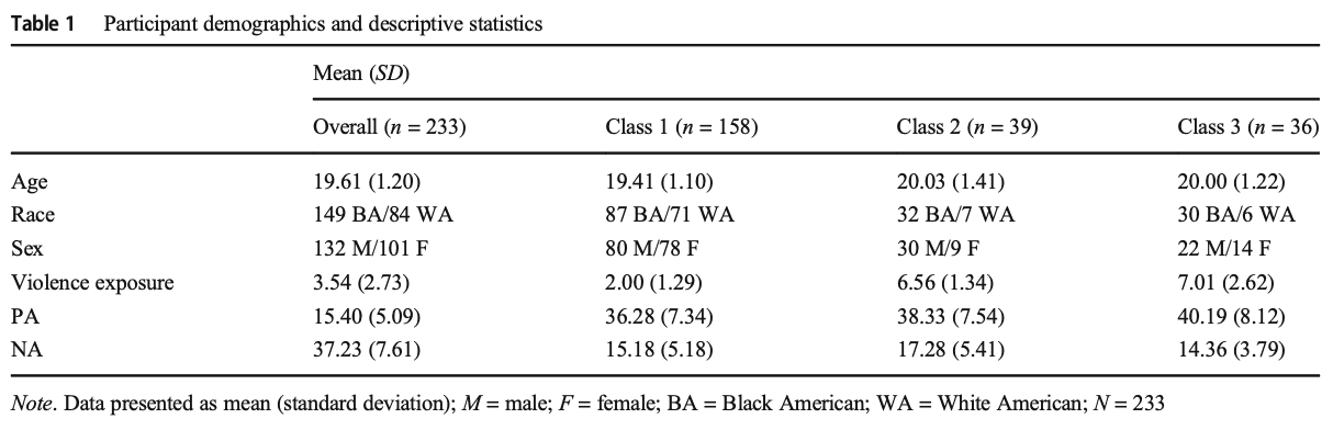
A Condition × PA × NA × Violence Exposure interaction was observed in the rsFC of the right amygdala with the left IPL (see Supplemental Table S10). Differences in affective style modulated the relationship between violence exposure and right amygdala connectivity with the left IPL pre-stress to post-stress. Specifically, violence exposure varied positively with post-stress, but not pre-stress rsFC among those with low NA and low PA (see Fig. 1a; Supplemental Table S11). Among those with low NA and high PA, violence exposure varied positively with pre-stress, but not post-stress rsFC (see Fig. 1b; Supplemental Table S11).

Fig 1

Hippocampus (Condition × PA × NA × Violence Exposure)

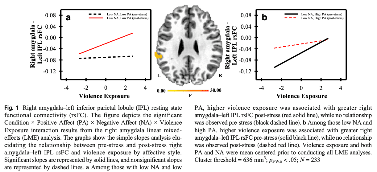
A Condition × PA × NA × Violence Exposure interaction was observed in the rsFC of the left hippocampus with the left mid cingulate gyrus and the left dmPFC (see Supplemental Table S12). Differences in affective style modulated the relationship between violence exposure and the rsFC of these regions from pre-stress to post-stress. Specifically, violence exposure varied positively with the rsFC of these regions pre-stress, but not post-stress, among those with low NA and low PA (see Fig. 2a; Supplemental Table S13). Violence exposure also varied positively with left hippocampus–left cingulate rsFC post-stress, but not pre-stress, among those with high NA and low PA (see Supplemental Table S13). Further, violence exposure varied positively with left hippocampus–left dmPFC rsFC post-stress, but not pre-stress, among those with low NA and high PA (see Fig. 2b; Supplemental Table S13).

Fig 2

VmPFC (Condition × PA × NA × Violence Exposure)

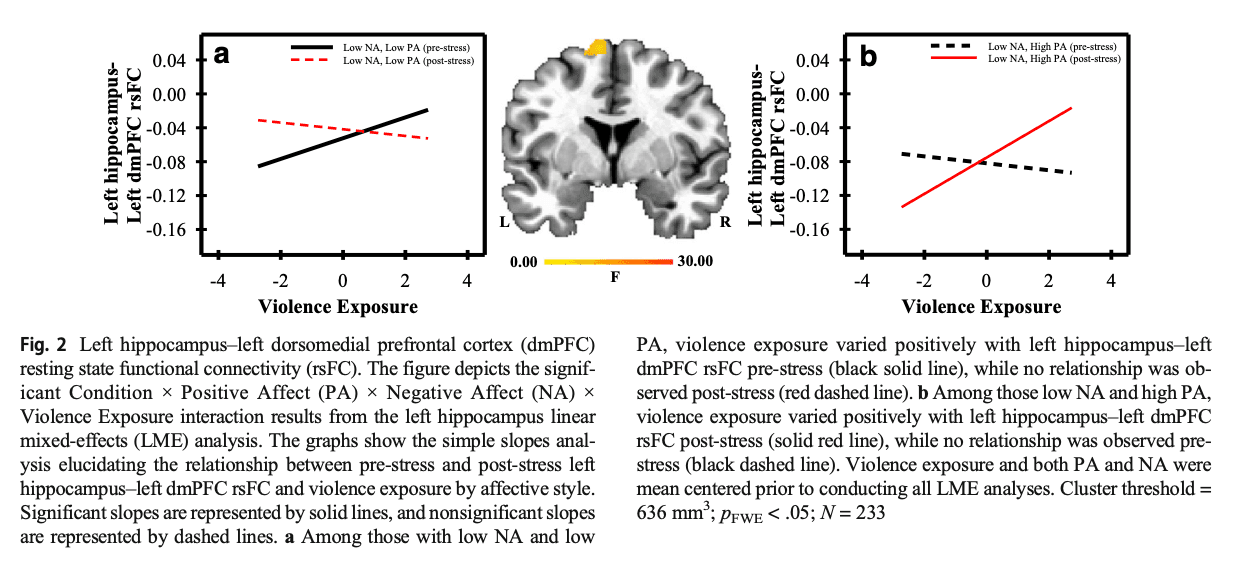
A Condition × PA × NA × Violence Exposure interaction was observed in the rsFC of the right vmPFC to the left dmPFC (see Supplemental Table S14). Differences in affective style modulated the relationship between violence exposure and right vmPFC connectivity with the left dmPFC pre-stress to post-stress. Specifically, violence exposure varied negatively with right vmPFC–left dmPFC rsFC post-stress among those with high NA and high PA (see Fig. 3a; Supplemental Table S15). Violence exposure also varied negatively with right vmPFC–left dmPFC rsFC pre-stress among those with high NA and low PA (see Fig. 3b; Supplemental Table S15).

Fig 3

Violence exposure class and rsFC

Results from the latent class analysis yielded a three-class solution. Each class referenced in the following section reflects a different trajectory of violence exposure experienced by participants in the current sample (see Supplemental Fig. S2). Participants in Class 1 reported experiencing relatively low and stable violence exposure across the four assessment waves. Participants in Class 2 initially (i.e., Wave 1; average age: 11.24 years) reported high levels of violence exposure that decreased across assessment Waves 2–4. Finally, participants in Class 3 reported violence exposure that increased across assessment waves (see Supplemental Fig. S2).

Amygdala

Left and right amygdala rsFC did not vary pre-stress to post-stress with violence exposure class.

Hippocampus

Left hippocampus–left dlPFC rsFC varied pre-stress to post-stress with violence exposure class, t(231) = –3.65, pFWE = .05 (see Supplemental Fig. S6). Participants in Class 3 demonstrated greater pre-stress (mean = .159, SEM = .024) than post-stress (mean = .047, SEM = .026) rsFC. Further, participants in Class 3 (mean = .159, SEM = .027) demonstrated greater pre-stress rsFC than participants in Class 1 (mean = .090, SEM = .011; see Supplemental Fig. S6). No pre-stress differences were observed between Classes 1 and 3 with Class 2 (mean = .124, SEM = .024; see Supplemental Fig. S6). There were no differences in post-stress rsFC. Right hippocampus rsFC did not vary pre-stress to post-stress with violence exposure class.

VmPFC

The left and right vmPFC did not vary pre-stress to post-stress with violence exposure class among hypothesized regions. However, left vmPFC rsFC varied with the right culmen (cerebellum; see Supplemental Results and Supplemental Fig. S7).

Violence exposure class and SCL

SCL did not vary pre-stress to post-stress with violence exposure classes, F(1,208) = .602, p = ns.

Violence exposure class and self-reported stress

There was a significant difference in Control versus Stress MIST self-reported stress by violence exposure class, F(1,218) = 5.63, p = .004. Participants in Classes 1–3 demonstrated greater self-reported stress to the Stress MIST compared with the Control MIST: Class 1 (Control MIST: mean = 14.63, SEM = 0.460; Stress MIST: mean = 26.74, SEM = 0.546), t(218) = −18.35, p < .001; Class 2 (Control MIST: mean = 15.42, SEM = 0.905; Stress MIST: mean = 23.55, SEM = 1.073), t(218) = −6.26, p < .001; Class 3: (Control MIST: mean = 15.86, SEM = 0.930; Stress MIST: mean = 24.31, SEM = 1.103), t(218) = −6.33, p < .001. Further, participants in Class 1 (mean = 26.74, SEM = .546) reported higher self-reported stress during the Stress MIST than participants in Class 2 (mean = 23.55, SEM = 1.073), t(218) = 2.64, p = .009, and participants in Class 3 (mean = 24.31, SEM = 1.103), t(218) = 1.97, p = .049 (see Supplemental Fig. S8).

Discussion

Repeated exposure to violence during childhood is linked to chronic emotion dysregulation and, in turn, greater susceptibility to internalizing psychopathology (Hanson et al., 2008; Mead et al., 2010; Mrug & Windle, 2010). Specifically, repeated exposure to violence appears to modify functional connectivity patterns within the brain that control emotion regulation processes (Saxbe et al., 2018; Thomason & Marusak, 2017; Thomason et al., 2015). Emotion regulation processes rely upon the connectivity of limbic, parietal, and prefrontal brain regions that may underlie internalizing symptomology by disrupting both the interpretation of and response to stressful events (Ochsner et al., 2012; Young & Koenigs, 2007). Therefore, determining the impact of violence exposure on functional brain connectivity may offer new insight into the neural processes that affect successful emotion regulation among those exposed to violence. Understanding the manner through which affective style modulates the relationship between violence exposure and brain connectivity would provide novel insight into individual differences in the development of emotional dysfunction. The present study examined the impact violence exposure and affective style have on stress-induced changes in functional brain connectivity. We found stress-induced changes in rsFC among prefrontal, fronto-limbic, and parieto-limbic regions that support the expression and regulation of emotion. Further, affective style moderated the relationship between violence exposure and functional brain connectivity. These findings suggest that the functional connectivity of prefrontal, fronto-limbic, and parieto-limbic regions that support emotion processes vary with childhood violence exposure and affective style.

Amygdala rsFC

In the present study, we found that amygdala–IPL rsFC varied with violence exposure as a function of affective style (see Fig. 1; Supplemental Tables S10 and S11). The amygdala is an important component of the neural circuity that underlies the peripheral expression of emotion (Cheng et al., 2006; Cheng et al., 2003; Klumpers, Kroes, Baas, & Fernández, 2017; Knight et al., 2005; Orem et al., 2019; Wood et al., 2014), while the IPL is important for the top-down attentional control of emotion (Sylvester et al., 2012). Among participants with low NA and low PA, post-stress amygdala–IPL rsFC varied positively with violence exposure (see Fig. 1a; Supplemental Tables S10 and S11). Prior work indicates that the amygdala and IPL are important for responding to emotion-related threats and coactivate during emotion regulation processes (Alarcón, Sauder, Teoh, Forbes, & Quevedo, 2019). Further, task-based functional connectivity research has found that amygdala–IPL connectivity increases during psychosocial stress (Fan et al., 2015). Thus, this prior work is generally consistent with the present findings that suggest high violence exposure is associated with greater coupling of the amygdala and IPL in those with both low NA and low PA (see Fig. 1a). The present findings suggest that violence exposure interacts with affect. More specifically, high violence exposure in combination with lower overall NA and PA may be associated with greater processing of emotional information in response to acute stress. In the present study, we hypothesized that the relationship between violence exposure and post-stress rsFC would be enhanced by high NA, and blunted by high PA. Our findings indicate that while high NA had limited impact on the relationship between violence exposure and amygdala–IPL rsFC, high PA diminished the apparent impact violence exposure has on amygdala–IPL rsFC post-stress (see Fig. 1b). This finding is generally consistent with the view that high PA may blunt the impact adolescent violence exposure has on stress-induced changes in amygdala–IPL connectivity.

Hippocampus rsFC

The hippocampus is important for the consolidation of emotionally salient memories (Phelps, 2004; Richter-Levin & Akirav, 2001) and stress-induced hippocampal activity decreases along with the activity of the medial orbitofrontal cortex (OFC) and ACC (Dedovic et al., 2009; Pruessner et al., 2008). Further, prior work shows that adult hippocampus–dmPFC rsFC varies as a function of childhood trauma (Birn, Patriat, Phillips, Germain, & Herringa, 2014). The present study advances this prior work, showing that hippocampus–dmPFC rsFC varied pre-stress to post-stress as a function of affective style and violence exposure. More specifically, post-stress hippocampus–dmPFC rsFC increased as violence exposure increased among those with low NA and high PA (see Fig. 2; Supplemental Tables S12 and S13). We hypothesized that the relationship between violence exposure and post-stress rsFC would be blunted by high PA. Instead, we only found a relationship between violence exposure and the rsFC of these regions when PA was high. Thus, PA did not attenuate the impact of violence exposure, and actually enhanced the effects on the rsFC of the hippocampus and dmPFC. Although the current findings do not precisely match our a priori hypothesis, they do suggest that PA may influence the connectivity and communication between the hippocampus and dmPFC. The dmPFC is important for the appraisal of threatening stimuli, while the hippocampus supports memory retrieval processes during acute stress and modulates HPA axis activity (Goodman, Harnett, & Knight, 2018; Kalisch, Wiech, Critchley, & Dolan, 2006; Kim & Diamond, 2002). Taken together with this prior work, the present findings suggest that among those with high PA and low NA, increased hippocampal-dmPFC rsFC may reflect differences in the appraisal of psychosocial stress and subsequent modulation of the stress response among those exposed to varying levels of violence.

VmPFC rsFC

The vmPFC is important for generating affective meaning as well as regulating behavioral and physiological responses (Roy et al., 2012). Further, connectivity between distinct regions of the PFC are important for emotion regulation (Hare et al., 2009; Ochsner et al., 2012). In the present study, vmPFC–dmPFC rsFC varied pre-stress to post-stress as a function of violence exposure and affective style. Specifically, among those with both high NA and PA, greater violence exposure was negatively associated with post-stress vmPFC–dmPFC rsFC (see Fig. 3; Supplemental Tables S14 and S15). These findings suggest that those with higher NA and PA and lower violence exposure, have greater functional coupling of brain regions (i.e., vmPFC and dmPFC) that are important for the regulation of emotion in response to psychosocial stress. Our findings, in combination with prior work (Kalisch et al., 2006; Morawetz et al., 2017; Phan et al., 2005), suggest that those with higher levels of NA and PA may be more attentive to emotional stimuli. Further, their ability to regulate the emotional response to psychosocial stress may vary as a function of violence exposure. Taken together, these findings suggest that individual differences in emotion regulation may explain variability in stress reactivity among young adults exposed to childhood violence.

SCL, self-reported stress, and rsFC

Secondary analyses were completed to determine whether SCL and self-reported stress were associated with rsFC. We found that differential rsFC between the vmPFC and insula varied positively with differential SCL. This finding suggests the functional coupling of these brain regions may underlie stress-related changes in autonomic activity. The vmPFC is important for assigning value to stimuli, guiding adaptive behavior, and regulating the emotional response, while the insula is important for interoceptive awareness (Damasio, 1994; Hare et al., 2009; Hiser & Koenigs, 2018; Sinha, Lacadie, Constable, & Seo, 2016). Prior research indicates that stress-related tasks activate the vmPFC and insula (Sinha et al., 2016), and that activity within these brain regions varies with skin conductance responses (Nagai et al., 2004). Therefore, the increased stress-induced rsFC observed among these brain regions in the present study provides support for the view that vmPFC and insula connectivity plays an important role in the modulation of the peripheral emotional response (Nagai et al., 2004; Thayer & Lane, 2000). In addition, we found that differential rsFC between the amygdala and dlPFC varied positively with differential self-reported stress. While prior work indicates the amygdala mediates the peripheral emotional response (Cheng et al., 2006; Cheng et al., 2003; Knight et al., 2005; Orem et al., 2019; Wood et al., 2014), the present study advances this prior work by demonstrating that amygdala connectivity with the dlPFC also appears to be important for the subjective experience of stress. The dlPFC supports working memory and attentional processes that play an important role in the top-down control of the amygdala (Comte et al., 2016; Delgado et al., 2008; Ochsner et al., 2009; Ochsner et al., 2012; Sylvester et al., 2012). Therefore, increased rsFC between the amygdala and dlPFC may reflect greater recruitment of these regions in response to acute stress. The findings from these secondary analyses suggest that changes in differential rsFC among these brain regions may reflect greater stress reactivity.

Violence exposure class and rsFC

The pre-stress to post-stress rsFC of the left hippocampus and left dlPFC appears to differ among the three violence exposure classes identified in the present study (see Supplemental Figs. S2 and S6). Specifically, we observed greater positive hippocampus–dlPFC rsFC among participants in Class 3 (i.e., moderate violence exposure in early adolescence that subsequently increased through late adolescence) compared with participants in Class 1 (i.e., low violence exposure throughout adolescence), which suggests greater baseline coupling of these regions (see Supplemental Fig. S6). Specifically, those who experienced moderate levels of violence that increased throughout adolescence (i.e., Class 3) may exhibit greater changes in the connectivity between the dlPFC and hippocampus in comparison to those who experienced consistently low violence exposure throughout adolescence. These findings suggest that the pattern of violence experienced during adolescence differentially affects the rsFC of the dlPFC and hippocampus. Further, acute stress decreased the positive rsFC among those in Class 3, but not those in Classes 1 or 2 (see Supplemental Fig. S6). This finding suggests that violence exposure that increases throughout adolescence may negatively impact the neural response to stress. The dlPFC is important for top-down emotion processes and modulates hippocampal activity (Benoit & Anderson, 2012; Benoit, Hulbert, Huddleston, & Anderson, 2015). Thus, decreased connectivity among these brain regions is consistent with the view that psychosocial stress reduces the dlPFC’s inhibitory control over the hippocampus among participants in Class 3. Prior work suggests that childhood violence exposure often becomes a chronic environmental condition rather than a series of isolated incidents (Finkelhor et al., 2007; Hooven et al., 2012; Mrug et al., 2008; Schwab-Stone et al., 1995). Thus, the experience of participants in Class 3 may more closely reflect this chronic environmental condition than the experiences of participants in Class 1, who have experienced consistently low levels of violence, as well as participants in Class 2, who encountered initial high levels of violence exposure that subsequently decreased across adolescence. The present study suggests that low or decreasing levels of violence during adolescence may have a minimal impact on the connectivity of the hippocampus and dlPFC, regions that underlie important aspects of the stress response (Ochsner et al., 2012; Phelps, 2004; Richter-Levin & Akirav, 2001). Additionally, Class 3 showed greater pre-stress to post-stress changes in the rsFC of these brain regions. Changes in the connectivity among these regions may negatively impact the stress response and subsequently lead to increased susceptibility to internalizing disorders. Prior work suggests that cumulative violence exposure is more important for predicting internalizing symptomology than the type of violence experienced (Mrug et al., 2008). Results from the present study advance this prior work, suggesting that the trajectory of violence exposure may also impact the connectivity of brain regions that influence internalizing symptoms. The present findings suggest that moderate levels of violence exposure in early adolescence that subsequently increase through late adolescence may result in greater susceptibility to the effects of acute stress. Future studies should consider both the trajectory as well as cumulative violence exposure when examining the impact of violence exposure on negative psychological outcomes.

Limitations

The present study focused on emerging adult participants. Emerging adulthood is an important stage of life that is marked by increased risk for psychopathology (Arnett, 2014). However, the functional brain connectivity demonstrated during this period may not yet reflect adult functional brain connectivity, as the brain continues to develop into young adulthood (Taber-Thomas & Pérez-Edgar, 2015). Specifically, neurodevelopmental changes in fronto-limbic connections as well as cortical structures may not have reached full maturation during emerging adulthood, and thus may not reflect fully developed adult brain function (Taber-Thomas & Pérez-Edgar, 2015). Additionally, participants in the present study completed one neuroimaging session. It is possible that preexisting connectivity differences or environmental factors during development contributed to differences in functional connectivity observed in the present study. Future studies should employ longitudinal neuroimaging designs to assess functional changes in the brain over time. Finally, the present study found that the relationship between pre-stress to post-stress changes in rsFC and violence exposure varied with affective style. This relationship is consistent with our general hypothesis that affective style modulates stress-elicited changes in rsFC. However, it is also possible that childhood violence exposure modulates the relationship between affective style and rsFC. Additional studies are need to fully disentangle these possible explanations.

Conclusion

Violence exposure during childhood and adolescence alters rsFC patterns in emerging adulthood among brain regions involved in emotion expression and regulation. Changes in the rsFC of brain regions that support emotion regulation processes may alter the ability to effectively regulate the emotional response to stress. Stress-induced changes in fronto-limbic, prefrontal, and parieto-limbic rsFC varied with violence exposure, suggesting that exposure to violence alters the functional connectivity of brain regions that support emotion-related processes. Furthermore, the relationship between violence exposure and the functional networks that support emotion regulation was moderated by PA and NA, indicating that individual differences in affective style play a role in how emerging adults exposed to violence during development respond to stress.

Open Article as PDF

Abstract

Chronic childhood stress is linked to greater susceptibility to internalizing disorders in adulthood. Specifically, chronic stress leads to changes in brain connectivity patterns, and, in turn, affects psychological functioning. Violence exposure, a chronic stressor, increases stress reactivity and disrupts emotion regulation processes. However, it is unclear to what extent violence exposure affects the neural circuitry underlying emotion regulation. Individual differences in affective style also moderate the impact of stress on psychological function and can thus alter the relationship between violence exposure and brain function. Resting-state functional connectivity (rsFC) is an index of intrinsic brain activity. Stress-induced changes in rsFC between the amygdala, hippocampus, and prefrontal cortex (PFC) are associated with emotion dysregulation and may elucidate how affective style modulates the relationship between violence exposure and brain connectivity. Therefore, the present study examined the impact of violence exposure and affective style on stress-induced changes in rsFC. Participants (n = 233) completed two 6-minute resting-state functional magnetic resonance imaging scans, one before (pre-stress) and one after (post-stress) a psychosocial stress task. The bilateral amygdala, hippocampus, and ventromedial prefrontal cortex (vmPFC) were used as seed regions for rsFC analyses. Significant stress-induced changes in the prefrontal, fronto-limbic, and parieto-limbic rsFC were observed. Further, pre-stress to post-stress differences in rsFC varied with violence exposure and affective style. These findings suggest that prefrontal, fronto-limbic, and parieto-limbic connectivity is associated with the emotional response to stress and provide new insight into the neural mechanisms through which affective style moderates the impact violence exposure has on the brain.

Introduction

Children exposed to violence often experience lasting psychological problems as teenagers and adults. These harmful effects may come from stress-related changes in how the brain works. Specific brain areas, such as the amygdala, hippocampus, and prefrontal cortex, are crucial for managing emotions. If these connections are disrupted by childhood violence, it can impair the body's ability to respond to stress healthily. This can lead to more symptoms of conditions like depression and anxiety. Understanding how childhood violence affects these brain changes may reveal new insights into emotional difficulties.

More than half of children in the United States encounter violence, either as victims or witnesses, before they turn 17. This violence often occurs repeatedly and in various parts of a child's life, such as at home, school, or in the community. It typically involves intentional actions, like threats or physical attacks, rather than accidental events. Research suggests that intentional acts of violence predict more severe negative mental health issues. When violence is a persistent part of a child's environment, rather than a single event, it can prevent them from developing effective coping strategies.

Ongoing exposure to violence during childhood and adolescence is associated with increased emotional reactivity and a state of hypervigilance, altering how individuals respond to stressful situations. This repeated exposure also correlates with higher rates of internalizing disorders, such as depression and anxiety, across different life stages. These observations indicate that violence exposure during critical developmental periods may interfere with the brain systems that manage stress responses.

Previous studies have shown that childhood maltreatment, including violence, causes both physical and functional changes in key brain regions involved in stress reactions and internalizing symptoms, such as the prefrontal cortex, amygdala, and hippocampus. Specifically, higher levels of maltreatment are linked to smaller volumes in these brain areas, which in turn are associated with symptoms of depression, anxiety, and post-traumatic stress disorder. These findings suggest that early life stress can alter brain function and contribute to the development of mental health symptoms in adulthood.

The current study aimed to investigate how childhood violence exposure affects changes in brain connectivity caused by stress. It also explored whether a person's individual emotional style, including positive and negative emotions, influences this relationship. By understanding these connections, researchers can gain new knowledge about how early life violence impacts how adults respond to stress and the development of emotional difficulties.

Method

The study involved recruiting participants and collecting various physiological and psychological data to examine the impact of childhood violence exposure on brain connectivity and stress response.

Participants

The study included 233 young adults from the Birmingham site of the Healthy Passages Study, a long-term project on adolescent health. Participants were originally recruited in 5th grade, with data collected at four time points between 2003 and 2017. The average age at the time of MRI data collection was about 19.61 years. The sample included a similar proportion of Black American and White American participants as the larger study, but had a higher proportion of males. There were no significant differences in violence exposure between the current sample and those who did not participate in this specific study. Exclusion criteria included standard MRI contraindications and various medical or psychological conditions.

Procedure

Upon arrival, participants gave informed consent. They completed questionnaires and underwent two 6-minute resting-state fMRI scans, during which they were asked to remain still with their eyes open. One scan was completed before (pre-stress) and one after (post-stress) a modified version of the Montreal Imaging Stress Task (MIST). The MIST is a computerized task involving mental arithmetic challenges and social evaluation, designed to induce psychological stress. Participants completed a control MIST scan followed by a stress MIST scan, with this fixed order used to minimize variability and carryover effects.

Measures

This section describes the tools used to collect data on violence exposure, emotional states, and stress levels.

Violence Exposure Measurement

Violence exposure was assessed at four points throughout adolescence using a specific questionnaire. Participants reported witnessing or being a victim of different types of violence in the past year, rating frequency on a 4-point scale. Responses were averaged and summed across all time points to create a total violence exposure score. A statistical analysis also identified three patterns of violence exposure over time: consistently low, high decreasing, and increasing.

Positive and Negative Affect Schedule (PANAS)

The PANAS is a self-report measure used to gauge general positive and negative emotional states. Participants rated 10 positive and 10 negative emotions on a 5-point scale. Scores for positive and negative emotions were calculated separately. This measure was completed at the MRI session before the scan.

Self-Reported Stress Measurement

Participants retrospectively reported their stress levels for both the control and stress MIST conditions after the post-stress fMRI scan. They rated eight statements on a 5-point scale, with scores summed for each condition.

Skin Conductance Level (SCL) Measurement

SCL data, a physiological measure of arousal, were collected using specialized equipment during the MRI scans. Electrodes were placed on the hand to measure skin conductance. Data were processed to obtain average SCL amplitude for both pre-stress and post-stress resting-state scans.

Functional MRI (fMRI)

Brain imaging data were collected using a 3T Siemens Allegra scanner. High-resolution anatomical images were first acquired, followed by resting-state fMRI scans which measure blood oxygen level-dependent (BOLD) signals. These signals indicate brain activity. The fMRI data were then preprocessed to minimize movement artifacts, correct for timing differences, and spatially smooth the images, making them suitable for analysis. Specific tissue-based signals (cerebrospinal fluid and white matter) were also removed.

Data Analysis

Statistical analyses were conducted using specialized software. For SCL, a paired t-test compared pre-stress and post-stress levels, and a linear mixed-effects (LME) model assessed how SCL changed with violence exposure and emotional style. For self-reported stress, similar paired t-tests and LME models were used to compare control and stress conditions, also considering violence exposure and emotional style.

For fMRI data, individual brain connectivity analyses were performed by correlating activity in specific brain regions (amygdala, hippocampus, and ventromedial prefrontal cortex, or vmPFC) with activity across the rest of the brain, generating whole-brain connectivity maps. These correlation values were then converted for statistical normalization. At the group level, paired t-tests identified differences in brain connectivity between pre-stress and post-stress scans. An LME model then explored how brain connectivity varied from pre-stress to post-stress based on violence exposure and its interaction with positive and negative emotional styles. Follow-up analyses using multiple regression were conducted to further investigate significant interactions. Additionally, analyses examined how different patterns of childhood violence exposure (identified as "classes") affected SCL, self-reported stress, and brain connectivity changes from pre-stress to post-stress.

Results

The study yielded several findings regarding physiological responses, self-reported stress, and brain connectivity.

Descriptive Statistics

General statistical summaries of the study participants and measures are presented in a separate table.

Behavioral Results

Skin conductance level (SCL), a physiological indicator of arousal, significantly increased from pre-stress to post-stress, confirming that the stress task was effective. SCL was also positively related to negative emotional style. An interaction between positive and negative emotional styles showed that among those with high negative emotion, SCL decreased as positive emotion increased.

Self-reported stress was significantly higher after the stress task compared to the control task, further confirming the effectiveness of the stress induction. Self-reported stress was negatively related to positive emotional style. A significant interaction was observed, where stress levels for the control task increased with violence exposure, but not for the stress task. Another interaction revealed that the relationship between positive emotional style and self-reported stress during both control and stress conditions varied depending on the level of negative emotional style.

Brain Connectivity (rsFC) Results

Analyses revealed pre- to post-stress differences in the functional connectivity of the amygdala, hippocampus, and vmPFC with other brain regions. For instance, the difference in SCL (post-stress minus pre-stress) was positively associated with the difference in vmPFC-insula connectivity. The difference in self-reported stress also showed positive associations with the difference in right amygdala-left dlPFC connectivity and negative associations with right vmPFC-left dlPFC connectivity. Various other regions also showed altered connectivity with SCL and self-reported stress both before and after stress.

Amygdala Connectivity Findings

A complex interaction involving stress condition, positive emotional style, negative emotional style, and violence exposure was found for the connectivity between the right amygdala and the left inferior parietal lobule (IPL). This indicated that a person's emotional style influenced how violence exposure related to amygdala-IPL connectivity before and after stress. Specifically, for individuals with both low negative and low positive emotional styles, higher violence exposure was linked to greater amygdala-IPL connectivity after stress but not before. In contrast, for those with low negative and high positive emotional styles, higher violence exposure was associated with greater connectivity before stress, but not after.

Hippocampus Connectivity Findings

A similar four-way interaction was observed for the connectivity of the left hippocampus with the left mid-cingulate gyrus and the left dorsomedial prefrontal cortex (dmPFC). This means emotional style modified the relationship between violence exposure and hippocampus-dmPFC connectivity from pre-stress to post-stress. Specifically, among individuals with low negative and low positive emotional styles, violence exposure positively correlated with connectivity in these regions before stress, but not after. For those with high negative and low positive emotional styles, violence exposure was positively linked to left hippocampus-left cingulate connectivity after stress. When individuals had low negative and high positive emotional styles, violence exposure showed a positive link with left hippocampus-left dmPFC connectivity after stress, but not before.

VmPFC Connectivity Findings

A four-way interaction was also present for the connectivity between the right vmPFC and the left dmPFC. Emotional style influenced how violence exposure related to this connectivity before and after stress. Specifically, for individuals with high negative and high positive emotional styles, higher violence exposure was negatively associated with vmPFC-dmPFC connectivity after stress. For those with high negative and low positive emotional styles, violence exposure was negatively associated with this connectivity before stress.

Violence Exposure Class Effects on Brain Connectivity

The study also analyzed brain connectivity based on different patterns of violence exposure over time. Three classes were identified: Class 1 (consistently low violence exposure), Class 2 (high violence exposure in early adolescence, decreasing later), and Class 3 (violence exposure increasing through adolescence). No differences in amygdala connectivity were found across these classes. However, left hippocampus-left dlPFC connectivity changed from pre-stress to post-stress based on violence exposure class. Participants in Class 3 showed greater connectivity before stress compared to after stress. They also had greater pre-stress connectivity than those in Class 1. No post-stress differences were observed. The vmPFC connectivity with other regions also showed some variation, though not in the originally hypothesized areas.

Violence Exposure Class Effects on Physiological Arousal

There were no significant differences in SCL changes from pre-stress to post-stress across the three violence exposure classes.

Violence Exposure Class Effects on Self-Reported Stress

Self-reported stress differences between the control and stress MIST conditions varied by violence exposure class. All three classes reported higher stress during the stress MIST compared to the control MIST. Notably, Class 1 (consistently low violence exposure) reported higher self-reported stress during the stress MIST than both Class 2 and Class 3.

Discussion

Repeated exposure to violence in childhood has been linked to long-term emotional regulation difficulties and a higher risk for mental health problems like depression and anxiety. These issues may stem from changes in brain connectivity patterns that govern how emotions are managed. Effective emotion regulation relies on connections between brain regions involved in emotion, attention, and executive control, which can be disrupted by stressful experiences. This study aimed to understand how violence exposure affects functional brain connectivity and how individual emotional styles might influence this relationship. The findings indicate that stress-induced changes in brain connectivity, especially in prefrontal, fronto-limbic, and parieto-limbic regions, vary with childhood violence exposure and a person's emotional style.

Amygdala Connectivity Insights

The study found that connectivity between the amygdala (involved in emotional expression) and the inferior parietal lobule (IPL, involved in attentional control of emotion) varied based on violence exposure and emotional style. For individuals with low levels of both negative and positive emotions, higher violence exposure was linked to greater amygdala-IPL connectivity after stress. This suggests that more processing of emotional information might occur in response to acute stress in this group. While it was hypothesized that high negative emotion would enhance this relationship and high positive emotion would lessen it, the findings instead showed that high positive emotion seemed to reduce the observed impact of violence exposure on amygdala-IPL connectivity after stress, aligning with a protective role for positive emotions.

Hippocampus Connectivity Insights

The hippocampus plays a key role in emotional memory and stress responses. The study showed that hippocampus-dmPFC connectivity (dmPFC is involved in appraising threats) changed from pre-stress to post-stress depending on emotional style and violence exposure. Specifically, for those with low negative emotion and high positive emotion, increased violence exposure was associated with increased hippocampus-dmPFC connectivity after stress. This finding, while not precisely matching the initial hypothesis about positive emotion's attenuating effect, suggests that positive emotional style may influence how the hippocampus and dmPFC communicate during stress. This altered communication might reflect differences in how individuals appraise psychosocial stress and regulate their stress response based on their past violence exposure and current emotional style.

VmPFC Connectivity Insights

The ventromedial prefrontal cortex (vmPFC) is crucial for assigning emotional meaning and regulating responses. The study found that vmPFC-dmPFC connectivity varied pre-stress to post-stress as a function of violence exposure and emotional style. Specifically, among individuals with both high negative and positive emotional styles, greater violence exposure was negatively associated with vmPFC-dmPFC connectivity after stress. This suggests that those with higher negative and positive emotional styles and lower violence exposure might have stronger functional coupling in these brain regions, which are important for emotion regulation during psychosocial stress. These findings indicate that individual differences in emotional regulation may contribute to variations in how young adults, exposed to childhood violence, react to stress.

Physiological and Self-Reported Stress Insights

Secondary analyses revealed that changes in vmPFC-insula connectivity were positively associated with changes in skin conductance levels (SCL), suggesting that the functional coupling of these regions underpins stress-related changes in autonomic activity. Both the vmPFC (evaluating stimuli) and insula (interoceptive awareness) are active during stress, and their connectivity may be important for regulating peripheral emotional responses. Additionally, changes in amygdala-dlPFC connectivity were positively linked to changes in self-reported stress. The amygdala handles emotional responses, while the dorsolateral prefrontal cortex (dlPFC) is involved in working memory and attention, providing top-down control over the amygdala. Increased connectivity here might signify greater recruitment of these regions during acute stress, reflecting enhanced stress reactivity.

Insights on Violence Exposure Classes

The study identified three patterns of violence exposure during adolescence: consistently low, decreasing from high, and increasing over time. The connectivity between the left hippocampus and left dlPFC changed from pre-stress to post-stress differently across these groups. Participants who experienced increasing violence exposure throughout adolescence (Class 3) showed greater baseline connectivity between these regions compared to those with consistently low violence exposure (Class 1). Furthermore, acute stress decreased this connectivity in Class 3, but not in the other classes, suggesting that increasing violence exposure may negatively impact the brain's response to stress. This indicates that the trajectory of violence exposure, not just cumulative exposure, can uniquely affect brain regions vital for the stress response.

Limitations

This study focused on young adults, a developmental period still marked by brain maturation. Therefore, the observed brain connectivity might not fully represent adult brain function. The study also used a single neuroimaging session, meaning pre-existing differences or developmental environmental factors could contribute to the findings. Future research with longitudinal neuroimaging designs could better track brain changes over time. Additionally, while the study found that emotional style moderated the relationship between violence exposure and stress-induced brain changes, further studies are needed to fully understand the intricate interactions.

Conclusion

Childhood and adolescent violence exposure influences brain connectivity patterns in young adulthood, particularly in regions involved in emotion expression and regulation. These changes may impact an individual's ability to effectively manage emotional responses to stress. Stress-induced alterations in connectivity across fronto-limbic, prefrontal, and parieto-limbic regions varied with violence exposure, indicating that such exposure reshapes the functional organization of emotion-related brain areas. Moreover, positive and negative emotional styles moderated this relationship, highlighting the role of individual emotional differences in how young adults, who experienced violence in their developmental years, cope with stress.

Open Article as PDF

Abstract

Chronic childhood stress is linked to greater susceptibility to internalizing disorders in adulthood. Specifically, chronic stress leads to changes in brain connectivity patterns, and, in turn, affects psychological functioning. Violence exposure, a chronic stressor, increases stress reactivity and disrupts emotion regulation processes. However, it is unclear to what extent violence exposure affects the neural circuitry underlying emotion regulation. Individual differences in affective style also moderate the impact of stress on psychological function and can thus alter the relationship between violence exposure and brain function. Resting-state functional connectivity (rsFC) is an index of intrinsic brain activity. Stress-induced changes in rsFC between the amygdala, hippocampus, and prefrontal cortex (PFC) are associated with emotion dysregulation and may elucidate how affective style modulates the relationship between violence exposure and brain connectivity. Therefore, the present study examined the impact of violence exposure and affective style on stress-induced changes in rsFC. Participants (n = 233) completed two 6-minute resting-state functional magnetic resonance imaging scans, one before (pre-stress) and one after (post-stress) a psychosocial stress task. The bilateral amygdala, hippocampus, and ventromedial prefrontal cortex (vmPFC) were used as seed regions for rsFC analyses. Significant stress-induced changes in the prefrontal, fronto-limbic, and parieto-limbic rsFC were observed. Further, pre-stress to post-stress differences in rsFC varied with violence exposure and affective style. These findings suggest that prefrontal, fronto-limbic, and parieto-limbic connectivity is associated with the emotional response to stress and provide new insight into the neural mechanisms through which affective style moderates the impact violence exposure has on the brain.

Introduction

Growing up with exposure to violence can have lasting negative effects on how adolescents and adults think and feel. This adverse impact on psychological well-being may be partly due to changes in brain function caused by stress. Specifically, connections between crucial brain regions like the amygdala, hippocampus, and parts of the prefrontal cortex (PFC) help manage emotions. When these connections are disrupted by stress related to childhood maltreatment or violence, it can lead to problems with emotional expression and regulation in adulthood. Such disruptions can also impair the body's healthy response to stress, potentially leading to increased symptoms of conditions like depression and anxiety. Therefore, understanding how childhood violence exposure affects these stress-induced brain changes could offer new insights into what causes emotional difficulties.

More than half of all children in the United States experience violence, either as victims or witnesses, before the age of 17. This violence rarely occurs in isolation; instead, children often encounter it repeatedly in multiple environments, such as at home, school, and in their community. The violence usually involves intentional harm or threats from other people, rather than accidental events. Intentional acts of violence are known to be stronger predictors of negative mental health outcomes. Furthermore, children exposed to a violent incident are more likely to face repeated violence throughout their childhood and adolescence. This consistent exposure to violence across different settings deprives children of a safe environment, making it harder for them to cope.

This ongoing exposure to violence functions as a chronic environmental stressor, potentially leading to specific challenges as adolescents transition into adulthood. Children and teenagers who have experienced maltreatment and violence often show heightened emotional reactions and hypervigilance, which changes how they respond to stressful situations. Repeated violence during these crucial developmental years is also linked to higher rates of internalizing disorders, such as depression and anxiety, across all ages into adulthood. This suggests that violence exposure during childhood and adolescence interferes with the brain systems that control the body's stress response.

Research has shown that childhood maltreatment and violence exposure lead to both structural and functional changes in brain regions vital for stress response and internalizing symptoms, including the PFC, amygdala, and hippocampus. For instance, high levels of early life maltreatment are associated with smaller volumes in these brain areas. Similarly, smaller amygdala and hippocampal volumes are found in adolescents and adults with depression, anxiety, or post-traumatic stress disorder (PTSD), indicating a link between these brain changes and emotional symptoms. Studies also reveal altered brain function, such as increased amygdala activity in response to threats, in those with a history of childhood maltreatment. This suggests that early violence may change amygdala function and influence the development of future psychiatric symptoms.

Both early life stress and immediate stress can affect how different parts of the brain connect and communicate. Childhood maltreatment, for example, has been associated with reduced connectivity between the amygdala and a specific part of the PFC (ventromedial prefrontal cortex, vmPFC) during adolescence. Such changes can interfere with the PFC's ability to regulate the amygdala, disrupting healthy emotional responses to stress. The vmPFC is crucial for evaluating emotional information and controlling amygdala activity, while other PFC regions direct attention to stressors. Thus, childhood violence exposure may disrupt the function of these brain areas, potentially leading to problems with acute stress-related emotional processes.

This study aimed to understand the relationship between childhood violence exposure and how stress changes brain connectivity, and whether an individual's emotional style (positive and negative affect) influences this relationship. It was hypothesized that changes in the brain's connectivity following stress would vary depending on a person's history of violence exposure. The study also predicted that both positive and negative emotional styles would modify how violence exposure impacts these stress-induced brain changes. By exploring these connections, the research sought to provide new insights into the brain mechanisms through which childhood violence exposure can affect stress reactivity in adulthood.

Study Methods

Two hundred thirty-three young adults, who were part of a larger longitudinal study called the Healthy Passages Study, participated in this research. They were recruited from public schools and followed across four time points from childhood into emerging adulthood. Participants underwent a neuroimaging session where they provided written consent, completed questionnaires, and had two 6-minute resting state functional magnetic resonance imaging (fMRI) brain scans. One scan was taken before and one after a modified version of the Montreal Imaging Stress Task (MIST), a computer-based mental math challenge combined with social evaluation designed to induce psychosocial stress.

Key measures included:

  • Violence exposure: Assessed at four time points, participants reported whether they had witnessed or been a victim of various types of physical violence or threats over the past year. Responses were combined to create an overall index of violence exposure. Different patterns of violence exposure over time were also identified.

  • Positive and Negative Affect Schedule (PANAS): A self-report questionnaire used to assess participants' general experience of positive and negative emotions.

  • Self-reported stress: Participants rated their subjective stress levels during the MIST's control and stress conditions.

  • Skin conductance level (SCL): Physiological data measuring changes in skin conductivity, reflecting the body's autonomic (stress) response.

  • Functional MRI (fMRI): Brain imaging data was collected to observe blood oxygen level-dependent (BOLD) signals, indicating brain activity. Images were processed to correct for movement, align slices, smooth data, and normalize them to a standard brain template.

For data analysis, paired-sample t-tests and linear mixed-effects (LME) models were used to examine differences in SCL and self-reported stress, and how these related to violence exposure and emotional style. For fMRI data, individual brain connectivity maps were created by correlating activity in specific regions of interest (amygdala, hippocampus, and vmPFC) with activity across the entire brain. LME models were then applied to these connectivity maps to investigate how violence exposure, emotional style, and their interactions influenced brain connectivity changes before and after stress. Follow-up analyses further explored significant interactions. Comparisons were also made between different patterns (classes) of violence exposure and their effects on brain connectivity and stress responses.

Key Findings

Behavioral and physiological results confirmed the effectiveness of the stress induction. Skin conductance levels were higher after the stress task compared to before, indicating a physiological stress response. Negative emotional style was associated with higher skin conductance. Self-reported stress was significantly greater during the stress condition of the MIST than the control condition. A more positive emotional style was linked to lower self-reported stress. Interestingly, self-reported stress during the control task varied positively with violence exposure, but this relationship was not observed during the stress task itself.

Brain connectivity analyses revealed complex interactions. The way the amygdala connected with the inferior parietal lobule (IPL), a region involved in attentional control, changed based on violence exposure and emotional style. For individuals with low negative and low positive emotional styles, higher violence exposure was linked to greater amygdala-IPL connectivity after stress. However, for those with low negative but high positive emotional styles, higher violence exposure was associated with greater pre-stress connectivity in these regions.

Similarly, connectivity between the hippocampus (important for memory and stress response) and the dorsomedial prefrontal cortex (dmPFC, involved in threat appraisal) also varied. For individuals with low negative and high positive emotional styles, higher violence exposure was associated with increased hippocampus-dmPFC connectivity after stress. The ventromedial prefrontal cortex (vmPFC), crucial for emotional regulation, showed that for individuals with both high negative and high positive emotional styles, greater violence exposure was negatively related to vmPFC-dmPFC connectivity after stress. This suggests that those with more negative and positive emotional traits, but lower violence exposure, had stronger connections in these emotion-regulating brain areas after stress.

When examining different patterns of violence exposure over time, specific differences emerged. Connectivity between the left hippocampus and the left dorsolateral prefrontal cortex (dlPFC) varied depending on a person's violence exposure trajectory. Participants who experienced moderate violence exposure in early adolescence that then increased through late adolescence (Class 3) showed greater baseline connectivity between these regions compared to those with consistently low violence exposure (Class 1). For Class 3 participants, this connectivity then decreased after acute stress, suggesting a potentially negative impact on their neural stress response. While physiological stress (SCL) did not differ among these classes, self-reported stress during the stress task was higher in the low violence exposure group (Class 1) compared to the other groups.

Discussion of Findings

The present study demonstrates that repeated exposure to violence during childhood alters brain connectivity patterns in young adulthood, particularly in regions involved in emotional expression and regulation. These changes can affect a person's ability to effectively manage emotional responses to stress. The findings indicate that connectivity in prefrontal, limbic, and parietal brain regions, which support emotional processes, varies with both childhood violence exposure and an individual's emotional style. This new insight highlights how individual differences in affect influence stress-elicited changes in brain connectivity.

Specific findings revealed that amygdala-IPL connectivity, crucial for emotion expression and attentional control, varied with violence exposure and emotional style. For instance, among individuals with low negative and low positive emotional styles, high violence exposure was linked to stronger amygdala-IPL coupling after stress, aligning with previous research on how these regions respond to threats and regulate emotions during psychosocial stress. Conversely, a high positive emotional style appeared to lessen the apparent impact of violence exposure on amygdala-IPL connectivity after stress. Furthermore, hippocampus-dmPFC connectivity, important for memory consolidation and threat appraisal, showed that among those with low negative and high positive emotional styles, increased violence exposure was associated with greater connectivity after stress. While it was hypothesized that positive affect might blunt the impact of violence, it instead appeared to enhance the effects on this connectivity, potentially reflecting differences in how individuals appraise psychosocial stress and regulate their stress response. Lastly, vmPFC-dmPFC connectivity, vital for generating affective meaning and regulating behavior, varied with violence exposure and emotional style. High negative and positive emotional styles, in combination with lower violence exposure, were associated with stronger functional coupling in these emotion-regulating brain regions after stress. These findings collectively suggest that individual differences in emotion regulation capabilities may explain variations in how young adults with a history of childhood violence respond to stress. The study also found that increased connectivity between the vmPFC and insula was associated with greater physiological stress (SCL), and increased connectivity between the amygdala and dlPFC was associated with greater self-reported stress, suggesting these brain connections play a role in both physiological and subjective stress responses.

The trajectory of violence exposure also plays a significant role in brain connectivity. Participants who experienced moderate violence exposure that increased throughout adolescence showed greater baseline connectivity between the left hippocampus and left dlPFC compared to those with consistently low violence exposure. Moreover, this connectivity decreased after acute stress in the increasing-violence group, but not in the low-violence groups. This suggests that a pattern of increasing violence exposure during adolescence may negatively impact the brain's response to stress by reducing the dlPFC's inhibitory control over the hippocampus. These results underscore that the pattern of violence exposure, not just its occurrence, is important for understanding how it affects the brain's stress response and susceptibility to emotional disorders.

Overall, the study's findings demonstrate that childhood violence exposure influences the functional connectivity of brain regions that support emotion processes. These alterations can lead to prolonged stress responses in adulthood. The interplay between violence exposure and individual emotional styles highlights that personal traits can either buffer or exacerbate the neural impact of early life trauma, influencing how emerging adults regulate their emotions and respond to stress.

Limitations

This study focused on young adults, a period marked by increased risk for mental health issues. However, the brain continues to develop into young adulthood, meaning the observed brain connectivity might not yet reflect fully mature adult brain function, particularly concerning fronto-limbic connections and cortical structures. The study used a single neuroimaging session, making it difficult to determine whether observed differences in functional connectivity were pre-existing or directly caused by the factors studied. Future research using longitudinal brain imaging designs could provide clearer insights into how brain function changes over time. Finally, while the study found that emotional style modulated the relationship between violence exposure and stress-elicited brain changes, it is also possible that childhood violence exposure influences the relationship between emotional style and brain connectivity. Further studies are needed to fully clarify these complex interactions.

Conclusion

Childhood and adolescent exposure to violence alters brain connectivity patterns in young adults, specifically in regions that control emotional expression and regulation. These changes in brain connectivity can affect a person's ability to effectively manage emotional responses to stress. Stress-induced changes in the connections between fronto-limbic, prefrontal, and parieto-limbic brain areas varied with violence exposure, suggesting that such exposure fundamentally reshapes the functional connectivity of brain regions involved in emotional processes. Furthermore, an individual's positive and negative emotional styles influenced the relationship between violence exposure and these functional brain networks. This indicates that personal emotional traits play a role in how young adults, who experienced violence during their development, respond to stress.

Open Article as PDF

Abstract

Chronic childhood stress is linked to greater susceptibility to internalizing disorders in adulthood. Specifically, chronic stress leads to changes in brain connectivity patterns, and, in turn, affects psychological functioning. Violence exposure, a chronic stressor, increases stress reactivity and disrupts emotion regulation processes. However, it is unclear to what extent violence exposure affects the neural circuitry underlying emotion regulation. Individual differences in affective style also moderate the impact of stress on psychological function and can thus alter the relationship between violence exposure and brain function. Resting-state functional connectivity (rsFC) is an index of intrinsic brain activity. Stress-induced changes in rsFC between the amygdala, hippocampus, and prefrontal cortex (PFC) are associated with emotion dysregulation and may elucidate how affective style modulates the relationship between violence exposure and brain connectivity. Therefore, the present study examined the impact of violence exposure and affective style on stress-induced changes in rsFC. Participants (n = 233) completed two 6-minute resting-state functional magnetic resonance imaging scans, one before (pre-stress) and one after (post-stress) a psychosocial stress task. The bilateral amygdala, hippocampus, and ventromedial prefrontal cortex (vmPFC) were used as seed regions for rsFC analyses. Significant stress-induced changes in the prefrontal, fronto-limbic, and parieto-limbic rsFC were observed. Further, pre-stress to post-stress differences in rsFC varied with violence exposure and affective style. These findings suggest that prefrontal, fronto-limbic, and parieto-limbic connectivity is associated with the emotional response to stress and provide new insight into the neural mechanisms through which affective style moderates the impact violence exposure has on the brain.

Summary

Childhood exposure to violence can have harmful effects on adult mental well-being. These negative impacts may be partly due to how stress changes brain function. Specific brain areas, such as the amygdala, hippocampus, and prefrontal cortex, work together to control emotions. When these connections are disrupted by childhood violence, it can lead to problems managing stress and an increased likelihood of developing conditions like depression and anxiety. Children and teenagers exposed to violence often show more emotional over-reactivity and hyper-alertness, which changes how they respond to stressful situations. Repeated violence exposure during these key developmental years seems to disrupt the brain systems that manage stress.

More than half of children in the United States experience violence as victims or witnesses before age 17. This violence rarely happens just once; instead, it often occurs repeatedly across many settings, such as at home, school, or in the community. It usually involves intentional acts of violence, rather than accidental events. Studies suggest that intentional violence predicts worse mental health outcomes. When violence is a persistent part of a child's life, rather than a single traumatic event, it can become a chronic stressor, which has significant implications for how these individuals adapt in adulthood.

Research shows that childhood abuse and violence lead to both physical and functional changes in brain regions vital for stress response and emotional problems. For example, high levels of childhood violence are linked to smaller volumes in the prefrontal cortex, amygdala, and hippocampus in both teenagers and young adults. Smaller volumes in these areas are also found in adults with depression, post-traumatic stress disorder, and anxiety. Studies have also revealed that early life stress changes how these brain regions function, often leading to increased activity in the amygdala when faced with threats. These findings collectively suggest that early life stress alters brain function and can influence the development of mental health challenges in adulthood.

Both early life stress and acute (short-term) stress can affect functional brain connectivity, which refers to how different parts of the brain communicate with each other. For instance, childhood abuse is linked to reduced communication between the amygdala and the ventromedial prefrontal cortex (vmPFC). This reduced communication may hinder the prefrontal cortex's ability to control the amygdala, making it harder to regulate emotional responses to stress. The amygdala processes emotional information, while the vmPFC helps evaluate this information and manage the amygdala's activity. Childhood abuse has also been linked to changes in connectivity between the amygdala and insula, which is important for recognizing emotional cues. These disruptions may affect acute stress-related emotional processes, but prior research has not specifically looked at how childhood violence impacts these brain changes in response to immediate stress.

A person's general mood, or "affective style," can either protect them from or make them more vulnerable to the negative impacts of stress. For example, a positive mood can help individuals interpret stressful events more favorably, potentially reducing stress reactivity and the risk of developing emotional problems. In contrast, a negative mood may act as a vulnerability factor, making individuals more prone to internalizing symptoms after childhood violence exposure. This study explored the link between childhood violence exposure and acute stress-induced changes in how brain regions communicate, and how a person's affective style might influence this relationship. The goal was to better understand the brain processes through which childhood violence affects stress responses in adulthood.

Method

Participants

Two hundred eighty-two people volunteered for this study. Forty-nine were excluded because of too much movement during scans, poor data quality, or incomplete information. Therefore, data from 233 young adults from the Birmingham site of the Healthy Passages Study were included. The Healthy Passages Study was a long-term project designed to identify factors that help or harm adolescent health. Participants were originally recruited from 5th-grade classrooms in public schools. Data was collected at four different times between 2003 and 2017. The average age of the participants at each time point was about 11 years at Time 1, 13 years at Time 2, 16 years at Time 3, and 19 years at Time 4. Brain imaging data was collected when participants were, on average, 19.61 years old, after the fourth Healthy Passages time point. The proportion of Black American and White American participants in the current study was similar to the larger Healthy Passages sample. However, there were more males in the current study compared to the larger sample. There was no difference in violence exposure levels between the current group and those from the Healthy Passages study who did not participate. People were excluded from this study if they had standard MRI issues (like metallic implants), were left-handed, had a previous head injury, had lost consciousness, had spinal cord problems, were pregnant, or had a history of claustrophobia, seizures, psychotic symptoms, or blood/circulation disorders.

Procedure

Upon arriving at the lab, participants gave their written consent, which was approved by the University of Alabama at Birmingham Institutional Review Board. The original Healthy Passages study had been approved by the Centers for Disease Control and Prevention and the original study sites. Participants filled out questionnaires and completed two six-minute resting state brain scans, during which they were told to lie still with their eyes open and not focus on anything specific. These resting state scans were done before and after a modified version of the Montreal Imaging Stress Task (MIST). The MIST is a psychological stress test used in brain imaging, involving computerized math challenges and social pressure. Participants completed two MIST scans: a Control scan followed by a Stress scan. The conditions were presented in a fixed order to better address the study questions and reduce variability. The specific version of the MIST used in this study has been described in other research.

Measures

Violence exposure

Violence exposure was measured using the Healthy Passages Violence Exposure tool at each of the four time points. Participants reported if they witnessed (1) a threat of physical violence, (2) actual physical violence, and (3) a threat or actual violence involving a weapon. They also reported if they were a victim of (1) a threat of physical violence, (2) actual physical violence, (3) a threat or actual violence involving a weapon, and (4) physical violence that caused an injury needing medical care, all within the past 12 months. Participants rated each item on a 4-point scale from "never" to "many times." Responses to each item were averaged and then summed across all time points to create a combined score for violence exposure. Violence exposure scores were adjusted before all analyses. A separate analysis was also done to identify patterns of violence exposure over the four time points, which resulted in three distinct groups. These three groups were then used in brain analyses to understand how different patterns of violence exposure affect brain connectivity.

Positive and Negative Affect Schedule (PANAS)

The PANAS is a self-report measure that assesses a person's general tendency for positive and negative moods. Participants rated how much they felt each of 10 positive and 10 negative emotions in general, using a 5-point scale from "very slightly or not at all" to "extremely." The scores for positive and negative emotions were added separately. Both positive and negative mood scores were adjusted before all analyses. Participants completed the PANAS during the brain imaging session, before the scan.

Self-reported stress

Participants reported their stress levels retrospectively (looking back) after the post-stress resting state brain scan. The self-reported stress measure included eight statements for both the Control and Stress conditions of the MIST. Participants rated each item on a 5-point scale from "not at all" to "extremely." The scale included four positively worded items (e.g., "I felt I had control") and four negatively worded items (e.g., "I felt overwhelmed"). Responses were summed separately for the Stress and Control conditions, with total scores ranging from 8 to 40 for each condition. Self-reported stress data was not collected for 12 participants.

Skin conductance level (SCL)

Skin conductance level (SCL) data was collected using equipment compatible with MRI. SCL measures how much the skin conducts electricity, which is an indicator of stress or arousal. Data was collected from the non-dominant hand. The data was filtered, re-sampled, and adjusted. Separate average SCL readings were taken for the pre-stress and post-stress resting-state scans. Data from 22 participants were not analyzed due to equipment problems or unmeasurable readings.

Functional MRI (fMRI)

Image acquisition

Brain imaging data was collected using a 3T Siemens Allegra scanner. High-resolution structural images were collected before the first resting-state scan to provide an anatomical reference for the functional data. Functional brain activity during resting state was measured using a specific pulse sequence.

Preprocessing

Images were prepared for analysis using specialized software packages. The data from the pre-stress and post-stress scans were processed to minimize movement artifacts and create motion correction parameters for later use. Images were then corrected for timing differences between slices and smoothed spatially. Data for tissue-based measures, including cerebrospinal fluid and white matter, were extracted from the functional data set.

Data analyses

SCL

A statistical test was conducted to see if SCL differed between the pre-stress and post-stress scans. Another analysis was performed to determine if SCL changes from pre-stress to post-stress were affected by violence exposure, positive mood, and negative mood. The analysis considered the "condition" (pre-stress vs. post-stress) as a factor within individuals, and violence exposure, positive mood, and negative mood as continuous factors. Race/ethnicity and sex were included as control variables. All possible two-way, three-way, and four-way interactions among violence exposure, positive mood, negative mood, and condition were tested.

Self-reported stress

A statistical test was conducted to see if self-reported stress differed between the Control and Stress conditions of the MIST. Another analysis was performed to determine if self-reported stress changes between the Control and Stress conditions were affected by violence exposure, positive mood, and negative mood. The analysis considered the "condition" (Control MIST vs. Stress MIST) as a factor within individuals, and violence exposure, positive mood, and negative mood as continuous factors. Race/ethnicity and sex were included as control variables. All possible two-way, three-way, and four-way interactions among violence exposure, positive mood, negative mood, and condition were tested.

FMRI

First-level analyses

Individual participant analyses used multiple linear regression to account for variables that were not of interest, such as average cerebrospinal fluid and white matter activity, six motion parameters, and various time courses. These variables were removed from the gray matter activity for each participant. Time points where too many brain pixels showed extreme values were excluded. On average, about 1-3 volumes (about 1%) were excluded from each scan. Participants with less than 80% usable data were excluded. The functional data was then adjusted to a standard brain coordinate system. For each participant, a small sphere (6mm) was placed in six specific brain regions of interest (the right and left amygdala, hippocampus, and vmPFC) based on a brain atlas. This resulted in one average time course for each of the six regions. Six brain-wide correlation analyses were performed to link the activity of each region of interest with the activity of all other brain areas. This produced one pre-stress and one post-stress brain connectivity map for each region of interest. Each correlation value was then converted mathematically to normalize its distribution for each participant, and each map was re-sampled.

Group-level analyses

(1) Six statistical tests were performed to examine the difference in brain connectivity between pre-stress and post-stress states for the amygdala, hippocampus, and vmPFC across the whole brain. To minimize errors, a specific statistical simulation was used, resulting in a minimum cluster size for significant findings. (2) A sophisticated statistical model was used to determine whether connectivity for the amygdala, hippocampus, and vmPFC throughout the brain differed between pre-stress and post-stress as a result of violence exposure, and also as a result of the interaction between violence exposure and both positive and negative mood. Both positive and negative mood were included in each analysis. A full model testing all main effects and two-way, three-way, and four-way interactions was conducted. The brain connectivity maps were used as the dependent variable for each analysis. Pre-stress and post-stress scans were entered into the model as a repeated factor within individuals. Race and sex were included as control variables in both analyses.

Follow-up analyses

After completing the main analyses for each brain region, additional follow-up analyses were conducted to further examine important interactions. For each interaction, average connectivity values were obtained for significant areas of activity for both pre-stress and post-stress scans. If the interaction involved the "condition" (pre-stress to post-stress difference), two separate follow-up analyses were done: one using pre-stress connectivity and another using post-stress connectivity as the outcome, to see how the interaction of violence exposure and mood varied before versus after stress. For main effects and interactions that did not include the "condition," pre-stress and post-stress connectivity data were averaged and used as the outcome. Multiple regression analysis was used to calculate how violence exposure affected connectivity at different levels of mood (one standard deviation below the mean for "low" and one standard deviation above the mean for "high"). Additionally, partial correlation was used to follow up on a significant interaction between condition and violence exposure. This analysis compared violence exposure with both pre-stress and post-stress connectivity, while controlling for mood, race, and sex, to understand the relationship during each scan.

Violence exposure class and brain connectivity

Differences in pre-stress to post-stress brain connectivity among the three violence exposure classes were assessed using statistical software, controlling for the violence exposure class. A minimum cluster size was also applied to this analysis for significant findings.

Violence exposure class and SCL

A statistical test was conducted to determine if there were differences in SCL from pre-stress to post-stress among the three violence exposure classes.

Violence exposure class and self-reported stress

A statistical test was conducted to determine if there were differences in self-reported stress among the three violence exposure classes between the Control and Stress conditions of the MIST.

Results

Descriptive statistics

General information about the participants is provided in a table.

Behavioral results

Skin conductance level (SCL)

Results showed that SCL was higher after stress than before stress. This indicates that the psychosocial stress task successfully caused a physical response. Further analysis revealed that SCL increased with negative mood. There was also a significant interaction between positive and negative mood. Specifically, among those with high negative mood, SCL decreased with positive mood, but there was no relationship between SCL and positive mood for those who reported low negative mood. No other significant effects were found.

Self-reported stress

Results showed that self-reported stress was much higher during the Stress condition of the MIST compared to the Control condition, indicating that the study successfully created a stressful situation. Further analysis revealed that self-reported stress decreased with positive mood. There was also a significant interaction between the condition and violence exposure. Specifically, self-reported stress for the Control MIST increased with violence exposure, while self-reported stress for the Stress MIST did not change with violence exposure. Finally, there was a significant interaction involving condition, positive mood, and negative mood. Among those with low negative mood, there was a negative relationship between self-reported stress and positive mood for the Control MIST, but no relationship for the Stress MIST. Among those with high negative mood, there was no relationship between self-reported stress and positive mood for the Control MIST, but self-reported stress decreased with positive mood for the Stress MIST. No other significant effects were found.

Resting state functional connectivity (rsFC) results

Pre- to post-stress differences in amygdala-, hippocampus-, and vmPFC-whole brain brain connectivity

Six statistical tests were conducted to see if there were differences in brain connectivity for the amygdala, hippocampus, and vmPFC. The results are presented in figures and tables.

Relationship between SCL and brain connectivity

Analyses were done to compare the change in SCL (post-stress minus pre-stress) with the change in brain connectivity (post-stress minus pre-stress). Changes in SCL increased with changes in connectivity between the left vmPFC and right insula. No other significant correlations were found. Additional analyses looked at whether pre-stress and post-stress SCL were related to pre-stress and post-stress brain connectivity, respectively. SCL was related to brain connectivity in many regions during both pre-stress and post-stress scans.

Relationship between self-reported stress and brain connectivity

Analyses were done to compare the change in self-reported stress (Stress minus Control MIST) with the change in brain connectivity (post-stress minus pre-stress). Changes in self-reported stress increased with changes in connectivity between the right amygdala and left dorsolateral prefrontal cortex (dlPFC). Additionally, changes in self-reported stress varied with changes in connectivity between the right vmPFC and left dlPFC. Changes in self-reported stress also increased with changes in connectivity between the right vmPFC and right superior temporal gyrus. No other significant correlations were found. Additional analyses looked at whether self-reported stress for the Control and Stress conditions of the MIST were related to pre-stress and post-stress brain connectivity. Self-reported stress for both conditions of the MIST was related to pre-stress and post-stress brain connectivity in several brain regions.

Linear mixed-effects (LME) analysis

Many results were obtained from the brain connectivity data analysis. The results discussed here focus on the main goals of the study: examining the relationships among violence exposure, affective style, and connectivity in specific brain regions, and are included in the Discussion.

Amygdala (Condition × Positive Mood × Negative Mood × Violence Exposure)

An interaction was observed in the brain connectivity of the right amygdala with the left inferior parietal lobule (IPL). Differences in mood influenced the relationship between violence exposure and right amygdala connectivity with the left IPL from before to after stress. Specifically, violence exposure increased with post-stress, but not pre-stress, connectivity among those with low negative mood and low positive mood. Among those with low negative mood and high positive mood, violence exposure increased with pre-stress, but not post-stress, connectivity.

Hippocampus (Condition × Positive Mood × Negative Mood × Violence Exposure)

An interaction was observed in the brain connectivity of the left hippocampus with the left mid cingulate gyrus and the left dorsomedial prefrontal cortex (dmPFC). Differences in mood influenced the relationship between violence exposure and the connectivity of these regions from before to after stress. Specifically, violence exposure increased with the connectivity of these regions before stress, but not after stress, among those with low negative mood and low positive mood. Violence exposure also increased with left hippocampus–left cingulate connectivity after stress, but not before stress, among those with high negative mood and low positive mood. Furthermore, violence exposure increased with left hippocampus–left dmPFC connectivity after stress, but not before stress, among those with low negative mood and high positive mood.

VmPFC (Condition × Positive Mood × Negative Mood × Violence Exposure)

An interaction was observed in the brain connectivity of the right vmPFC to the left dmPFC. Differences in mood influenced the relationship between violence exposure and right vmPFC connectivity with the left dmPFC from before to after stress. Specifically, violence exposure decreased with right vmPFC–left dmPFC connectivity after stress among those with high negative mood and high positive mood. Violence exposure also decreased with right vmPFC–left dmPFC connectivity before stress among those with high negative mood and low positive mood.

Violence exposure class and brain connectivity

The analysis of violence exposure patterns resulted in three distinct groups. Each group represents a different history of violence exposure experienced by participants. Participants in Class 1 reported consistently low and stable violence exposure throughout the four assessment periods. Participants in Class 2 initially reported high levels of violence exposure (around age 11) that decreased over time. Finally, participants in Class 3 reported violence exposure that increased over the assessment periods.

Amygdala

Connectivity of the left and right amygdala did not change from pre-stress to post-stress based on violence exposure class.

Hippocampus

Connectivity between the left hippocampus and left dlPFC changed from pre-stress to post-stress depending on the violence exposure class. Participants in Class 3 showed greater connectivity before stress than after stress. Additionally, participants in Class 3 showed greater connectivity before stress compared to participants in Class 1 (who had low violence exposure). No differences in connectivity after stress were observed. Connectivity of the right hippocampus did not change from pre-stress to post-stress based on violence exposure class.

VmPFC

The left and right vmPFC connectivity did not change from pre-stress to post-stress with violence exposure class in the expected regions. However, left vmPFC connectivity did change with a part of the cerebellum.

Violence exposure class and SCL

SCL did not change from pre-stress to post-stress based on violence exposure classes.

Violence exposure class and self-reported stress

There was a significant difference in self-reported stress between the Control and Stress MIST conditions depending on violence exposure class. Participants in all three classes reported greater self-reported stress during the Stress MIST compared to the Control MIST. Furthermore, participants in Class 1 reported higher self-reported stress during the Stress MIST than participants in Class 2 and Class 3.

Discussion

Repeated exposure to violence during childhood is linked to ongoing difficulty managing emotions, which in turn increases the risk of developing mental health problems like depression and anxiety. Specifically, repeated violence exposure seems to change how different brain parts connect and communicate, especially those involved in emotion regulation. These emotion regulation processes depend on the connections between brain regions in the limbic system, parietal lobe, and prefrontal cortex. Disruptions in these connections might lead to mental health problems by affecting how individuals interpret and respond to stressful events. Therefore, understanding how violence exposure affects functional brain connectivity can offer new insights into the brain processes that impact successful emotion regulation in those exposed to violence. Understanding how a person's general mood ("affective style") influences the link between violence exposure and brain connectivity would provide new information about individual differences in developing emotional problems. This study examined the impact of violence exposure and affective style on stress-induced changes in functional brain connectivity. The study found that stress caused changes in connectivity among prefrontal, fronto-limbic, and parieto-limbic brain regions that support emotion expression and regulation. Furthermore, affective style influenced the relationship between violence exposure and functional brain connectivity. These findings suggest that the functional connectivity of brain regions involved in emotion processes varies depending on childhood violence exposure and affective style.

Amygdala brain connectivity

In this study, amygdala–IPL brain connectivity changed with violence exposure depending on a person's affective style. The amygdala is a key part of the brain circuits that control the physical signs of emotion, while the IPL is important for consciously directing attention to control emotions. Among participants with low negative mood and low positive mood, post-stress amygdala–IPL brain connectivity increased with violence exposure. Other research shows that the amygdala and IPL are important for responding to emotional threats and work together during emotion regulation. Also, brain connectivity between the amygdala and IPL increases during psychological stress. This prior work generally supports the current findings, which suggest that high violence exposure is linked to stronger coupling between the amygdala and IPL in those with both low negative and low positive mood. The current findings suggest that violence exposure interacts with mood. More specifically, high violence exposure combined with lower overall negative and positive mood might be linked to greater processing of emotional information in response to acute stress. The study hypothesized that the link between violence exposure and post-stress brain connectivity would be reduced by high positive mood and increased by high negative mood. The findings indicated that while high negative mood had a limited effect on the relationship between violence exposure and amygdala–IPL connectivity, high positive mood lessened the impact violence exposure seemed to have on amygdala–IPL connectivity after stress. This finding generally aligns with the idea that high positive mood can reduce the impact of adolescent violence exposure on stress-induced changes in amygdala–IPL connectivity.

Hippocampus brain connectivity

The hippocampus plays a crucial role in forming emotionally significant memories. Stress-induced hippocampal activity decreases along with activity in the medial orbitofrontal cortex and anterior cingulate cortex. Furthermore, past research shows that adult hippocampus–dmPFC brain connectivity varies depending on childhood trauma. This study builds on that work by showing that hippocampus–dmPFC connectivity changed from before to after stress based on a person's affective style and violence exposure. More specifically, post-stress hippocampus–dmPFC connectivity increased as violence exposure increased among those with low negative mood and high positive mood. The study had hypothesized that high positive mood would reduce the impact of violence exposure on post-stress connectivity. Instead, a relationship between violence exposure and the connectivity of these regions was only found when positive mood was high. This means positive mood did not weaken the impact of violence exposure, but actually strengthened its effects on the connectivity of the hippocampus and dmPFC. Although these findings do not exactly match the initial hypothesis, they do suggest that positive mood can influence the connectivity and communication between the hippocampus and dmPFC. The dmPFC is important for evaluating threatening situations, while the hippocampus supports memory retrieval during acute stress and helps regulate the body's stress response system. Considering this previous research, the current findings suggest that among those with high positive mood and low negative mood, increased hippocampus–dmPFC connectivity may reflect differences in how psychological stress is evaluated and how the stress response is managed among those who experienced different levels of violence.

VmPFC brain connectivity

The vmPFC is important for understanding the emotional meaning of things and for controlling behavioral and physical responses. Additionally, connections between different parts of the prefrontal cortex are important for regulating emotions. In this study, vmPFC–dmPFC brain connectivity changed from before to after stress depending on violence exposure and affective style. Specifically, among those with both high negative and positive mood, greater violence exposure was negatively linked to post-stress vmPFC–dmPFC connectivity. These findings suggest that those with higher negative and positive mood and lower violence exposure have stronger functional coupling between brain regions (the vmPFC and dmPFC) that are important for regulating emotions in response to psychological stress. The findings, combined with previous research, suggest that those with higher levels of negative and positive mood may be more attentive to emotional cues. Furthermore, their ability to regulate emotional responses to psychological stress might vary depending on their violence exposure. Overall, these findings suggest that individual differences in emotion regulation may explain variations in how young adults respond to stress if they were exposed to childhood violence.

SCL, self-reported stress, and brain connectivity

Additional analyses were conducted to see if SCL and self-reported stress were related to brain connectivity. The study found that changes in brain connectivity between the vmPFC and insula increased with changes in SCL. This suggests that the functional coupling of these brain regions may be related to stress-induced changes in automatic bodily responses. The vmPFC is important for assigning value to stimuli, guiding behavior, and regulating emotional responses, while the insula is important for being aware of one's internal bodily states. Previous research indicates that stress-related tasks activate the vmPFC and insula, and activity in these regions changes with skin conductance responses. Therefore, the increased stress-induced connectivity observed between these brain regions in the current study supports the idea that vmPFC and insula connectivity plays an important role in managing the body's outward emotional response. Additionally, the study found that changes in brain connectivity between the amygdala and dlPFC increased with changes in self-reported stress. While previous work shows the amygdala helps control the body's physical emotional response, this study adds by demonstrating that amygdala connectivity with the dlPFC also appears to be important for the subjective experience of stress. The dlPFC supports working memory and attention processes that play a vital role in the prefrontal cortex's control over the amygdala. Therefore, increased connectivity between the amygdala and dlPFC may reflect greater use of these regions in response to acute stress. The findings from these additional analyses suggest that changes in brain connectivity among these regions may indicate greater reactivity to stress.

Violence exposure class and brain connectivity

Brain connectivity between the left hippocampus and left dlPFC from before to after stress appeared to differ among the three violence exposure classes identified in this study. Specifically, greater positive hippocampus–dlPFC connectivity was observed among participants in Class 3 (those with moderate violence exposure in early adolescence that later increased) compared to participants in Class 1 (those with consistently low violence exposure throughout adolescence). This suggests greater baseline communication between these regions. Specifically, those who experienced moderate levels of violence that increased throughout adolescence (Class 3) might show greater changes in connectivity between the dlPFC and hippocampus compared to those who experienced consistently low violence exposure. These findings suggest that the pattern of violence experienced during adolescence differently affects the brain connectivity of the dlPFC and hippocampus. Furthermore, acute stress decreased the positive connectivity among those in Class 3, but not those in Classes 1 or 2. This finding suggests that violence exposure that increases throughout adolescence may negatively impact the brain's response to stress. The dlPFC is important for conscious emotion processes and influences hippocampal activity. Thus, decreased connectivity among these brain regions is consistent with the idea that psychological stress reduces the dlPFC's control over the hippocampus among participants in Class 3. Previous research suggests that childhood violence exposure often becomes a chronic environmental condition rather than a series of isolated incidents. Thus, the experiences of participants in Class 3 may more closely reflect this chronic condition than the experiences of participants in Class 1, who had consistently low levels of violence, or participants in Class 2, who initially had high levels of violence that later decreased. This study suggests that low or decreasing levels of violence during adolescence may have minimal impact on the connectivity of the hippocampus and dlPFC, regions crucial for the stress response. Additionally, Class 3 showed greater changes in the connectivity of these brain regions from before to after stress. Changes in connectivity among these regions may negatively impact the stress response and subsequently lead to an increased risk of internalizing disorders. Prior work suggests that the total amount of violence experienced is more important for predicting internalizing symptoms than the specific type of violence. The results of this study build on that work, suggesting that the pattern of violence exposure over time may also impact the connectivity of brain regions that influence internalizing symptoms. The current findings suggest that moderate levels of violence exposure in early adolescence that later increase may result in greater susceptibility to the effects of acute stress. Future studies should consider both the pattern and total amount of violence exposure when examining its impact on negative mental health outcomes.

Limitations

This study focused on young adults. While young adulthood is an important life stage with increased risk for mental health problems, brain connectivity during this period might not yet fully reflect adult brain connectivity, as the brain continues to develop into young adulthood. Specifically, developmental changes in connections between frontal and limbic brain areas, as well as in cortical structures, might not have reached full maturity during young adulthood, and thus may not represent fully developed adult brain function. Additionally, participants in this study completed only one brain imaging session. It is possible that pre-existing differences in connectivity or environmental factors during development contributed to the observed differences in functional connectivity. Future studies should use long-term brain imaging designs to assess changes in brain function over time. Finally, this study found that the relationship between changes in brain connectivity from before to after stress and violence exposure varied with affective style. While this relationship is consistent with the general hypothesis that affective style influences stress-induced changes in brain connectivity, it is also possible that childhood violence exposure influences the relationship between affective style and brain connectivity. More research is needed to fully distinguish these possible explanations.

Conclusion

Exposure to violence during childhood and adolescence alters brain connectivity patterns in young adulthood among brain regions involved in expressing and regulating emotions. Changes in the connectivity of brain regions that support emotion regulation processes may affect a person's ability to effectively manage emotional responses to stress. Stress-induced changes in fronto-limbic, prefrontal, and parieto-limbic brain connectivity varied with violence exposure, suggesting that exposure to violence changes the functional connections of brain regions that support emotion-related processes. Furthermore, the relationship between violence exposure and the brain networks that support emotion regulation was influenced by positive and negative mood. This indicates that individual differences in a person's general emotional style play a role in how young adults exposed to violence during development respond to stress.

Open Article as PDF

Abstract

Chronic childhood stress is linked to greater susceptibility to internalizing disorders in adulthood. Specifically, chronic stress leads to changes in brain connectivity patterns, and, in turn, affects psychological functioning. Violence exposure, a chronic stressor, increases stress reactivity and disrupts emotion regulation processes. However, it is unclear to what extent violence exposure affects the neural circuitry underlying emotion regulation. Individual differences in affective style also moderate the impact of stress on psychological function and can thus alter the relationship between violence exposure and brain function. Resting-state functional connectivity (rsFC) is an index of intrinsic brain activity. Stress-induced changes in rsFC between the amygdala, hippocampus, and prefrontal cortex (PFC) are associated with emotion dysregulation and may elucidate how affective style modulates the relationship between violence exposure and brain connectivity. Therefore, the present study examined the impact of violence exposure and affective style on stress-induced changes in rsFC. Participants (n = 233) completed two 6-minute resting-state functional magnetic resonance imaging scans, one before (pre-stress) and one after (post-stress) a psychosocial stress task. The bilateral amygdala, hippocampus, and ventromedial prefrontal cortex (vmPFC) were used as seed regions for rsFC analyses. Significant stress-induced changes in the prefrontal, fronto-limbic, and parieto-limbic rsFC were observed. Further, pre-stress to post-stress differences in rsFC varied with violence exposure and affective style. These findings suggest that prefrontal, fronto-limbic, and parieto-limbic connectivity is associated with the emotional response to stress and provide new insight into the neural mechanisms through which affective style moderates the impact violence exposure has on the brain.

Experiencing violence as a child can cause lasting harm to how people think and feel as teenagers and adults. This bad effect may happen because stress changes how the brain works. Specific brain parts, like the amygdala and hippocampus, work together to control feelings. If a child experiences abuse or violence, the way these brain parts connect can be messed up later in life. This can make it harder for adults to handle stress in a healthy way. When these brain connections are not working right, it can lead to more sadness and worry. It is important to understand how childhood violence changes these brain connections during stress to learn why some adults struggle with their feelings.

Method

More than half of children in the United States experience violence before age 17. This violence often happens many times and in different places, like at home or school, leading to ongoing stress. This study looked at how childhood violence and a person's usual feelings (called 'affective style') impact brain connections during stress. The study included 233 young adults, mostly around 19 years old, who had been part of a larger health study since 5th grade. They answered questions about their past experiences with violence and completed brain scans. The brain scans were done before and after the young adults completed a computer task that made them feel stressed, like doing hard math problems under pressure.

Results

The study first showed that the stress task was effective: people reported feeling more stressed, and their bodies showed physical signs of stress. The main findings revealed that connections between certain brain parts changed when people were under stress. How these brain connections changed was linked to how much violence a person had experienced as a child and their usual feelings (their 'affective style'). For example, some brain areas involved in fear and emotion control connected differently after stress in adults who had high levels of childhood violence. This difference was also shaped by whether they generally had more positive or negative feelings. The study also found that the type of violence experienced in childhood – such as violence that got worse over time – also affected how certain brain parts connected during stress.

Discussion

These findings suggest that violence experienced in childhood changes how parts of the brain involved in emotions and managing stress are connected in young adults. These changes can alter how someone understands and reacts to stressful events. A person's usual feelings, whether positive or negative, also play a part in this. For example, having positive feelings might lessen the impact of past violence on how a person deals with stress. The study also showed that how the brain changes during stress is connected to both the body's physical stress responses and how much stress a person says they feel. This suggests that experiencing violence as a child, especially ongoing violence, can make adults more sensitive to stress later in life.

Conclusion

In summary, this study shows that experiencing violence as a child changes how brain parts that handle feelings and stress are connected in young adults. These changes can make it harder for adults to manage their emotions when under stress. A person's general mood, whether positive or negative, also plays a role in how these brain connections react to stress. It is important to remember that this study focused on young adults whose brains are still growing, so the findings might be different for older adults. Future research should follow people for longer to see how these brain changes develop over time. This study offers new understanding about how to help adults who faced violence in childhood to better handle stress and emotions.

Open Article as PDF

Footnotes and Citation

Cite

Dark, H.E., Harnett, N.G., Goodman, A.M. et al. Violence exposure, affective style, and stress-induced changes in resting state functional connectivity. Cogn Affect Behav Neurosci 20, 1261–1277 (2020). https://doi.org/10.3758/s13415-020-00833-1

    Highlights