The effects of trauma on feedback processing: an MEG study
Abdulrahman S. Sawalma
Christian M. Kiefer
Frank Boers
Jon Shah
Nibal Khudeish
SimpleOriginal

Summary

Traumatized people can learn from feedback just as well as others, but their brains process feedback differently—suggesting trauma may subtly change how people evaluate outcomes and emotions, even when behavior appears normal.

2023

The effects of trauma on feedback processing: an MEG study

Keywords psychological trauma; post-traumatic stress disorder; PTSD; post-traumatic stress symptoms; PTSS; feedback processing; reinforcement learning; magnetoencephalography; MEG; neural dynamics

Abstract

The cognitive impact of psychological trauma can manifest as a range of post-traumatic stress symptoms that are often attributed to impairments in learning from positive and negative outcomes, aka reinforcement learning. Research on the impact of trauma on reinforcement learning has mainly been inconclusive. This study aimed to circumscribe the impact of psychological trauma on reinforcement learning in the context of neural response in time and frequency domains. Two groups of participants were tested - those who had experienced psychological trauma and a control group who had not - while they performed a probabilistic classification task that dissociates learning from positive and negative feedback during a magnetoencephalography (MEG) examination. While the exposure to trauma did not exhibit any effects on learning accuracy or response time for positive or negative feedback, MEG cortical activity was modulated in response to positive feedback. In particular, the medial and lateral orbitofrontal cortices (mOFC and lOFC) exhibited increased activity, while the insular and supramarginal cortices showed decreased activity during positive feedback presentation. Furthermore, when receiving negative feedback, the trauma group displayed higher activity in the medial portion of the superior frontal cortex. The timing of these activity changes occurred between 160 and 600 ms post feedback presentation. Analysis of the time-frequency domain revealed heightened activity in theta and alpha frequency bands (4–10 Hz) in the lOFC in the trauma group. Moreover, dividing the two groups according to their learning performance, the activity for the non-learner subgroup was found to be lower in lOFC and higher in the supramarginal cortex. These differences were found in the trauma group only. The results highlight the localization and neural dynamics of feedback processing that could be affected by exposure to psychological trauma. This approach and associated findings provide a novel framework for understanding the cognitive correlates of psychological trauma in relation to neural dynamics in the space, time, and frequency domains. Subsequent work will focus on the stratification of cognitive and neural correlates as a function of various symptoms of psychological trauma. Clinically, the study findings and approach open the possibility for neuromodulation interventions that synchronize cognitive and psychological constructs for individualized treatment.

1. Introduction

According to the Diagnostic and Statistical Manual, 5th edition, (DSM-5), exposure to psychological trauma can induce four clusters of symptoms: intrusive re-experiencing of events related to the traumatic event, persistent avoidance of stimuli or memories associated with the traumatic event, negative alteration in cognition and mood and symptoms of arousal (American Psychiatric Association, 2013). For a diagnosis of post-traumatic stress disorder (PTSD) to be made, the severity and frequency of these post-traumatic stress symptoms (PTSS) must reach a threshold for diagnosis (Megías et al., 2007). However, it is also recognized that subthreshold PTSS are debilitating in their own right (Schnurr et al., 2000; Brancu et al., 2016; Kim et al., 2020). Compared to individuals without trauma exposure, people with PTSS may experience social and functional impairments, suicidal ideations, and other psychiatric problems, including anxiety and depression (Marshall et al., 2001; Zlotnick et al., 2002; Friedman et al., 2011; Morgan-López et al., 2020).

Classically, PTSS were attributed to impairments in Pavlovian conditioning, extinction, or recall of fear (Lissek and van Meurs, 2015). However, limited research has examined reinforcement learning and the associated neural dynamics following exposure to trauma. Traumatic experiences are known to reduce positive feedback expectancy and satisfaction (Hopper et al., 2008), decrease the ability of the person to exploit information about positive feedback in their environment (Hanson et al., 2017) and increase their sensitivity to negative stimuli (Sawyer et al., 2016). However, little is known about the dynamics of brain signals during feedback processing following traumatic experiences and how it relates to learning from positive and negative feedback.

Exposure to trauma has been shown to affect the neural circuitry for feedback processing, including multiple cortical and subcortical structures. Indeed, exposure to trauma has been associated with decreased activation of the medial prefrontal cortex (mPFC; encodes stimulus value; Sescousse et al., 2013; Purves et al., 2018), higher activation in the amygdala (important for fear conditioning; Greco and Liberzon, 2016), lower connectivity between the anterior cingulate cortex (ACC; assessing feedback based on choices; Purves et al., 2018) and hippocampus (memory formation; Alvarez and Squire, 1994), and increased connectivity between the insula (integration of internal and somatic states; Craig, 2002; Sescousse et al., 2013; Purves et al., 2018) and other regions in the salience network (Sripada et al., 2012). Furthermore, trauma exposure has also been associated with a decrease in the size of the amygdala, insula, ACC, and mPFC (Ganzel et al., 2008). However, although response to positive and negative feedback can explain some trauma-related symptoms, it is still unclear how trauma exposure affects online processing of feedback with temporal and spatial precision.

Studying the temporal and spectral aspects of feedback processing can provide a better understanding of the cognitive effects of trauma exposure. Given that feedback components follow reproducible patterns of activity that are time- and frequency-dependent (Bernat et al., 2015), studying these components can shed more light into the processes underlying them. Previous studies have relied on electroencephalography (EEG) to investigate the effect of trauma on the temporal dynamics of feedback processing following trauma (Pechtel and Pizzagalli, 2013; Lieberman et al., 2017). However, while EEG provides a high temporal resolution, when compared to other modalities, such as fMRI, it has very low spatial resolution (Glover, 2011). To bridge the gap between temporal and spatial resolution, magnetoencephalography (MEG) would be the ideal resort (Hämäläinen et al., 1993). To date, only a handful of studies have used MEG to characterize feedback signal components in healthy individuals and compared them to EEG literature (Miltner et al., 2003; Keil et al., 2010; Talmi et al., 2012). MEG studies addressing psychological trauma have mainly focused on studying resting state (Huang et al., 2014; James et al., 2021, 2022), face-processing tasks (Badura-Brack et al., 2018) or working-memory tasks (McDermott et al., 2016). However, to the best of the authors’ knowledge, no MEG study has investigated feedback processing in the context of psychological trauma.

We hypothesize that traumatic experiences affect feedback processing, which will be reflected as differences in activity of the involved regions in the temporal and frequency domains. To test this, we examine the spatio-temporal and spectro-temporal facets of feedback processing in the context of trauma exposure. A feedback-based learning task was administered to two groups of individuals - a group with a history of trauma exposure and a control group without such history - during an MEG examination. As a difference in the cortical brain regions involved in the processing of positive and negative feedback was expected between the two groups, feedback processing differences in the temporal and spectral domains were further investigated. This is the first study to examine the potential changes in spatial, spectral, and temporal aspects of feedback processing following traumatic events.

2. Methods

2.1. Experimental paradigm

Participants were administered a probabilistic classification task that dissociates learning from positive and negative feedback (Herzallah et al., 2017). On each trial, one of four fractal images was presented to the participant, and they were asked to guess whether a fractal image (stimulus) predicts weather as “sun” or “rain.” The stimulus remained visible until the participant responded. A red line below the “sun” or “rain” image indicated the chosen answer for 700 milliseconds, followed by a 300 ms blank screen (Figure 1). Feedback was presented for a variable time of between 900 and 1,000 ms as either a green smiley face with the text “+25,” representing positive feedback; a red frowny face with the text “-25,” representing negative feedback; or a gray circle without text, representing no feedback. Two of the stimuli had a 90% probability of predicting “sun” and a 10% chance of predicting “rain.” The other two stimuli predicted “rain” with a 90% probability and “sun” with a 10% probability. For feedback type, two stimuli (positive-feedback stimuli) were associated with positive feedback when answered optimally and no feedback when answered non-optimally. The other two stimuli (negative-feedback stimuli) were associated with negative when answered non-optimally and no feedback when answered optimally. Task structure of the task is shown in Table 1.

Figure 1
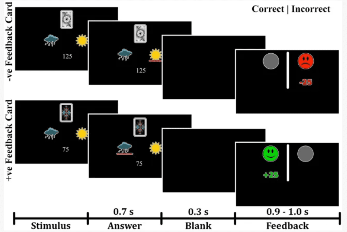
Figure 1

Experimental paradigm of the probabilistic task. The task paradigm for negative-feedback stimuli is illustrated in the upper portion. Participants received negative feedback when they provided an incorrect answer and no feedback when they gave a correct answer. The lower portion depicts the paradigm for positive-feedback stimuli, where participants received positive feedback for correct answers and no feedback for incorrect answers.

Table 1
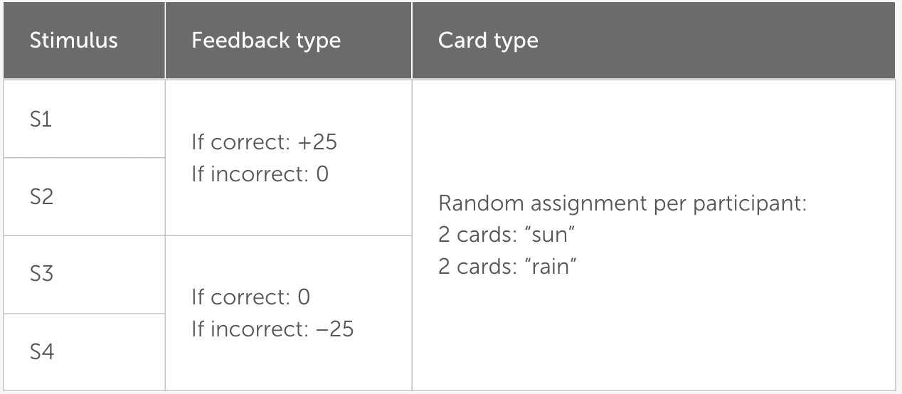
Table 1

Structure of the probabilistic classification task. Two stimuli give positive or no feedback, and the other two give negative or no feedback. The assignment of “sun” and “rain” to the four stimuli is done at random and is fixed per participant.

The task consisted of three blocks, each containing 160 trials, and the participants needed an average duration of 24.7 ± 3.8 min to finish the task. Stimulus presentation was performed using the Python package PsychoPy 2.0 (Peirce et al., 2019). The visual stimuli were presented on an MEG-compatible screen using a Barco FL35 WUXGA projector with a resolution of 1980 × 1,200 pixels and a frequency of 60 Hz.

2.2. Participants

Seventy-nine participants between the ages of 21 and 43 were recruited using flyers, social media, and snowball sampling. All participants were interviewed by a trained researcher who administered the mini-international neuropsychiatric interview (MINI; Sheehan et al., 1998). Using the MINI, the presence of trauma was determined, and participants were divided into two groups, the trauma-exposed group (Trauma) and the no-trauma-exposed control group (Control). The study was conducted according to the criteria of the Declaration of Helsinki and was approved by the ethics committee of RWTH Aachen University Hospital, Germany. Following an explanation of the procedure, written informed consent was collected from every participant at the beginning of the session.

For inclusion in the Trauma group, participants were required to have had a traumatic event (criterion A) as defined by the MINI. The rest were assigned to the Control group. Using the MINI, comorbid psychiatric conditions were determined. The presence of psychiatric disorders, as defined by the MINI, was only accepted in the Trauma group. From the sample taken, 14 participants in the Trauma group met the criteria of either major depressive disorder, dysthymia, suicidality, social phobia, obsessive-compulsive disorder, or generalized anxiety disorder. The inclusion criteria for the Control group constituted the absence of any traumatic event and the absence of any psychiatric disorders at the time of testing, which was confirmed using the MINI. The exclusion criteria for all groups were the current use of psychotropic drugs, left-handedness, inability to understand the computer-based task, and major neurological or medical illnesses, including endocrine disorders. After the exclusion of three participants, the sample included 76 participants: 56 in the Trauma group and 20 in the Control group. Participant demographics are presented in Table 2.

Table 2
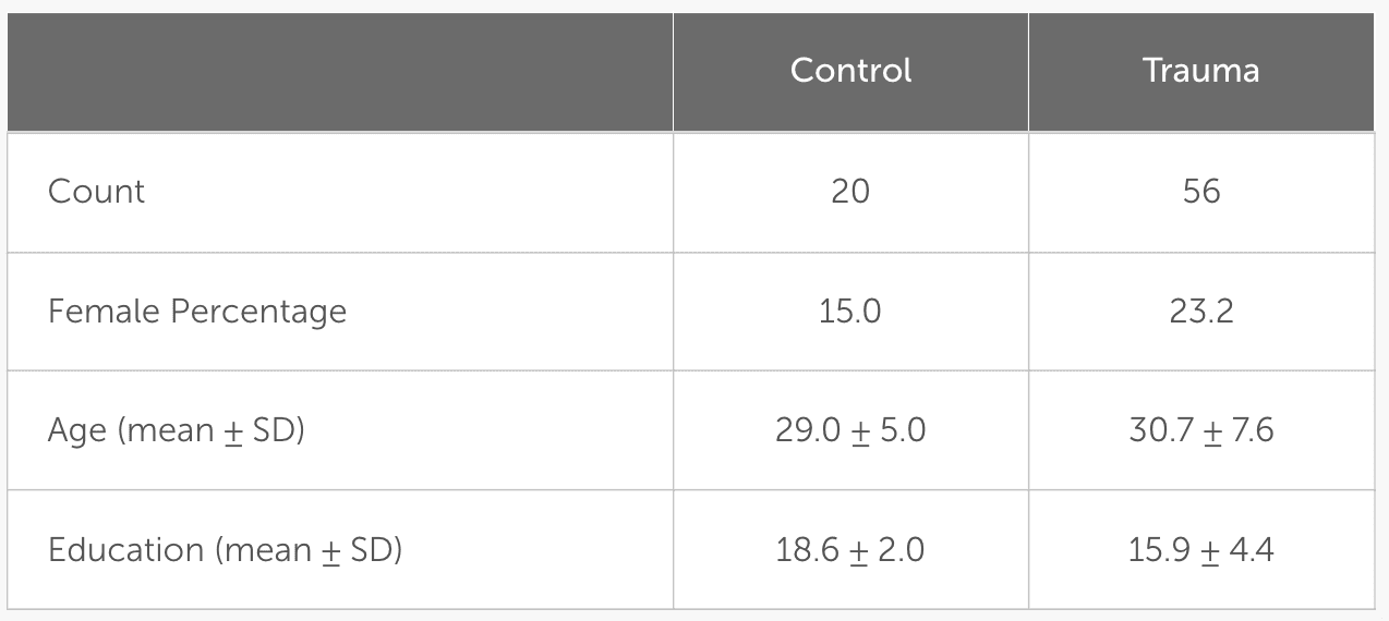
Table 2

Demographics of the tested sample. Age and education are shown as mean ± standard deviation (SD).

To improve data accuracy and reduce noise, any trials with high signal amplitudes (above 5 picotesla) were excluded from the MEG analysis. As a result, one participant was excluded from the positive-feedback analysis, and two participants were excluded from the negative-feedback analysis. The final number of participants included in each of the analyses is shown in Table 3.

Table 3
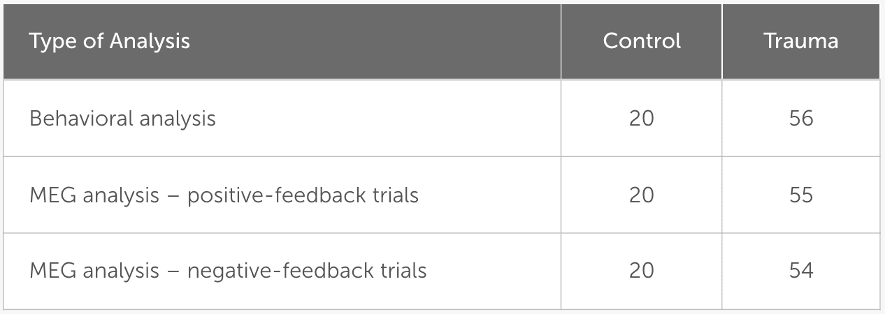
Table 3

Number of participants included in each of the analyses.

2.3. Behavioral analysis

In order to investigate whether trauma affects feedback processing and perception, learning accuracy and reaction time was analyzed for both positive and negative feedback. In order to avoid the multiple comparison problem, a one-way Multivariate analysis of variance (MANOVA) was initially performed with positive feedback accuracy, negative feedback accuracy, positive feedback reaction time, and negative feedback reaction time as dependent variables; and Group as the independent variable.

Due to time constraints for some participants and the tight schedule for testing, not all participants completed all 480 trials. Sixty-nine participants completed all 480 trials, one participant completed 416 trials, five participants completed 320 trials, and one participant only completed 160 trials. To compensate for the difference in the number of trials completed, behavioral analysis was performed once on the full data set for each participant and once on the first 160 trials only.

In order to obtain greater insight into the effect of trauma on feedback-based learning, the percentages of “learners” and “non-learners” were calculated for both positive- and negative-feedback trials. “Learners” of a particular feedback were defined as those who had more than 65% correct responses (Myers et al., 2013) for either of the feedback cards. The percentage of learners between the two groups was compared using the chi-square test.

2.4. MEG data acquisition

MEG data were collected using the Magnes-3600WH MEG system from 4d-Neuroimaging (San Diego, United States of America). Brain activity was measured with a sampling rate of 1017.25 Hz using 248 magnetometers. Additionally, cardiac and ocular activity were collected using the BrainAmp ExG MR amplifier (Brain Products, Gilching, Germany) with a sampling frequency of 5,000 Hz. This signal was down-sampled to 1017.25 Hz and merged with the MEG signal into a single recording. Electrooculography (EOG) signals were collected by attaching two electrodes lateral to the eyes and two electrodes above and below one of the eyes to record eye movements and eye blinks, respectively. Electrocarduigraphy (ECG) signals were collected by attaching an electrode at the middle of the right clavicle, one electrode at the left side below the apex of the heart, and a third electrode at the left leg. Finally, three head location coils were attached to the head to measure its relative position in space before and after each experiment.

2.5. MEG-MRI co-registration

To obtain anatomical information about the head and brain, MR measurements were taken from all participants at either a 3 T PRISMA scanner (Siemens, Erlangen, Germany) using the MPRAGE sequence (Mugler and Brookeman, 1990) or at a 7 T MAGNETOM Terra scanner (Siemens Healthineers, Erlangen, Germany) using the MP2RAGE sequence. As the participants of the Trauma group were also included in another study that used the 7 T MR system, images obtained at 7 T were used for this group to avoid further inconvenience. A sanity check was manually performed for each MEG file after the co-registration step to ensure that the use of the two MR systems did not affect the source localization of the signal. This was conducted by recording the mean error of co-registration and ensuring it was less than 3 mm.

MEG brain activity was aligned with the structural information by means of source (i.e., brain) space construction utilizing the FreeSurfer package (Dale et al., 1999; Fischl et al., 1999). For source localization, dynamic statistical parametric mapping (dSPM) was applied with a depth weighting of 0.8 (Dale et al., 2000). The individual source estimates were then projected onto the average template brain, as provided by FreeSurfer, using 8,196 vertices with an average distance between vertices of about 5 mm. Finally, the source activity was divided into anatomical regions based on the areas defined by the Desikan-Killiany atlas (Desikan et al., 2006). Co-registration from the MEG to the MRI coordinate space and the solving of the forward and inverse problems was performed using the MNE-Python library (Gramfort et al., 2014).

Three participants did not have an MR scan or had a corrupt file, so the average MR brain shape provided by Freesurfer (fsaverage template brain) was used instead. To ensure the quality of co-registration for all participants, including the three participants with missing files, the mean error of co-registration was recorded to be less than 3 mm using mne-coreg interface (Gramfort et al., 2013).

2.6. Preprocessing the MEG signal

Strong artifacts in MEG channels were identified using an in-house algorithm based on density-based spatial clustering of applications with noise (DBSCAN), as implemented in scikit- learn (Ester et al., 1996; Pedregosa et al., 2011). This was followed by visual inspection to identify noisy channels missed by this function. The signal of the identified “bad” channels was replaced by an interpolated signal from surrounding channels (Perrin et al., 1989). Environmental noise and powerline noise was removed by subtracting individually weighted reference channels from each of the MEG channels (Robinson, 1989).

Biological noise (i.e., ocular and cardiac signals) was removed using independent component analysis (ICA; Hyvärinen and Oja, 2000). The signal was split into segments with an average of 155.1 s per segment, then bandpass filtered (1–45 Hz) to improve the quality of the ICA decomposition (Winkler et al., 2015). Ocular activity was detected by finding components with a Pearson’s correlation of 0.3 or more with the EOG channel, while cardiac activity was detected using cross-trial phase statistics (CTPS; Dammers et al., 2008). A visual inspection of the results was finally performed to ensure all EOG and ECG signals were removed. The components were then projected back to the data. The average number of removed components was found to be 7.0 for all participants. Dividing the groups into Trauma and Control, the average number of removed components for the Trauma group is 7.1, while the average for the Control group is 6.5, with no significant difference between the two groups (p-value = 0.453).

2.7. Creating epochs

In order to analyze the data in the spatial, temporal and spectral domains, epochs were extracted around the event of interest (i.e., positive or negative feedback) starting 250 ms prior to the event and extending to 600 ms after the event, with the time 0 representing the feedback onset. The interval − 250 – 0 ms was chosen as a baseline. This means that, for each of the analyses, the mean of the signal in the baseline interval was subtracted from the entire signal of the epoch. The signal was then divided by the standard deviation of the baseline signal.

To ensure comparable signal-to-noise ratios between the two groups, the number of trials was equalized between the two groups by removing trials from the participants with larger trial counts until the density distribution of trials was equal. This was done by dividing the data into 20 bins, each containing one participant from the Control group and 2–3 participants from the Trauma group. Within each bin, the number of trials was equalized to match the participant with the lowest number of trials. For positive-feedback trials, the average number of trials became similar to those in the trauma group (Control: min = 24, max = 215, mean = 155; Trauma: min = 24, max = 215, mean = 154). The case was also similar for negative-feedback trials (Control: min = 15, max = 65 mean = 35 for both groups). The results of the density matching are illustrated in Figure 2.

Figure 2
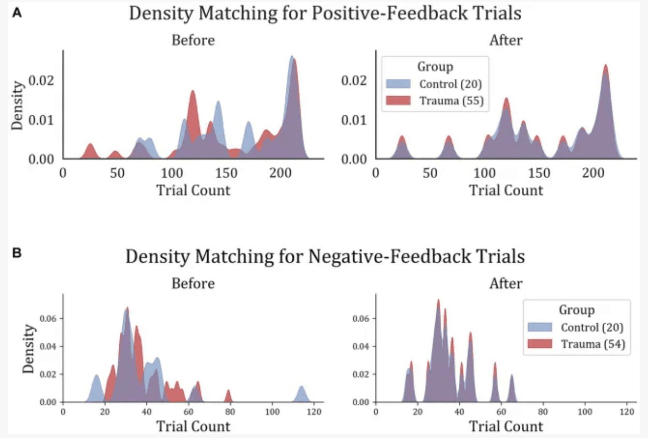
Figure 2

Trial density matching for (A) positive-feedback and (B) negative-feedback trials. Trauma and Control groups were matched in trials in order to remove the bias that might result from the difference in trial count per group.

2.8. Regions of interest

Regions of interest were defined as regions that showed significantly-different activity in space and time between the two groups (Control vs. Trauma) in positive and negative-feedback trials separately. The Monte-Carlo-based non-parametric spatio-temporal cluster permutation test (SCPT) was performed to extract differences between the two groups (Maris and Oostenveld, 2007). Since SCPT only performs a single statistical test on the entire data set, the multiple comparison problem does not apply. This means that differences across conditions directly reflect the significance level (Maris and Oostenveld, 2007).

SCPT was performed using a two-sample permutation t-test with 104 permutations. In order to choose the cluster threshold, the difference in trial numbers between the two conditions was taken into account. As the number of trials was much smaller for negative-feedback trials (35 vs. 154), the significant clusters were likely to have a shorter duration of activity. Therefore, the cluster threshold for positive-feedback trials was chosen to be equivalent to a t-value at an alpha level of 0.05 (corresponding to a threshold value of 1.99), while for negative-feedback trials, it was chosen to be equivalent to a t-value at an alpha level of 0.001 (corresponding to a threshold value of 3.43). The choice of cluster threshold does not affect the false alarm rate, but it does affect the sensitivity of the test (Maris and Oostenveld, 2007). Finally, the significance threshold for both types of comparisons was set to 0.05.

To ensure that the resulting areas did not include outliers in space or time, clusters with 15 vertices or less in the template brain (corresponding to an active cortical area of about 2.29 cm2) were excluded. Similarly, temporally short clusters with a duration of less than 20 ms were discarded from the analysis. Finally, vertices within the medial wall of the brain were also excluded from the analysis. As previously stated, this test was conducted for both positive-feedback and negative-feedback trials separately. The resulting regions of interest were used to further conduct the time-course analysis and the spectro-temporal analysis.

2.9. Time-course analysis

In order to investigate possible changes in the temporal dynamics of feedback processing after trauma, positive feedback-related activity for Control vs. Trauma was compared on positive-feedback trials. For this comparison, a representative source time course (rSTC) was computed for each of the ROIs identified in the previous step using the mean activation of all “significant” vertices within the respective ROI. These representative time courses represent the average of epochs and vertices for a given ROI. Afterwards, all rSTCs were z-scored using the mean and standard deviation from the pre-stimulus interval.

To identify differences in the temporal dynamics between the groups, a cluster permutation test was applied in a similar way to the SCPT described above, but only in the temporal domain, so as to investigate differences between the ROI activation time courses (Maris and Oostenveld, 2007). Cluster permutation tests were applied using a paired t-test with a clustering threshold using a critical alpha level of 0.05, which corresponds to a t-threshold of 1.99, and a significance level of p < 0.05. The number of permutations used was set to 104 permutations.

To study how the activity in brain regions affect learning, the two groups are divided into “learners,” with more than 65% accuracy, and “non-learners.” The signal within the significant time intervals (time intervals of interest; TOIs) of each rSTC was extracted for each subject to be compared between the two groups. This was conducted separately for Trauma and Control groups to control for trauma-related effects.

2.10. Spectro-temporal analysis

In addition to the statistical analysis in the temporal domain, differences between groups were also investigated in the time-frequency domain using a spectro-temporal cluster permutation test based on the previously identified ROIs. This test was conducted on the same principles as used in the SCPT, with the only change that clusters are built across time and frequency (Maris and Oostenveld, 2007). For this analysis, power spectral density (PSD) was computed following positive and negative feedback was compared between the two groups for each region of interest using spectro-temporal cluster permutation test.

The complex Morlet wavelet transform was applied using a frequency range from 4 to 45 Hz with a spectral resolution of 1 Hz. The number of cycles for each frequency (f) was set to f/3. The results were z-scored using the mean and standard deviation of the 250 ms preceding feedback. For both types of comparisons (positive feedback and negative feedback), a paired t-test was used to identify clusters, with a clustering threshold using a critical alpha level of 0.05, corresponding to a t-threshold of 2.0, and a significance level of p < 0.05. The number of permutations used was set to 104 permutations.

Similar to the previous section, in order to study how time-frequency clusters affect learning, the “learners” and “non-learners” in our sample were compared on the average activity within each significant cluster (time-frequency cluster of interest; TFOI). This was done by extracting the PSD within the significant time-frequency clusters for each participant and averaging it over time and frequency for each group (i.e., “learners” vs. “non-learners”). This was performed separately for Trauma and Control groups to understand the signal’s contribution to learning accuracy in time and frequency.

3. Results

3.1. Behavioral analysis

In order to test whether trauma affects feedback-based learning, the two groups (Control vs. Trauma) were compared in terms of their learning accuracy and reaction time. This analysis was done on the complete data set from all subjects. First, MANOVA was used to test the effect of group (Trauma vs. Control) on positive feedback accuracy, negative feedback accuracy, positive feedback reaction time, and negative feedback reaction time. Using Wilks’ lambda, the results showed no significant effect of group on the dependent variables (F(4,71) = 0.8567, p = 0.4943), leading to the conclusion that there is no difference between the Trauma and Control groups in terms of accuracy or reaction time for positive or negative feedback types. Secondly, to avoid any ceiling effect due to the seven participants who completed less than 480 trials (see Methods section), the analysis was repeated using only the first 160 trials, and the results were found to be similar, with no significant effect for the group on any of the dependent variables (results not shown).

To confirm the findings, the ratio of learners to non-learners was compared in the two groups of comparison. In the tested sample, all participants, except for one, learnt the negative feedback cards more than with more than 65%. Thus, negative feedback is excluded from this analysis. As for positive feedback trials, no significant difference in the learners’ proportions was found between the two groups (χ2(1) = 0.0019, p-value = 0.9651). This further supports the notion that feedback-based learning is not affected in trauma survivors. Behavioral results are shown in Figure 3.

Figure 3
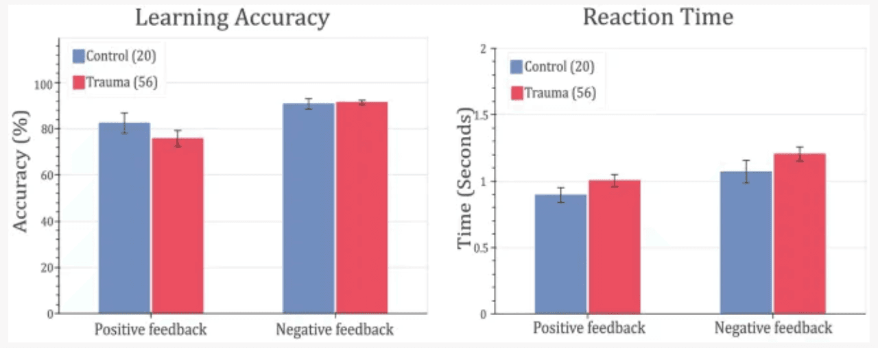
Figure 3

Feedback-learning accuracy and response time. Left: learning accuracy (left) and response time (right) for positive- and negative-feedback trials for Control vs. Trauma groups. Error bars represent the standard error of the mean.

To ensure the absence of confounding effects from other variables in subsequent analyses, the “learners” and “non-learners” groups were compared in terms of gender, age, education level and the 14 modules of the MINI interview. These include major depressive disorder, dysthymia, suicidality, mania, panic attacks, agoraphobia, social phobia, obsessive compulsive disorder, alcoholism, substance use, psychosis, anorexia nervosa, bulimia nervosa, and anxiety. There was no significant difference between the two groups in terms of the proportions of males and females (χ2 = 0.983, p-value = 0.3215), no significant difference in age (learners = 29.23 ± 6.44, non-learners = 32.23 ± 7.89, t = −1.6703, p-value = 0.0993) and years of education (learners = 17.05 ± 4.02, non-learners = 15.62 ± 4.47, t = 1.2895, p-value = 0.2018). As for the MINI modules, there were no significant differences between the two groups in any of the modules (smallest p-value = 0.2433).

3.2. ROI analysis

For positive feedback, results from the spatio-temporal cluster permutation test (SCPT) revealed two main clusters that showed significant differences between the two groups in space and time (Figure 4). The first cluster covers the right insula and part of the right supramarginal area as well as a small portion of the lateral part of the lateral orbitofrontal cortex. The activity in this cluster was higher for the Control group (p-value = 0.0024). The second cluster covered a large portion of the lateral orbitofrontal cortex as well as the medial orbitofrontal cortex. The activity in this cluster was higher for the Trauma group (p-value = 0.0294). By projecting the clusters to brain regions defined by the Desikan-Killiany atlas and eliminating small regions with areas equal to or smaller than 2.29 cm2 and regions with short activity duration, four regions were assigned to be the positive-feedback ROIs: supramarginal, lateral orbitofrontal cortex (lOFC) and medial orbitofrontal cortex (mOFC). One cluster was identified in the medial portion of the superior frontal gyrus for negative feedback. This cluster had higher activity in the Trauma group (p-value of 0.022). The identified region is considered to be the negative-feedback ROI.

Figure 4
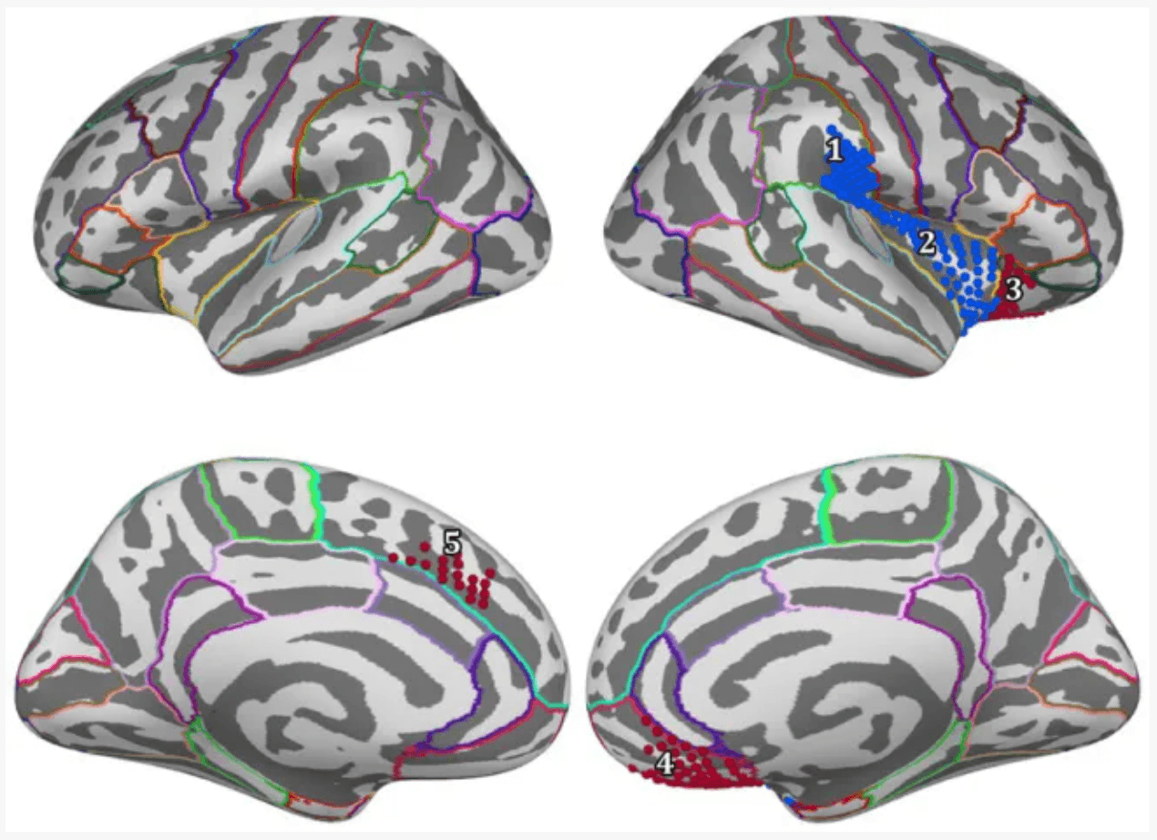
Figure 4

Regions of interest are areas that exhibit a significant difference between the Control group and the Trauma group during positive or negative-feedback trials. Vertices in red indicate higher activity in the Trauma group, while the blue color indicates higher activity in the Control group. Regions #1–4 pertain to positive-feedback clusters, while region #5 pertains to the negative-feedback cluster.

Table 4 details the location of each region and the percentage of the covered area according to the Desikan-Killiany atlas.

Table 4
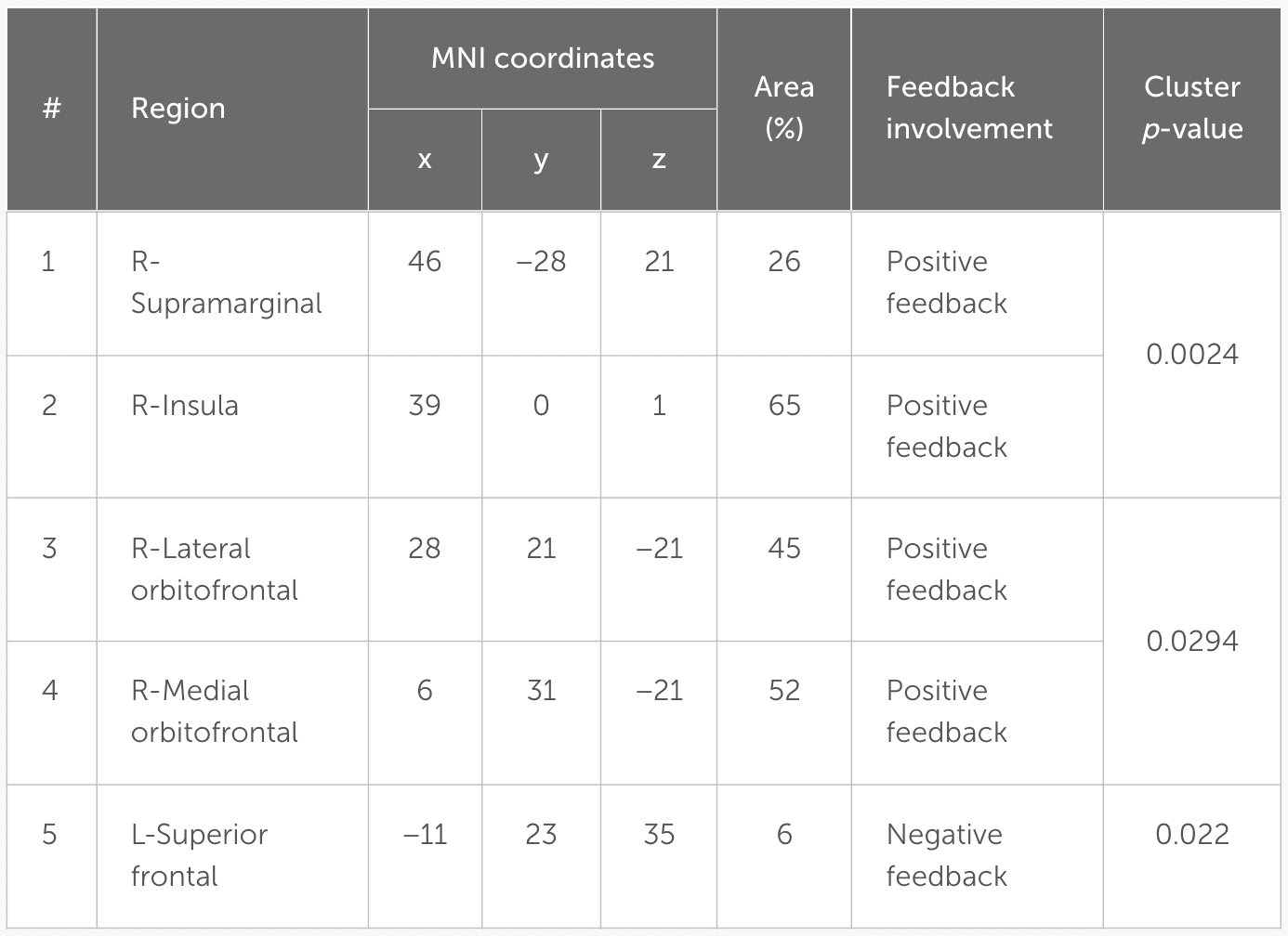
Table 4

Regions of interest. The MNI coordinates represent the center of mass for each label. Region (%) refers to the percentage of the active part of the label compared to the entire label.

3.3. Temporal dynamics of feedback-related activity

In order to gain more insight into the temporal aspect of the differences in signal processing, representative time courses were extracted from the five ROIs and compared between the two groups. The cluster-permutation test in the temporal domain revealed significant clusters in all five ROIs. For positive-feedback trials, the differences span the time interval 160–600 ms after feedback onset. The midway time point for positive-feedback trials was 390 ms (SD = 35 ms). The results of the temporal analysis showed higher activity for the Control group in the insula and the supramarginal cortices, while the Trauma group exhibited higher activity in the lOFC, mOFC, and the medial portion of the superior frontal cortex. This direction of differences is similar to that found using SCPT in the previous section, which confirms the findings reported above. Differences in the temporal dynamics between the two groups and the five brain areas are depicted in Figure 5. The results are also summarized in Table 5.

Figure 5
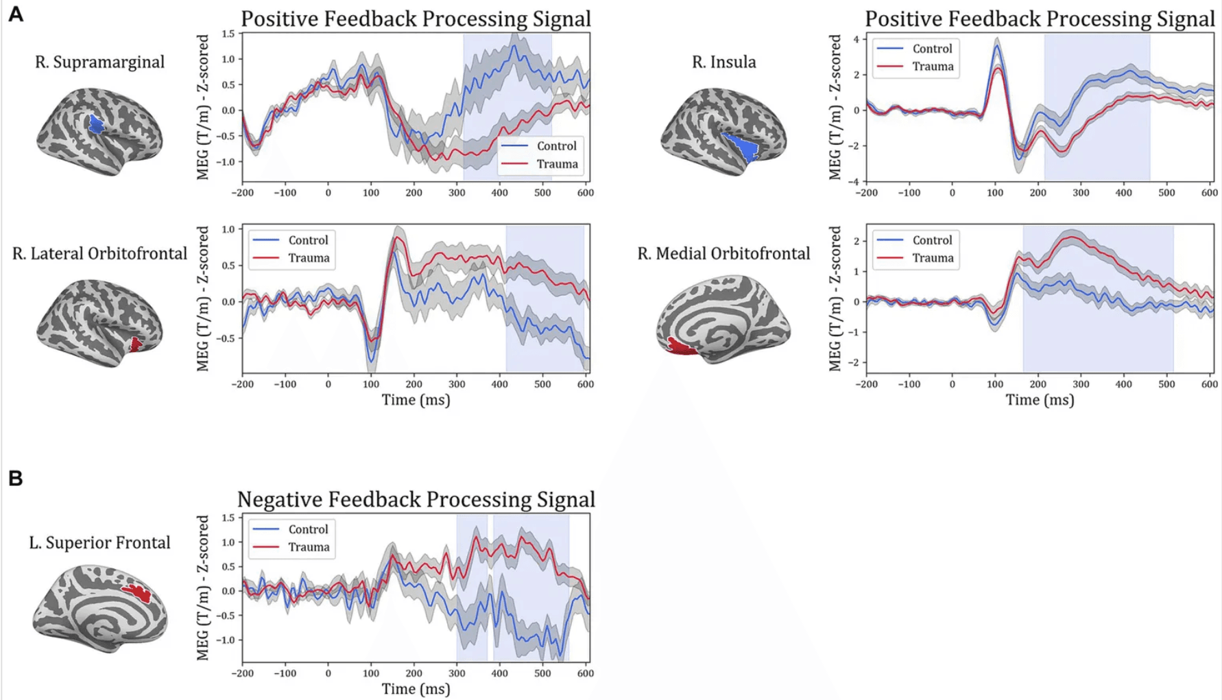
Figure 5

Temporal course of feedback processing of Trauma vs. Control groups. The figure illustrates brain activity in regions that showed significant differences in the time domain for (A) positive-feedback trials and (B) negative-feedback trials. The light blue shaded area represents the time interval with a significant difference. The gray shaded area around the timeseries represents the standard error of the mean.

To understand the potential correlations between brain activity within the selected ROIs and learning from positive or negative feedback, the MEG activity within each of the identified time intervals (times of interest; TOIs) was extracted and was used to compare “learners” and “non-learners” in the Trauma and Control groups separately. For the Trauma group, when comparing “learners” and “non-learners,” the results indicate significant differenes in lateral orbitofrontal cortex in the time period 415–600 ms (t(53) = −2.664, mean learners = 0.08, mean non-learners = 0.827, p-value = 0.01) and the supramarginal cortex in the time period 315–525 ms (t(53) = 2.559, mean learners = −0.077, mean non-learners = −1.102, p-value = 0.013). Taking the absolute values, we find that the “learners” group have lower activity in both regions compared to the “non-learners” group. No significant differences were found for any of the regions in the Control group (p-values >0.18). The results are shown in Figure 6.

Figure 6
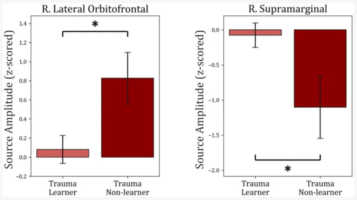
Figure 6

Comparison between “learners” and “non-learners” subgroups of the Trauma group. The figure shows the average source amplitude for the significant time periods found in Figure 5 for both “learners” and “non-learners.” For the supramarginal region, the time period tested was 315–525 ms, and for the lOFC, the period tested was 415–600 ms. The comparison shows significant differences in lOFC and supramarginal cortex. No significant differences were found between “learners” and “non-learners” of the Control group. * p < 0.05.

3.4. Time-frequency analysis

In order to investigate differences between subject groups in the time-frequency domain, the five regions of interest were included to test for differences between Trauma and Control groups for both positive and negative-feedback ROIs. Cluster permutation tests done in the spectro-temporal domain for positive-feedback trials revealed one significant cluster in the lateral temporal lobe that covers the theta band while spanning the interval of 185–555 ms following feedback presentation. Running the analysis on the negative-feedback ROIs did not yield any significant cluster. An illustration of the spectro-temporal test can be found in Figure 7.

Figure 7
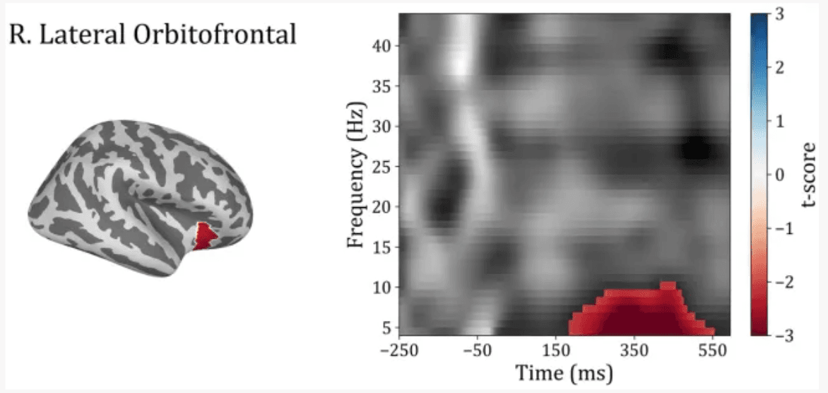
Figure 7

Time-frequency representations of Controls vs. Trauma group comparison. The cluster permutation test revealed a significant cluster (highlighted in red) with higher power in theta (4–8 Hz) and alpha (8–10) bands in the Trauma group. The cluster was found in the right lateral orbitofrontal region in the time window between approximately 185–555 ms where time zero represents the feedback presentation time.

The average power within identified TFOIs was compared with positive-feedback scores in for “learners” and “non-learners” in both groups. Trauma group showed a significant difference between “learners” (36) and “non-learners” (19) (t(53) = −3.953, mean learners = 9.964, mean non-learners = 14.16, p-value <0.001). No significant difference was found in the Control group (p-value = 0.353). The results are shown in Figure 8.

Figure 8
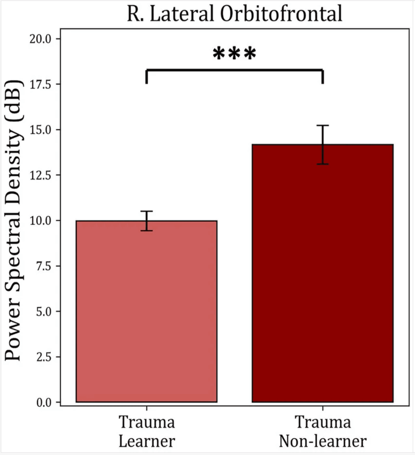
Figure 8

Average power comparison between “learners” and “non-learners” subgroups of the Trauma group. The figure shows the average PSD within the significant time-frequency cluster found in Figure 7 for both the “learners” and “non-learners” groups. The time period tested was 185–555 ms in the frequency range (4–10 Hz). The results show a significantly higher power in the “non-learner” group. No significant differences were found for the healthy controls. *** p < 0.001.

4. Discussion

The aim of this study was to understand the effect of trauma on the ability to learn from feedback by utilizing MEG to compare brain activity in individuals with a history of trauma with a control group. No significant differences were found in behavioral performance measures, such as accuracy or reaction time, between the two groups. However, by analyzing the brain activity related to feedback processing, significant differences were discovered in the spatial, temporal, and frequency domains. Specifically, when compared to the control group, individuals with a history of trauma displayed increased brain activity in regions such as the mOFC and lOFC and decreased activity in the supramarginal and insular cortices during positive feedback presentation. For negative feedback, the Trauma group exhibited increased activity in the medial part of the superior frontal cortex. These differences occurred relatively late after the presentation of feedback and were characterized by activity within the theta and alpha frequency ranges.

To study the cognitive effects of trauma, learning accuracy was compared between the two groups. The results showed that both groups learned similarly from both positive and negative feedback and exhibited similar reaction times, leading to the conclusion that that response to feedback is not affected in the trauma-exposed group at the crude behavioral level (see Figure 3). Additionally, dividing the sample into learners and non-learners showed no significant difference between the two groups in the positive-feedback trials. On the other hand, almost all participants learnt negative-feedback trials with more than 65% accuracy. This is can be simply attributed to the design of the task, where negative-feedback trials are associated with negative feedback when answered incorrectly, while positive-feedback trials are associated with no feedback. Having no feedback can be also interpreted as absence of negative feedback in our study design, which makes positive-feedback trials more prone to mistakes in learning. This result was the same for both Trauma and Control groups.

These results are consistent with previous studies showing comparable learning and reaction times between trauma-exposed groups and controls (Vythilingam et al., 2009). Other studies have also shown equal learning between a PTSD group and a group of controls (Levy-Gigi et al., 2012; Boukezzi et al., 2020), which suggests that even when the effect of trauma is strong enough to cross the severity threshold for PTSD, it does not seem to affect learning from feedback significantly. Thus, one can conclude that traumatic experiences do not affect the behavioral processing of feedback associated with a stimulus. However, response to feedback may be affected by the trauma at the neural level.

To determine which brain areas respond differently to positive and negative feedback, brain activity was measured in both the Trauma and Control groups during feedback processing using MEG. SCPT was used to compare activity between the two groups while controlling for multiple comparisons. The analysis focused separately on trials when participants received positive or negative feedback. The results showed that there were four regions exhibiting distinct activity between the groups during positive-feedback trials, namely the right supramarginal gyrus, the insula, mOFC, and lOFC. However, during negative-feedback trials, only the medial part of the superior frontal cortex showed a difference in activity.

Previous reports have shown that the aforementioned ROIs are also involved in positive feedback-related processes (see Figure 4 and Table 4). For example, the mOFC and lOFC are involved in forming associations between the stimulus and the feedback (Rolls, 2004) and encoding values to stimuli (Padoa-Schioppa and Assad, 2006; Purves et al., 2018), which is exhibited as higher activity when receiving positive feedback (Sescousse et al., 2013; Oldham et al., 2018; Nguyen et al., 2021). These areas receive input from various sensory regions that provide information about previous experiences to improve value estimation (Radcliffe Hospital et al., 2005; Purves et al., 2018). The insula is more generally activated when positive feedback is received (Wittmann et al., 2010) and is usually involved in the affective processing of positive stimuli and feedback (Sescousse et al., 2013; Nguyen et al., 2021). As part of a larger network, the insula sends information to the OFC to create a representation of the hedonic valence of stimuli (Craig, 2002; Purves et al., 2018; Nguyen et al., 2021). In turn, the supramarginal gyrus connects with the OFC, especially the lOFC (Du et al., 2020), and potentially modulates learning by increasing activity during the retrieval of information acquired through physical enactment (Russ et al., 2003). Accordingly, the supramarginal gyrus may be involved in the process of integrating new information with previous knowledge for future recall. In sum, the processing of positive feedback is a multidimensional process that engages a myriad of neural systems, and our results shed new light on the spatial facets of processing positive feedback with psychological trauma.

Conversely, in terms of the negative-feedback trials, the medial portion of the superior frontal cortex showed increased activity. In general, as a part of the brain’s performance-monitoring system, the superior frontal cortex is activated in response to errors in judgment and helps in avoiding further errors by influencing activity in other relevant brain areas and reducing distracting information (Danielmeier et al., 2011). In essence, one could argue that the increased activity seen in the superior frontal cortex might be the basis of the increased avoidance of negative encounters observed after exposure to trauma. However, in order for this to be proven, an examination of variability in the expression of avoidance symptoms in the Trauma group would be necessary.

The involvement of the insula and orbitofrontal cortices is also supported by the current neuroanatomical theory of PTSD, which indicates that trauma causes hypoactivity in the orbitofrontal cortex and medial prefrontal cortex in response to affective stimuli (Giustino and Maren, 2015; Stark et al., 2015). This could decrease the top-down regulation of amygdala activity and exacerbate symptoms of hyperarousal (Giustino and Maren, 2015; Stark et al., 2015). On the other hand, it has been reported that people who have experienced trauma display an increase in insular activity in response to emotional stimuli (Stein et al., 2007) and that both the insula and medial prefrontal cortex show a decrease in gray matter size following psychological trauma, even in the absence of PTSD (Ganzel et al., 2008).

Interestingly, in the current study, the Trauma group showed decreased activity in the insula but higher activity in the prefrontal, mOFC, and lOFC in response to positive feedback, which may initially seem counterintuitive. However, given that the sample consisted of trauma survivors with no or minimal PTSD symptoms at the time of testing, these results might reflect higher-than-average levels of appraisal of positive stimuli, which might be associated with a better response to trauma. This is further supported by previous studies, which found an increased valuation of positive stimuli in trauma survivors when compared to individuals with PTSD (Myers et al., 2013). This evidence suggests that trauma can cause long-lasting changes to cognition, including more positive feedback reappraisal, which might counter the effects of fear-related and avoidance symptoms. These differences are found at the neural activity level but do not seem to be prominent enough to affect crude measures of task performance at the behavioral level.

In order to further understand the differences in brain dynamics caused by trauma, activity in the regions of interest in the time domain were additionally analyzed (see Figure 5). Changes in processing positive feedback were evident between 165 and 600 ms, which covers two event-related potentials: the p300, and the late positive potential. The p300 is an event-related potential that is seen around 300 ms following feedback reported in EEG studies (Bernat et al., 2015) and usually correlates to secondary aspects of positive feedback, such as those requiring evaluation and comparison (Yeung and Sanfey, 2004; Bernat et al., 2011, 2015). Conversely, the late positive potential starts at 300 ms, which is usually sustained until 2000 ms following positive feedback (Dennis and Hajcak, 2009), is linked to the selective processing of emotional stimuli and activation of emotional systems in response to positive stimuli (Cuthbert and Kozak, 2013). Thus, it is possible to conclude that the differences between the two groups are due to differences in the processing of secondary aspects of positive feedback as well as the emotional processing of positive stimuli. However, since our study design does not directly correlate the processing of secondary emotional aspects or emotional processing with the activity difference between the two groups, we suggest replicating our findings using experiments designed differently before definitively concluding such a correlation.

Analyzing the signal in the frequency domain provided more insight into feedback processing. Feedback processing consists of multiple components, each of which runs in a particular frequency band (Bernat et al., 2015). Using permutation-based clustering tests for the temporo-spectral domain, a cluster of differences was identified in the right lOFC (Figure 7). This cluster of activity spans the time interval of roughly 185–555 ms in the theta frequency band. Although the time interval should not be taken literally due to the nature of the permutation-based clustering test (Sassenhagen and Draschkow, 2019; Meyer et al., 2021), it should at least give an idea about the timing and frequency in which the significant differences were found.

Finally, the activity within each of the identified TOIs and TFOIs was compared between the “learners” and “non-learners” subgroups separately for the Trauma group and the Control group (Figure 6, Figure 8). This comparison was performed to determine whether the difference in learning is reflected in the activity within the TOIs and TFOIs in question. The results show that the activity within the TOIs was different between the two groups in the supramarginal (lower in the non-learners) and the lOFC (higher in the non-learners) regions. Similarly, the average power was significantly higher in the TFOI of the lOFC in the non-learner group. This result can be explained by one of two hypotheses. First, it is possible that the activity within these two regions is correlated with learning from positive feedback. Specifically, the activity in the lOFC region may have a negative impact on learning from positive feedback, while the activity within the supramarginal cortex region may have a positive impact on learning in the trauma group. Meanwhile, this effect is not noticeable on the behavioral level. Secondly, when examining the absolute values, the “learners” group have lower activity in the supramarginal and lOFC TOIs, as well as in the lOFC TFOI. This suggests that they have more attenuated activity in both regions. This can be due to the habituation effect, which refers to a decrease in response to the repeated presentation of a stimulus with emotional valence (Wright et al., 2001). Previous research supports these findings, where repeated presentation of an emotional stimulus resulted in lower brain activity in certain brain regions (Wright et al., 2001). Regardless of the explanation, these results confirm the correlation between brain activity in the lOFC and the supramarginal gyrus with receiving positive feedback. Furthermore, the “non-learners” subgroup seems to be the main contributor to the effects seen between Controls and Trauma in lOFC and supramarginal regions. However, since this learning effect is missing in the “non-learners” subgroup of the Controls, this suggests that the contribution of lOFC and supramarginal regions to learning is potentially perturbed following exposure to psychological trauma.

Taken together, no evidence to show that trauma-exposed individuals differ from individuals with no history of trauma was found at the behavioral level. However, differences between the groups were found at the neural level in the space, time, and frequency domains of cortical activity. These findings provide a deeper understanding of the cognitive processes that are affected as a result of trauma and demonstrate a novel framework for studying the underlying cognitive mechanisms that contribute to psychiatric symptoms by assessing the contribution of different domains of the brain signal on the targeted behavior. To the best of the authors’ knowledge, this is the first study to combine the spatial, temporal, and spectral aspects of feedback processing in individuals with exposure to psychological trauma. It is anticipated that future work will build on these findings to focus on the potential of using cognitive and psychological constructs in assessing symptom improvement following trauma as part of individualized treatment plans.

A limitation of this study is that the impact of trauma was only assessed after exposure, which does not discount the presence of the reported differences before trauma exposure. To address this, future studies should employ a longitudinal design to identify any cognitive and neural differences that develop specifically as a result of exposure to trauma. Additionally, since the ROIs were determined using total signal and there is an inverse correlation between total signal and frequency, it is more likely that our ROIs will exhibit differences in low-frequency bands. This issue can be addressed in the future by selecting an alternative approach to identify the areas of interest.

Open Article as PDF

Abstract

The cognitive impact of psychological trauma can manifest as a range of post-traumatic stress symptoms that are often attributed to impairments in learning from positive and negative outcomes, aka reinforcement learning. Research on the impact of trauma on reinforcement learning has mainly been inconclusive. This study aimed to circumscribe the impact of psychological trauma on reinforcement learning in the context of neural response in time and frequency domains. Two groups of participants were tested - those who had experienced psychological trauma and a control group who had not - while they performed a probabilistic classification task that dissociates learning from positive and negative feedback during a magnetoencephalography (MEG) examination. While the exposure to trauma did not exhibit any effects on learning accuracy or response time for positive or negative feedback, MEG cortical activity was modulated in response to positive feedback. In particular, the medial and lateral orbitofrontal cortices (mOFC and lOFC) exhibited increased activity, while the insular and supramarginal cortices showed decreased activity during positive feedback presentation. Furthermore, when receiving negative feedback, the trauma group displayed higher activity in the medial portion of the superior frontal cortex. The timing of these activity changes occurred between 160 and 600 ms post feedback presentation. Analysis of the time-frequency domain revealed heightened activity in theta and alpha frequency bands (4–10 Hz) in the lOFC in the trauma group. Moreover, dividing the two groups according to their learning performance, the activity for the non-learner subgroup was found to be lower in lOFC and higher in the supramarginal cortex. These differences were found in the trauma group only. The results highlight the localization and neural dynamics of feedback processing that could be affected by exposure to psychological trauma. This approach and associated findings provide a novel framework for understanding the cognitive correlates of psychological trauma in relation to neural dynamics in the space, time, and frequency domains. Subsequent work will focus on the stratification of cognitive and neural correlates as a function of various symptoms of psychological trauma. Clinically, the study findings and approach open the possibility for neuromodulation interventions that synchronize cognitive and psychological constructs for individualized treatment.

Introduction

Exposure to psychological trauma can lead to four main types of symptoms: re-experiencing the event, avoiding things linked to the event, changes in thinking and mood, and increased arousal. For a diagnosis of post-traumatic stress disorder (PTSD), these symptoms must meet a certain level of severity. However, even less severe symptoms, known as subthreshold post-traumatic stress symptoms (PTSS), can be very difficult to manage. Individuals with PTSS may experience problems in social settings, difficulties with daily functioning, thoughts of suicide, and other mental health issues like anxiety and depression, compared to those who have not experienced trauma.

Traditionally, PTSS were understood as problems with how the brain learns and forgets fear. However, there has been limited research on how trauma affects learning through rewards and punishments, and the brain activity involved. Traumatic experiences are known to reduce expectations and satisfaction from positive feedback, decrease a person's ability to use positive information from their environment, and increase their sensitivity to negative experiences. Despite these observations, little is understood about the brain's signals during feedback processing after trauma, and how these signals relate to learning from both positive and negative feedback.

Trauma exposure has been shown to impact brain networks involved in processing feedback, including several areas in the brain's outer layers and deeper structures. For example, trauma has been linked to reduced activity in the medial prefrontal cortex (an area that assesses value), increased activity in the amygdala (important for fear learning), reduced connections between the anterior cingulate cortex (which evaluates choices) and the hippocampus (involved in memory), and increased connections between the insula (which integrates internal and bodily states) and other parts of the "salience network" (which identifies important stimuli). Additionally, trauma exposure has been associated with smaller sizes in the amygdala, insula, anterior cingulate cortex, and medial prefrontal cortex. While how people respond to positive and negative feedback can explain some trauma-related symptoms, it is still unclear how trauma affects the real-time processing of feedback with precise timing and location.

Studying the timing and frequency aspects of feedback processing can offer a better understanding of how trauma affects thinking. Since different components of feedback processing show consistent patterns of brain activity that depend on both time and frequency, examining these patterns can reveal more about the underlying processes. Previous studies have used electroencephalography (EEG) to investigate how trauma affects the timing of feedback processing. However, while EEG offers excellent temporal detail, its ability to pinpoint exact locations in the brain is limited compared to other methods like fMRI. To bridge this gap between temporal and spatial resolution, magnetoencephalography (MEG) is an ideal tool. To date, only a few MEG studies have looked at feedback signals in healthy individuals and compared them to EEG findings. MEG studies on psychological trauma have mostly focused on brain activity when the brain is at rest, during face-processing tasks, or during working-memory tasks. However, no MEG study has investigated feedback processing in the context of psychological trauma.

It is hypothesized that traumatic experiences affect how feedback is processed, and this will be seen as differences in brain activity in the involved regions across different time points and frequency ranges. To test this, the spatial, temporal, and frequency aspects of feedback processing are examined in relation to trauma exposure. A learning task based on feedback was given to two groups of individuals: one with a history of trauma and a control group without such history, all while undergoing an MEG scan. Since differences in brain regions involved in processing positive and negative feedback were expected between the two groups, feedback processing differences in terms of timing and frequency were further investigated. This is the first study to examine potential changes in the spatial, frequency, and temporal aspects of feedback processing after traumatic events.

Methods

Experimental Design

Participants completed a task designed to separate learning from positive and negative feedback. In each trial, one of four fractal images appeared, and participants had to guess if it predicted "sun" or "rain." The image stayed on screen until a response was made. A red line below "sun" or "rain" showed the chosen answer for 700 milliseconds, followed by a 300 ms blank screen. Feedback was then displayed for 900 to 1,000 ms: a green smiley face with "+25" for positive feedback, a red frowny face with "-25" for negative feedback, or a gray circle for no feedback. Two stimuli had a 90% chance of predicting "sun" and 10% for "rain," while the other two had the opposite probabilities. For feedback type, two stimuli were linked to positive feedback when the correct answer was chosen and no feedback for incorrect answers. The other two stimuli were linked to negative feedback for incorrect answers and no feedback for correct answers.

The task included three blocks, each with 160 trials. Participants took about 24.7 ± 3.8 minutes to finish. Stimuli were presented using the PsychoPy 2.0 software on an MEG-compatible screen with specific resolution and refresh rate.

Participants

Seventy-nine individuals between 21 and 43 years old were recruited. A trained researcher interviewed all participants using the mini-international neuropsychiatric interview (MINI) to identify trauma exposure. Participants were then placed into a trauma-exposed group (Trauma) or a no-trauma control group (Control). The study followed the Declaration of Helsinki guidelines and was approved by the RWTH Aachen University Hospital ethics committee. All participants provided written informed consent.

To be in the Trauma group, individuals needed to have experienced a traumatic event as defined by the MINI. The Control group consisted of those without trauma exposure. The MINI also identified other mental health conditions; these were only accepted in the Trauma group. Fourteen trauma participants had other conditions such as major depressive disorder, anxiety, or obsessive-compulsive disorder. The Control group required no traumatic events and no mental health disorders at the time of testing. Exclusion criteria for all participants included current use of psychiatric medications, left-handedness, inability to understand the task, and major medical or neurological illnesses. After excluding three participants, the final sample included 76 participants: 56 in the Trauma group and 20 in the Control group.

To improve data quality, trials with very strong signal amplitudes (above 5 picotesla) were removed from the MEG analysis. This led to one participant being excluded from the positive-feedback analysis and two from the negative-feedback analysis.

Behavioral Analysis

To see if trauma impacts feedback processing and perception, learning accuracy and reaction time were analyzed for both positive and negative feedback. To avoid issues with multiple comparisons, a Multivariate Analysis of Variance (MANOVA) was first performed. The dependent variables were positive and negative feedback accuracy and reaction times, with "Group" (Trauma vs. Control) as the independent variable.

Some participants did not complete all 480 trials due to time constraints. To account for this, behavioral analysis was done once using each participant's full data and again using only the first 160 trials.

To better understand trauma's effect on feedback-based learning, the percentages of "learners" and "non-learners" were calculated for both positive and negative feedback. "Learners" were defined as those with more than 65% correct responses for a given feedback type. The percentages of learners between the two groups were compared using a chi-square test.

MEG Data Acquisition

MEG data were collected using a Magnes-3600WH MEG system with 248 magnetometers, sampling at 1017.25 Hz. Cardiac and ocular (eye) activity were also recorded using a BrainAmp ExG MR amplifier, sampled at 5,000 Hz, then down-sampled to 1017.25 Hz and combined with the MEG signal. Electrodes were placed around the eyes to record eye movements (EOG) and on the chest for heart activity (ECG). Three head location coils tracked head position before and after each experiment.

MEG-MRI Co-registration

To gather anatomical details of the head and brain, MRI scans were performed on all participants using either a 3 T PRISMA scanner or a 7 T MAGNETOM Terra scanner. Images from the 7 T system were used for the Trauma group, as they were part of another study using this system. A manual check ensured that using two different MRI systems did not affect the accuracy of source localization, with a mean co-registration error of less than 3 mm.

MEG brain activity was aligned with structural MRI information using the FreeSurfer software to create a brain space. Dynamic statistical parametric mapping (dSPM) was applied for source localization. Individual source estimates were then projected onto an average brain template with 8,196 points, spaced about 5 mm apart. Finally, brain activity was divided into anatomical regions based on the Desikan-Killiany atlas. Co-registration and solving of forward and inverse problems were done using the MNE-Python library.

Three participants lacked MRI scans or had corrupted files; for these, the average brain shape from Freesurfer (fsaverage template) was used. The mean co-registration error remained below 3 mm for all participants, confirming quality.

Preprocessing the MEG Signal

Strong artifacts in MEG channels were identified using a custom algorithm based on DBSCAN, followed by visual inspection. Signals from "bad" channels were replaced by interpolation from surrounding channels. Environmental and powerline noise was removed by subtracting weighted reference channels.

Biological noise, such as eye and heart signals, was removed using independent component analysis (ICA). The signal was divided into segments, bandpass filtered (1–45 Hz) to improve ICA quality. Eye activity was detected by components with a high correlation (0.3 or more) with the EOG channel. Heart activity was detected using cross-trial phase statistics. A visual check confirmed that all EOG and ECG signals were removed. The components were then reintegrated into the data. An average of 7.0 components were removed per participant, with no significant difference between the Trauma (7.1) and Control (6.5) groups.

Creating Epochs

To analyze data in terms of location, time, and frequency, "epochs" (short segments of data) were extracted around the feedback event. Each epoch started 250 ms before the feedback and extended to 600 ms after, with 0 ms marking feedback onset. The interval from -250 ms to 0 ms was used as a baseline. For each analysis, the average signal during this baseline was subtracted from the entire epoch, and then the result was divided by the baseline's standard deviation.

To ensure similar signal-to-noise ratios between the groups, the number of trials was balanced. This involved removing trials from participants with higher counts until the distribution of trials was equal. For positive-feedback trials, the average number of trials became similar in both groups (Control: mean = 155; Trauma: mean = 154). The same was done for negative-feedback trials (Control: mean = 35; Trauma: mean = 35).

Regions of Interest

Regions of interest (ROIs) were defined as areas that showed significantly different activity in space and time between the two groups (Control vs. Trauma), separately for positive and negative feedback trials. A Monte-Carlo-based non-parametric spatio-temporal cluster permutation test (SCPT) was used to identify these differences. This test performs a single statistical comparison on the entire dataset, so the problem of multiple comparisons does not apply.

SCPT was performed using a two-sample permutation t-test with 10,000 permutations. The cluster threshold was adjusted based on the number of trials; since negative-feedback trials had fewer trials (35 vs. 154), their significant clusters were expected to be shorter. Therefore, the cluster threshold for positive-feedback trials corresponded to a t-value at an alpha level of 0.05 (threshold 1.99), while for negative-feedback trials, it corresponded to an alpha level of 0.001 (threshold 3.43). The significance threshold for both comparisons was set to 0.05.

To ensure robustness, clusters with 15 or fewer vertices (about 2.29 cm2 active cortical area) and temporally short clusters (less than 20 ms duration) were excluded. Vertices within the brain's medial wall were also excluded. This test was conducted separately for positive and negative feedback trials. The identified ROIs were then used for further time-course and spectro-temporal analyses.

Time-course Analysis

To explore changes in the timing of feedback processing after trauma, activity related to positive feedback for the Control group was compared to the Trauma group. A representative source time course (rSTC) was calculated for each ROI identified in the previous step, using the average activation of all significant points within that ROI. These time courses represent the average across epochs and points for a given ROI. All rSTCs were then standardized using the mean and standard deviation from the pre-stimulus interval.

To find differences in timing between the groups, a cluster permutation test was applied. This was similar to the SCPT but focused only on the temporal domain, comparing the activation time courses of the ROIs. Cluster permutation tests used a paired t-test with a clustering threshold at an alpha level of 0.05 (t-threshold 1.99) and a significance level of p < 0.05, with 10,000 permutations.

To understand how brain activity in these regions affects learning, the two groups were divided into "learners" (over 65% accuracy) and "non-learners." The MEG signal within the significant time intervals (times of interest; TOIs) of each rSTC was extracted for each person. These were then compared between "learners" and "non-learners" within the Trauma and Control groups separately, to isolate trauma-related effects.

Spectro-temporal Analysis

In addition to timing, differences between groups were also investigated in the time-frequency domain using a spectro-temporal cluster permutation test, based on the previously identified ROIs. This test followed the same principles as the SCPT, but clusters were formed across both time and frequency. For this analysis, the power spectral density (PSD) after positive and negative feedback was compared between the two groups for each ROI using the spectro-temporal cluster permutation test.

A complex Morlet wavelet transform was applied across a frequency range of 4 to 45 Hz with a 1 Hz resolution. The number of cycles for each frequency was set to f/3. The results were standardized using the mean and standard deviation of the 250 ms before feedback. For both positive and negative feedback comparisons, a paired t-test was used to identify clusters, with a clustering threshold at an alpha level of 0.05 (t-threshold 2.0) and a significance level of p < 0.05, using 10,000 permutations.

Similar to the time-course analysis, to study how time-frequency clusters affect learning, the "learners" and "non-learners" in the sample were compared based on the average activity within each significant cluster (time-frequency cluster of interest; TFOI). This involved extracting the PSD within the significant time-frequency clusters for each participant and averaging it over time and frequency for each subgroup (learners vs. non-learners). This was done separately for the Trauma and Control groups to understand the signal's contribution to learning accuracy in terms of time and frequency.

Results

Behavioral Analysis

To determine if trauma affects learning based on feedback, the two groups (Control vs. Trauma) were compared regarding their learning accuracy and reaction time using the complete dataset. A MANOVA initially assessed the effect of group on positive feedback accuracy, negative feedback accuracy, positive feedback reaction time, and negative feedback reaction time. The results showed no significant effect of group on these variables, indicating no differences between the Trauma and Control groups in accuracy or reaction time for either feedback type. To ensure these findings were not due to a ceiling effect from participants completing fewer trials, the analysis was repeated using only the first 160 trials, yielding similar results with no significant group effect.

To confirm these findings, the ratio of learners to non-learners was compared between the two groups. Almost all participants learned the negative feedback cards with more than 65% accuracy, so negative feedback was excluded from this specific analysis. For positive feedback trials, no significant difference was found in the proportions of learners between the two groups. This further supports the conclusion that feedback-based learning, at a basic behavioral level, is not affected in individuals who have experienced trauma.

To rule out other influencing factors in subsequent analyses, the "learners" and "non-learners" groups were compared across several demographic variables (gender, age, education level) and 14 MINI interview modules (including depression, anxiety, suicidality, etc.). No significant differences were found between these subgroups for any of these variables.

Region of Interest (ROI) Analysis

For positive feedback, the spatio-temporal cluster permutation test (SCPT) identified two main brain regions that showed significant differences in activity between the two groups over time and space. The first cluster included the right insula, part of the right supramarginal area, and a small portion of the lateral orbitofrontal cortex. Activity in this cluster was higher in the Control group. The second cluster covered a large part of the lateral orbitofrontal cortex and the medial orbitofrontal cortex, with higher activity in the Trauma group. By projecting these clusters onto brain regions defined by the Desikan-Killiany atlas and removing small or short-duration active regions, four positive-feedback ROIs were identified: supramarginal, lateral orbitofrontal cortex (lOFC), and medial orbitofrontal cortex (mOFC). For negative feedback, one cluster was identified in the medial portion of the superior frontal gyrus, showing higher activity in the Trauma group; this was considered the negative-feedback ROI.

Temporal Dynamics of Feedback-Related Activity

To gain more insight into the timing of differences in brain signal processing, representative time courses were extracted from the five identified ROIs and compared between the two groups. A cluster-permutation test in the temporal domain showed significant clusters in all five ROIs. For positive-feedback trials, differences appeared between 160 and 600 ms after feedback onset, with an average midpoint of 390 ms. The Control group showed higher activity in the insula and supramarginal cortices, while the Trauma group exhibited higher activity in the lOFC, mOFC, and the medial part of the superior frontal cortex. These directions of difference mirrored the SCPT findings, confirming the previous results.

To understand how brain activity within these ROIs relates to learning from positive or negative feedback, MEG activity within each identified time interval (times of interest; TOIs) was extracted for each participant. This was then used to compare "learners" and "non-learners" within the Trauma and Control groups separately. In the Trauma group, significant differences were found between "learners" and "non-learners" in the lateral orbitofrontal cortex (415–600 ms) and the supramarginal cortex (315–525 ms). "Learners" showed lower absolute activity in both regions compared to "non-learners." No significant differences were found in any regions within the Control group.

Time-Frequency Analysis

To investigate differences between subject groups in the time-frequency domain, the five identified regions of interest were examined for differences between Trauma and Control groups for both positive and negative feedback ROIs. Cluster permutation tests in the spectro-temporal domain for positive-feedback trials revealed one significant cluster in the right lateral temporal lobe. This cluster covered the theta band and spanned the interval of 185–555 ms after feedback presentation. No significant clusters were found when running the analysis on the negative-feedback ROIs.

The average power within the identified time-frequency clusters of interest (TFOIs) was compared with positive-feedback scores for "learners" and "non-learners" in both groups. The Trauma group showed a significant difference between "learners" and "non-learners," with "non-learners" having significantly higher power. No significant difference was found in the Control group.

Discussion

This study aimed to understand how trauma affects the ability to learn from feedback by using MEG to compare brain activity in individuals with a history of trauma versus a control group. No significant differences were found in behavioral measures like accuracy or reaction time between the two groups. However, by analyzing brain activity related to feedback processing, significant differences were found in the location, timing, and frequency of brain signals. Specifically, compared to the control group, individuals with a history of trauma showed increased brain activity in regions such as the medial orbitofrontal cortex (mOFC) and lateral orbitofrontal cortex (lOFC), and decreased activity in the supramarginal and insular cortices during positive feedback. For negative feedback, the Trauma group exhibited increased activity in the medial part of the superior frontal cortex. These differences appeared relatively late after feedback was presented and involved activity within the theta and alpha frequency ranges.

To study the cognitive effects of trauma, learning accuracy was compared between the two groups. The results showed that both groups learned similarly from positive and negative feedback and had similar reaction times, suggesting that, at a basic behavioral level, responses to feedback are not affected in the trauma-exposed group. Additionally, dividing the sample into "learners" and "non-learners" showed no significant difference between the two groups in positive-feedback trials. Most participants learned negative-feedback trials with high accuracy. This might be due to the task design, where negative-feedback trials involved negative feedback for incorrect answers, while positive-feedback trials involved no feedback for incorrect answers, potentially making positive-feedback learning more prone to errors. This pattern was consistent across both Trauma and Control groups.

These findings align with previous studies showing similar learning and reaction times between trauma-exposed individuals and controls. Other research has also shown equal learning between PTSD patients and controls, suggesting that even severe trauma may not significantly affect learning from feedback. Thus, it can be concluded that traumatic experiences do not affect the behavioral processing of feedback associated with a stimulus, but may impact brain activity at a neural level.

To identify which brain areas respond differently to positive and negative feedback, brain activity was measured in both groups during feedback processing using MEG. SCPT was used to compare activity while controlling for multiple comparisons, focusing separately on positive and negative feedback trials. The analysis revealed four regions with distinct activity between groups during positive-feedback trials: the right supramarginal gyrus, the insula, mOFC, and lOFC. During negative-feedback trials, only the medial part of the superior frontal cortex showed a difference in activity.

Previous reports support the involvement of these identified ROIs in positive feedback processes. For instance, the mOFC and lOFC are known for forming stimulus-feedback associations and assigning value to stimuli, showing higher activity with positive feedback. These areas receive input from sensory regions to improve value estimation. The insula is generally active during positive feedback, involved in the emotional processing of positive stimuli, and sends information to the OFC to create an emotional representation of stimuli. The supramarginal gyrus connects with the OFC and may modulate learning by increasing activity during information retrieval. In essence, positive feedback processing is a complex process engaging many neural systems, and these results offer new insights into how psychological trauma affects the spatial aspects of this process.

Conversely, for negative-feedback trials, the medial portion of the superior frontal cortex showed increased activity in the Trauma group. As part of the brain's performance-monitoring system, the superior frontal cortex activates in response to errors, helping to prevent future mistakes by influencing other brain areas and reducing distractions. This increased activity might underlie the heightened avoidance of negative experiences observed after trauma, though further examination of avoidance symptoms in the Trauma group would be needed to confirm this.

The involvement of the insula and orbitofrontal cortices is also consistent with current theories of PTSD, which suggest trauma leads to reduced activity in the orbitofrontal and medial prefrontal cortex in response to emotional stimuli. This could reduce the brain's top-down control over amygdala activity, worsening hyperarousal symptoms. Conversely, individuals who have experienced trauma have been reported to show increased insular activity in response to emotional stimuli. Both the insula and medial prefrontal cortex have also been observed to decrease in gray matter size after psychological trauma, even without a PTSD diagnosis.

Interestingly, this study found decreased activity in the insula but higher activity in the prefrontal, mOFC, and lOFC in response to positive feedback in the Trauma group, which might seem contradictory at first. However, since the sample included trauma survivors with no or minimal PTSD symptoms, these results could indicate a higher-than-average evaluation of positive stimuli, potentially linked to better trauma recovery. This is supported by studies showing increased valuation of positive stimuli in trauma survivors compared to individuals with PTSD. This evidence suggests that trauma can cause long-lasting cognitive changes, including a more positive reinterpretation of feedback, which might counteract fear and avoidance symptoms. These differences are seen at the neural activity level, but not strongly enough to affect basic behavioral task performance.

To further understand the differences in brain dynamics caused by trauma, activity in the regions of interest was also analyzed in the time domain. Changes in positive feedback processing were apparent between 165 and 600 ms, covering two event-related potentials: the p300 and the late positive potential. The p300, occurring around 300 ms after feedback, is linked to secondary aspects of positive feedback like evaluation. The late positive potential, starting at 300 ms and often lasting longer, is associated with selective processing of emotional stimuli and activation of emotional systems in response to positive stimuli. Thus, it is possible that differences between the groups stem from variations in processing these secondary and emotional aspects of positive feedback. However, since the study design does not directly link these specific processes to the observed activity differences, further experiments with different designs would be needed to draw definitive conclusions.

Analyzing the signal in the frequency domain provided more insights into feedback processing. Feedback processing involves multiple components, each operating within a specific frequency band. Using permutation-based clustering tests for the temporo-spectral domain, a cluster of differences was identified in the right lOFC. This activity cluster spanned roughly 185–555 ms in the theta frequency band. While the exact time interval should be interpreted with caution due to the nature of permutation-based clustering tests, it provides an idea of the timing and frequency of the significant differences.

Finally, activity within each identified time interval (TOI) and time-frequency cluster of interest (TFOI) was compared between "learners" and "non-learners" subgroups, separately for the Trauma and Control groups. This comparison aimed to determine if learning differences were reflected in activity within these brain regions. The results showed that in the Trauma group, activity within the TOIs differed between the two subgroups in the supramarginal (lower in non-learners) and lOFC (higher in non-learners) regions. Similarly, the average power was significantly higher in the lOFC TFOI for the non-learner group. This could mean that activity in the lOFC negatively impacts learning from positive feedback, while activity in the supramarginal cortex positively impacts it, an effect not noticeable behaviorally. Alternatively, "learners" in the Trauma group showed more attenuated (reduced) activity in both supramarginal and lOFC TOIs, as well as the lOFC TFOI. This could be due to a habituation effect, where repeated exposure to an emotional stimulus leads to decreased brain activity. Regardless of the explanation, these results confirm a correlation between brain activity in the lOFC and supramarginal gyrus and positive feedback processing. Furthermore, the "non-learners" subgroup appears to be the primary contributor to the observed differences between Controls and Trauma in the lOFC and supramarginal regions. However, since this learning effect is absent in the "non-learners" subgroup of the Controls, it suggests that the contribution of the lOFC and supramarginal regions to learning might be disrupted after psychological trauma.

In summary, no evidence was found to suggest that individuals exposed to trauma behave differently from those without a trauma history at a behavioral level. However, differences between the groups were observed at the neural level in the spatial, temporal, and frequency domains of cortical activity. These findings deepen the understanding of cognitive processes affected by trauma and present a new approach for studying the underlying cognitive mechanisms contributing to psychiatric symptoms by assessing how different aspects of brain signals relate to targeted behavior. This is the first study to combine spatial, temporal, and spectral aspects of feedback processing in individuals exposed to psychological trauma. Future work is expected to build on these findings to explore the potential of using cognitive and psychological constructs to assess symptom improvement as part of individualized treatment plans after trauma.

A limitation of this study is that trauma's impact was only assessed after exposure, which means the reported differences cannot be confirmed to be absent before the trauma occurred. Future studies should use a longitudinal design to identify cognitive and neural differences that specifically develop as a result of trauma exposure. Additionally, because ROIs were determined using total signal, and total signal is inversely correlated with frequency, it is more likely that ROIs will show differences in low-frequency bands. This could be addressed in the future by choosing an alternative approach to identify areas of interest.

Open Article as PDF

Abstract

The cognitive impact of psychological trauma can manifest as a range of post-traumatic stress symptoms that are often attributed to impairments in learning from positive and negative outcomes, aka reinforcement learning. Research on the impact of trauma on reinforcement learning has mainly been inconclusive. This study aimed to circumscribe the impact of psychological trauma on reinforcement learning in the context of neural response in time and frequency domains. Two groups of participants were tested - those who had experienced psychological trauma and a control group who had not - while they performed a probabilistic classification task that dissociates learning from positive and negative feedback during a magnetoencephalography (MEG) examination. While the exposure to trauma did not exhibit any effects on learning accuracy or response time for positive or negative feedback, MEG cortical activity was modulated in response to positive feedback. In particular, the medial and lateral orbitofrontal cortices (mOFC and lOFC) exhibited increased activity, while the insular and supramarginal cortices showed decreased activity during positive feedback presentation. Furthermore, when receiving negative feedback, the trauma group displayed higher activity in the medial portion of the superior frontal cortex. The timing of these activity changes occurred between 160 and 600 ms post feedback presentation. Analysis of the time-frequency domain revealed heightened activity in theta and alpha frequency bands (4–10 Hz) in the lOFC in the trauma group. Moreover, dividing the two groups according to their learning performance, the activity for the non-learner subgroup was found to be lower in lOFC and higher in the supramarginal cortex. These differences were found in the trauma group only. The results highlight the localization and neural dynamics of feedback processing that could be affected by exposure to psychological trauma. This approach and associated findings provide a novel framework for understanding the cognitive correlates of psychological trauma in relation to neural dynamics in the space, time, and frequency domains. Subsequent work will focus on the stratification of cognitive and neural correlates as a function of various symptoms of psychological trauma. Clinically, the study findings and approach open the possibility for neuromodulation interventions that synchronize cognitive and psychological constructs for individualized treatment.

Introduction

Experiencing psychological trauma can lead to four main types of symptoms. These include repeatedly reliving the event, actively avoiding anything connected to the trauma, negative changes in thoughts and mood, and increased arousal. A diagnosis of post-traumatic stress disorder (PTSD) is made when these symptoms are severe and frequent enough. However, even less severe symptoms, known as subthreshold post-traumatic stress symptoms (PTSS), can be very disruptive. Individuals with PTSS may struggle socially and functionally, have thoughts of self-harm, and experience other mental health issues like anxiety and depression, more so than those who have not experienced trauma.

Traditionally, PTSS were thought to come from problems with how fear is learned, unlearned, or remembered. However, there has been limited research on how trauma affects reinforcement learning and the related brain activity. Traumatic experiences are known to reduce expectations and satisfaction from positive feedback, decrease a person's ability to use positive information from their environment, and increase their sensitivity to negative experiences. Yet, little is known about how brain signals change during feedback processing after trauma and how this relates to learning from positive and negative feedback.

Trauma exposure has been shown to affect the brain circuits involved in processing feedback, including various parts of the brain's outer layer (cortex) and deeper structures. For instance, trauma has been linked to less activity in the medial prefrontal cortex (a brain area that processes stimulus value), more activity in the amygdala (important for fear learning), reduced connections between the anterior cingulate cortex (which assesses feedback based on choices) and the hippocampus (involved in memory), and increased connections between the insula (which combines internal and body sensations) and other brain regions. Additionally, trauma exposure has been associated with a decrease in the size of the amygdala, insula, anterior cingulate cortex, and medial prefrontal cortex. While how individuals respond to positive and negative feedback can explain some trauma-related symptoms, it is still unclear how trauma affects the real-time, precise processing of feedback in terms of timing and location in the brain.

Studying the timing and frequency aspects of feedback processing can help better understand the mental effects of trauma. Since feedback components show consistent patterns of brain activity that depend on time and frequency, examining these components can provide more insight into the processes behind them. Previous studies have used electroencephalography (EEG) to investigate how trauma affects the timing of feedback processing. While EEG offers excellent time resolution, its spatial resolution (pinpointing where in the brain activity occurs) is very low compared to other methods like fMRI. To combine good time and spatial resolution, magnetoencephalography (MEG) is an ideal tool. To date, only a few studies have used MEG to describe feedback signal components in healthy individuals and compare them to EEG findings. MEG studies on psychological trauma have mostly focused on brain activity at rest, face-processing tasks, or working-memory tasks. However, no MEG study has yet investigated feedback processing in the context of psychological trauma.

It is hypothesized that traumatic experiences affect how feedback is processed, which will be evident as differences in brain activity in the involved regions over time and across different brainwave frequencies. To test this, the spatial, temporal, and frequency aspects of feedback processing were examined in relation to trauma exposure. A learning task based on feedback was given to two groups of individuals—one group with a history of trauma and a control group without—while undergoing an MEG scan. Since differences in brain regions involved in processing positive and negative feedback were expected between the two groups, feedback processing differences in the time and frequency domains were further investigated. This is the first study to examine potential changes in the spatial, frequency, and temporal aspects of feedback processing after traumatic events.

Methods

Experimental Paradigm

Participants completed a probabilistic classification task designed to separate learning from positive and negative feedback. In each trial, one of four fractal images appeared, and participants had to guess if it predicted "sun" or "rain." The image remained visible until a response was made. A red line beneath "sun" or "rain" indicated the chosen answer for 700 milliseconds, followed by a 300 ms blank screen. Feedback was displayed for 900 to 1,000 ms as either a green smiley face with "+25" (positive feedback), a red frowny face with "-25" (negative feedback), or a gray circle with no text (no feedback). Two stimuli had a 90% chance of predicting "sun" and a 10% chance of "rain." The other two stimuli had a 90% chance of predicting "rain" and a 10% chance of "sun." For feedback, two stimuli (positive-feedback stimuli) provided positive feedback for optimal answers and no feedback for non-optimal answers. The other two stimuli (negative-feedback stimuli) provided negative feedback for non-optimal answers and no feedback for optimal answers.

The task involved three blocks, each with 160 trials. Participants took about 24.7 ± 3.8 minutes to complete it. Stimuli were shown on an MEG-compatible screen using specific projection equipment.

Participants

Seventy-nine individuals aged 21 to 43 were recruited. All participants were interviewed by a trained researcher using the Mini-International Neuropsychiatric Interview (MINI) to identify the presence of trauma. Participants were then divided into a trauma-exposed group (Trauma) and a control group without trauma exposure (Control). The study followed ethical guidelines and received approval from the ethics committee. Written informed consent was obtained from each participant.

To be included in the Trauma group, individuals needed to have experienced a traumatic event as defined by the MINI. The remaining participants formed the Control group. The MINI also identified other mental health conditions. Psychiatric disorders were allowed in the Trauma group; 14 participants in this group met criteria for conditions such as major depressive disorder, dysthymia, suicidality, social phobia, obsessive-compulsive disorder, or generalized anxiety disorder. The Control group required no traumatic events and no psychiatric disorders at the time of testing. Exclusion criteria for all groups included current use of psychiatric medications, left-handedness, inability to understand the computer task, and major neurological or medical conditions. After excluding three participants, the final sample included 76 participants: 56 in the Trauma group and 20 in the Control group.

To improve data accuracy, trials with high signal amplitudes (above 5 picotesla) were removed from MEG analysis. As a result, one participant was excluded from the positive-feedback analysis, and two participants were excluded from the negative-feedback analysis.

Behavioral Analysis

To investigate if trauma affects feedback processing and perception, learning accuracy and reaction time were analyzed for both positive and negative feedback. To avoid problems with multiple comparisons, a multivariate analysis of variance (MANOVA) was initially performed. This analysis used positive feedback accuracy, negative feedback accuracy, positive feedback reaction time, and negative feedback reaction time as outcome variables, and Group (Trauma vs. Control) as the independent variable.

Due to time limits for some participants, not all completed all 480 trials. To account for different numbers of completed trials, behavioral analysis was performed once on each participant's full dataset and once using only the first 160 trials.

To further understand how trauma affects feedback-based learning, the percentages of "learners" and "non-learners" were calculated for both positive and negative feedback trials. "Learners" were defined as those with more than 65% correct responses for a particular feedback type. The percentage of learners between the two groups was compared using the chi-square test.

MEG Data Acquisition

MEG data were collected using the Magnes-3600WH MEG system, which measured brain activity at a sampling rate of 1017.25 Hz using 248 magnetometers. Heart and eye activity were also recorded using a separate amplifier, then down-sampled and combined with the MEG signal. Electrodes were placed near the eyes to record eye movements and blinks, and on the chest and leg to record heart activity. Three head location coils were attached to measure the head's position before and after each experiment.

MEG-MRI Co-registration

To obtain anatomical brain information, MRI scans were performed on all participants using either a 3 T PRISMA scanner or a 7 T MAGNETOM Terra scanner. Images from the 7 T system were used for the Trauma group to avoid additional inconvenience, as they were also part of another study. A manual check ensured that using two MRI systems did not affect the accuracy of signal source localization, with a mean co-registration error of less than 3 mm.

MEG brain activity was aligned with structural MRI information using the FreeSurfer package to create a brain space. Dynamic statistical parametric mapping (dSPM) was used for source localization. Individual source estimates were then projected onto an average brain template and divided into anatomical regions based on the Desikan-Killiany atlas. Co-registration and solving of the forward and inverse problems were performed using the MNE-Python library.

Three participants lacked an MRI scan or had a corrupted file, so an average brain template was used for them. The mean co-registration error remained below 3 mm for all participants, confirming data quality.

Preprocessing the MEG Signal

Strong artifacts in MEG channels were identified using a density-based clustering algorithm and then visually inspected. Signals from "bad" channels were replaced by interpolated signals from surrounding channels. Environmental and powerline noise was removed by subtracting weighted reference channel signals from each MEG channel.

Biological noise, such as eye and heart activity, was removed using independent component analysis (ICA). The signal was segmented and bandpass filtered (1–45 Hz) to improve ICA quality. Eye activity was detected by components with a Pearson's correlation of 0.3 or more with the EOG channel, while heart activity was detected using cross-trial phase statistics. A visual inspection confirmed the removal of all EOG and ECG signals. The components were then projected back to the data. An average of 7.0 components were removed for all participants, with no significant difference between the Trauma (7.1) and Control (6.5) groups.

Creating Epochs

To analyze data in terms of space, time, and frequency, data segments (epochs) were extracted around the feedback events, starting 250 ms before and extending to 600 ms after the event, with time 0 being feedback onset. The -250 to 0 ms interval was used as a baseline. For each analysis, the mean of the signal in the baseline interval was subtracted from the entire epoch signal, and then the signal was divided by the standard deviation of the baseline signal.

To ensure comparable signal-to-noise ratios between the two groups, the number of trials was made equal by removing trials from participants with higher trial counts until the trial density distribution was uniform. This involved dividing the data into 20 bins, each containing one Control participant and 2–3 Trauma participants. Within each bin, the number of trials was matched to the participant with the fewest trials. For positive-feedback trials, the average number of trials became similar in both groups (Control: mean = 155; Trauma: mean = 154). The same was true for negative-feedback trials (Control and Trauma: mean = 35).

Regions of Interest

Regions of interest (ROIs) were defined as brain areas showing significantly different activity in space and time between the two groups (Control vs. Trauma) for positive and negative feedback trials separately. A Monte-Carlo-based non-parametric spatio-temporal cluster permutation test (SCPT) was performed to identify these differences. Since SCPT performs a single statistical test on the entire dataset, it inherently addresses the multiple comparison problem.

SCPT used a two-sample permutation t-test with 10,000 permutations. The cluster threshold was chosen based on the difference in trial numbers between conditions. Because negative-feedback trials had significantly fewer trials (35 vs. 154), their significant clusters were expected to be shorter in duration. Therefore, a t-value equivalent to an alpha level of 0.05 (threshold of 1.99) was used for positive-feedback trials, while a t-value equivalent to an alpha level of 0.001 (threshold of 3.43) was used for negative-feedback trials. The significance threshold for both comparisons was set to 0.05.

To ensure robust results, clusters with 15 or fewer vertices (about 2.29 cm2 of active cortical area) were excluded. Similarly, clusters with a duration of less than 20 ms were discarded. Vertices within the brain's medial wall were also excluded. This test was conducted separately for positive and negative-feedback trials. The resulting ROIs were used for further time-course and spectro-temporal analyses.

Time-Course Analysis

To investigate changes in the timing of feedback processing after trauma, representative source time courses (rSTCs) were calculated for each ROI identified in the previous step. These rSTCs represent the average activity across epochs and vertices for a given ROI. All rSTCs were then standardized (z-scored) using the mean and standard deviation from the pre-stimulus interval.

A cluster permutation test, similar to the SCPT but only in the temporal domain, was applied to identify differences in temporal dynamics between the groups within the ROI activation time courses. Paired t-tests were used with a clustering threshold corresponding to a critical alpha level of 0.05 (t-threshold of 1.99) and a significance level of p < 0.05, with 10,000 permutations.

To understand how brain activity in these regions affects learning, the two groups were divided into "learners" (more than 65% accuracy) and "non-learners." The signal within the identified significant time intervals (times of interest; TOIs) of each rSTC was extracted for each participant and compared between the two subgroups, separately for the Trauma and Control groups, to account for trauma-related effects.

Spectro-temporal Analysis

In addition to temporal analysis, group differences were also investigated in the time-frequency domain using a spectro-temporal cluster permutation test based on the previously identified ROIs. This test followed the same principles as SCPT, but clusters were formed across time and frequency. For this analysis, power spectral density (PSD) after positive and negative feedback was compared between the two groups for each ROI using the spectro-temporal cluster permutation test.

The complex Morlet wavelet transform was applied using a frequency range of 4 to 45 Hz with a spectral resolution of 1 Hz. The number of cycles for each frequency (f) was set to f/3. Results were z-scored using the mean and standard deviation of the 250 ms preceding feedback. For both positive and negative feedback comparisons, a paired t-test was used to identify clusters, with a clustering threshold corresponding to a critical alpha level of 0.05 (t-threshold of 2.0) and a significance level of p < 0.05, with 10,000 permutations.

Similar to the time-course analysis, to study how time-frequency clusters affect learning, "learners" and "non-learners" were compared based on the average activity within each significant cluster (time-frequency cluster of interest; TFOI). This involved extracting the PSD within the significant time-frequency clusters for each participant and averaging it over time and frequency for each subgroup ("learners" vs. "non-learners"). This was done separately for the Trauma and Control groups to understand the signal's contribution to learning accuracy across time and frequency.

Results

Behavioral Analysis

To determine if trauma affects feedback-based learning, the two groups (Control vs. Trauma) were compared on learning accuracy and reaction time using the full dataset. A MANOVA showed no significant effect of group on positive feedback accuracy, negative feedback accuracy, positive feedback reaction time, or negative feedback reaction time. This indicates no difference between the Trauma and Control groups in these behavioral measures for either positive or negative feedback. Repeating the analysis using only the first 160 trials (to account for participants who did not complete all trials) yielded similar results, with no significant group effect.

To further confirm these findings, the ratio of learners to non-learners was compared between the two groups. Negative feedback trials were excluded from this analysis as almost all participants (all but one) learned these cards with over 65% accuracy. For positive feedback trials, no significant difference was found in the proportion of learners between the two groups. These results suggest that trauma survivors' feedback-based learning is not affected at a basic behavioral level.

To ensure no confounding factors influenced subsequent analyses, "learners" and "non-learners" groups were compared on gender, age, education level, and 14 MINI interview modules (including major depressive disorder, dysthymia, suicidality, mania, panic attacks, agoraphobia, social phobia, obsessive compulsive disorder, alcoholism, substance use, psychosis, anorexia nervosa, bulimia nervosa, and anxiety). No significant differences were found between the groups for any of these variables.

ROI Analysis

For positive feedback, the spatio-temporal cluster permutation test (SCPT) revealed two main brain activity clusters that significantly differed between the two groups. The first cluster, showing higher activity in the Control group, included the right insula, part of the right supramarginal area, and a small part of the lateral orbitofrontal cortex. The second cluster, showing higher activity in the Trauma group, covered a large portion of the lateral orbitofrontal cortex and the medial orbitofrontal cortex. By mapping these clusters to brain regions defined by the Desikan-Killiany atlas and removing small or short-duration regions, four positive-feedback ROIs were identified: supramarginal cortex, lateral orbitofrontal cortex (lOFC), and medial orbitofrontal cortex (mOFC). For negative feedback, one cluster was identified in the medial superior frontal gyrus, which showed higher activity in the Trauma group. This region was considered the negative-feedback ROI.

Temporal Dynamics of Feedback-Related Activity

To further understand the timing of signal processing differences, representative time courses were extracted from the five ROIs and compared between the two groups. A cluster-permutation test in the temporal domain found significant clusters in all five ROIs. For positive-feedback trials, differences occurred between 160–600 ms after feedback onset, with an average midpoint of 390 ms. Temporal analysis showed higher activity for the Control group in the insula and supramarginal cortices, while the Trauma group exhibited higher activity in the lOFC, mOFC, and the medial superior frontal cortex. This pattern of differences aligns with the SCPT findings, confirming the results.

To study how activity in these brain regions affects learning, MEG activity within each identified time interval (times of interest; TOIs) was extracted and compared between "learners" and "non-learners" within the Trauma and Control groups separately. In the Trauma group, significant differences were found between "learners" and "non-learners" in the lateral orbitofrontal cortex (415–600 ms, learners had lower activity) and the supramarginal cortex (315–525 ms, learners had lower activity). No significant differences were found in any regions for the Control group.

Time-Frequency Analysis

To investigate group differences in the time-frequency domain, the five regions of interest were examined for both positive and negative-feedback ROIs. Cluster permutation tests in the spectro-temporal domain for positive-feedback trials revealed one significant cluster in the right lateral temporal lobe. This cluster showed activity in the theta band (4-8 Hz) between approximately 185–555 ms after feedback presentation. Analysis of the negative-feedback ROIs did not yield any significant clusters.

The average power within the identified time-frequency clusters of interest (TFOIs) was compared with positive-feedback scores for "learners" and "non-learners" in both groups. The Trauma group showed a significant difference between "learners" and "non-learners," with "non-learners" exhibiting significantly higher average power. No significant difference was found in the Control group.

Discussion

This study aimed to understand how trauma affects the ability to learn from feedback by comparing brain activity in individuals with a trauma history to a control group using MEG. No significant differences were found in behavioral measures like accuracy or reaction time between the two groups. However, by analyzing brain activity related to feedback processing, significant differences were observed in the spatial, temporal, and frequency domains. Specifically, compared to the control group, individuals with a trauma history showed increased brain activity in regions such as the medial orbitofrontal cortex (mOFC) and lateral orbitofrontal cortex (lOFC), and decreased activity in the supramarginal and insular cortices during positive feedback. For negative feedback, the trauma group exhibited increased activity in the medial superior frontal cortex. These differences occurred relatively late after feedback presentation and were characterized by activity in the theta and alpha frequency ranges.

To investigate the mental effects of trauma, learning accuracy was compared between the two groups. The results indicated that both groups learned similarly from positive and negative feedback and had similar reaction times. This suggests that, at a basic behavioral level, responses to feedback are not affected in the trauma-exposed group. Additionally, dividing the sample into learners and non-learners showed no significant difference between the two groups in positive-feedback trials. Conversely, almost all participants learned negative-feedback trials with over 65% accuracy. This may be due to the task design, where negative-feedback trials provided negative feedback for incorrect answers, while positive-feedback trials provided no feedback for incorrect answers. "No feedback" could be interpreted as the absence of negative feedback, making positive-feedback trials more prone to learning mistakes. This pattern was consistent for both the Trauma and Control groups.

These findings are consistent with previous studies that reported similar learning and reaction times between trauma-exposed individuals and controls. Other research has also shown comparable learning between groups with PTSD and control groups, suggesting that even severe trauma (meeting PTSD diagnostic criteria) may not significantly affect learning from feedback. Thus, it can be concluded that traumatic experiences do not affect the behavioral processing of feedback associated with a stimulus. However, responses to feedback might be affected by trauma at the neural level.

To identify which brain areas responded differently to positive and negative feedback, brain activity was measured in both the Trauma and Control groups during feedback processing using MEG. The spatio-temporal cluster permutation test (SCPT) was used to compare activity between the groups while controlling for multiple comparisons. The analysis separately focused on trials where participants received positive or negative feedback. The results revealed four regions with distinct activity between the groups during positive-feedback trials: the right supramarginal gyrus, the insula, mOFC, and lOFC. However, during negative-feedback trials, only the medial superior frontal cortex showed a difference in activity.

Previous research supports the involvement of these identified ROIs in positive feedback-related processes. For example, the mOFC and lOFC are known to form associations between stimuli and feedback, and encode stimulus values, showing higher activity during positive feedback. These areas receive input from various sensory regions that provide information from past experiences to refine value estimation. The insula is generally activated by positive feedback and is involved in the emotional processing of positive stimuli and feedback. As part of a larger network, the insula sends information to the orbitofrontal cortex (OFC) to create a representation of the emotional value of stimuli. In turn, the supramarginal gyrus connects with the OFC, particularly the lOFC, and may modulate learning by increasing activity during the retrieval of information acquired through action. Therefore, the supramarginal gyrus might be involved in integrating new information with existing knowledge for future recall. In summary, positive feedback processing is a complex process engaging many neural systems, and these results offer new insights into the spatial aspects of positive feedback processing in individuals with psychological trauma.

Conversely, in negative-feedback trials, the medial superior frontal cortex showed increased activity. As part of the brain's performance-monitoring system, the superior frontal cortex is activated in response to judgment errors and helps prevent future errors by influencing activity in other relevant brain areas and reducing distracting information. One could argue that the increased activity in the superior frontal cortex might underpin the increased avoidance of negative experiences observed after trauma exposure. However, further examination of the variability in avoidance symptom expression within the Trauma group would be needed to confirm this.

The involvement of the insula and orbitofrontal cortices is also consistent with current theories of PTSD neuroanatomy, which suggest that trauma causes reduced activity in the orbitofrontal cortex and medial prefrontal cortex in response to emotional stimuli. This could lead to less top-down regulation of amygdala activity and worsen symptoms of hyperarousal. On the other hand, reports indicate that individuals who have experienced trauma display increased insular activity in response to emotional stimuli, and both the insula and medial prefrontal cortex show a decrease in gray matter size following psychological trauma, even without a PTSD diagnosis.

Interestingly, in the current study, the Trauma group showed decreased activity in the insula but higher activity in the prefrontal, mOFC, and lOFC in response to positive feedback, which might seem counterintuitive at first. However, given that the sample consisted of trauma survivors with no or minimal PTSD symptoms at the time of testing, these results might reflect higher-than-average levels of positive stimulus appraisal, potentially associated with a better response to trauma. This is further supported by previous studies that found an increased valuation of positive stimuli in trauma survivors compared to individuals with PTSD. This evidence suggests that trauma can cause long-lasting changes in cognition, including more positive feedback reappraisal, which might counteract the effects of fear-related and avoidance symptoms. These differences are evident at the neural activity level but do not appear pronounced enough to affect crude behavioral measures of task performance.

To further understand trauma-induced differences in brain dynamics, activity in the regions of interest in the time domain was additionally analyzed. Changes in positive feedback processing were evident between 165 and 600 ms, encompassing two event-related potentials: the P300 and the late positive potential. The P300, typically seen around 300 ms after feedback in EEG studies, usually correlates with secondary aspects of positive feedback that require evaluation and comparison. In contrast, the late positive potential, starting at 300 ms and often sustained for up to 2000 ms after positive feedback, is linked to the selective processing of emotional stimuli and the activation of emotional systems in response to positive stimuli. Thus, it is possible that the differences between the two groups stem from variations in processing secondary aspects of positive feedback and the emotional processing of positive stimuli. However, since the study design does not directly link these specific processes to the observed activity differences, replicating these findings with differently designed experiments is suggested before drawing definitive conclusions.

Analyzing the signal in the frequency domain provided deeper insight into feedback processing. Feedback processing involves multiple components, each operating within a particular frequency band. Using permutation-based clustering tests for the temporo-spectral domain, a cluster of differences was identified in the right lOFC. This activity cluster spans approximately 185–555 ms in the theta frequency band. While the exact time interval should be interpreted with caution due to the nature of permutation-based clustering tests, it provides an indication of the timing and frequency of significant differences.

Finally, activity within each of the identified time intervals (TOIs) and time-frequency clusters of interest (TFOIs) was compared between the "learners" and "non-learners" subgroups, separately for the Trauma and Control groups. This comparison aimed to determine if learning differences were reflected in the activity within these specific TOIs and TFOIs. The results showed that activity within the TOIs differed between the two groups in the supramarginal cortex (lower in non-learners) and the lOFC (higher in non-learners). Similarly, the average power was significantly higher in the lOFC TFOI in the non-learner group. This finding could be explained by two hypotheses. First, activity in these two regions might correlate with learning from positive feedback. Specifically, lOFC activity might negatively impact learning from positive feedback, while supramarginal cortex activity might positively impact learning in the trauma group. This effect, however, is not noticeable at the behavioral level. Second, when considering absolute values, the "learners" group displayed lower activity in both the supramarginal and lOFC TOIs, as well as in the lOFC TFOI. This suggests more attenuated activity in both regions for learners. This could be due to habituation, a decrease in response to repeated emotional stimuli. Previous research supports this, showing that repeated exposure to emotional stimuli leads to lower brain activity in certain regions. Regardless of the explanation, these results confirm a correlation between brain activity in the lOFC and supramarginal gyrus and receiving positive feedback. Furthermore, the "non-learners" subgroup appears to be the primary contributor to the observed effects between Controls and Trauma in the lOFC and supramarginal regions. However, since this learning effect is absent in the "non-learners" subgroup of the Controls, it suggests that the contribution of the lOFC and supramarginal regions to learning is potentially disrupted following psychological trauma.

In summary, no evidence was found that individuals exposed to trauma differ from those without a trauma history at the behavioral level. However, differences between the groups were observed at the neural level across spatial, temporal, and frequency domains of cortical activity. These findings provide a deeper understanding of the cognitive processes affected by trauma and present a new framework for studying the underlying cognitive mechanisms contributing to psychiatric symptoms by assessing how different brain signal domains contribute to targeted behavior. To the best of current knowledge, this is the first study to combine the spatial, temporal, and spectral aspects of feedback processing in individuals exposed to psychological trauma. Future work is expected to build on these findings to focus on the potential of using cognitive and psychological constructs in assessing symptom improvement after trauma as part of individualized treatment plans.

A limitation of this study is that the impact of trauma was assessed only after exposure, which does not rule out the possibility that reported differences existed before trauma exposure. To address this, future studies should employ a longitudinal design to identify any cognitive and neural differences that develop specifically as a result of trauma exposure. Additionally, since the ROIs were determined using the total signal, and there is an inverse correlation between total signal and frequency, it is more likely that the ROIs will show differences in low-frequency bands. This issue can be addressed in the future by selecting an alternative approach to identify the areas of interest.

Open Article as PDF

Abstract

The cognitive impact of psychological trauma can manifest as a range of post-traumatic stress symptoms that are often attributed to impairments in learning from positive and negative outcomes, aka reinforcement learning. Research on the impact of trauma on reinforcement learning has mainly been inconclusive. This study aimed to circumscribe the impact of psychological trauma on reinforcement learning in the context of neural response in time and frequency domains. Two groups of participants were tested - those who had experienced psychological trauma and a control group who had not - while they performed a probabilistic classification task that dissociates learning from positive and negative feedback during a magnetoencephalography (MEG) examination. While the exposure to trauma did not exhibit any effects on learning accuracy or response time for positive or negative feedback, MEG cortical activity was modulated in response to positive feedback. In particular, the medial and lateral orbitofrontal cortices (mOFC and lOFC) exhibited increased activity, while the insular and supramarginal cortices showed decreased activity during positive feedback presentation. Furthermore, when receiving negative feedback, the trauma group displayed higher activity in the medial portion of the superior frontal cortex. The timing of these activity changes occurred between 160 and 600 ms post feedback presentation. Analysis of the time-frequency domain revealed heightened activity in theta and alpha frequency bands (4–10 Hz) in the lOFC in the trauma group. Moreover, dividing the two groups according to their learning performance, the activity for the non-learner subgroup was found to be lower in lOFC and higher in the supramarginal cortex. These differences were found in the trauma group only. The results highlight the localization and neural dynamics of feedback processing that could be affected by exposure to psychological trauma. This approach and associated findings provide a novel framework for understanding the cognitive correlates of psychological trauma in relation to neural dynamics in the space, time, and frequency domains. Subsequent work will focus on the stratification of cognitive and neural correlates as a function of various symptoms of psychological trauma. Clinically, the study findings and approach open the possibility for neuromodulation interventions that synchronize cognitive and psychological constructs for individualized treatment.

Summary

Psychological trauma can lead to four main types of symptoms: re-experiencing the event, avoiding related memories or triggers, negative changes in thinking and mood, and increased arousal. While these symptoms can reach a level that results in a post-traumatic stress disorder (PTSD) diagnosis, even less severe symptoms can be harmful. People with these symptoms may struggle socially and functionally, experience suicidal thoughts, and develop other mental health issues like anxiety and depression.

Traditionally, these symptoms were linked to problems with how people learn or forget fear. However, less is known about how trauma affects the brain's reward learning systems. Traumatic experiences can reduce expectations for positive outcomes and satisfaction, make it harder to use positive information, and increase sensitivity to negative things. There is still much to learn about how brain signals change during feedback processing after trauma and how this affects learning from both good and bad experiences.

Trauma exposure is known to impact brain areas involved in processing feedback. For example, it has been linked to less activity in the medial prefrontal cortex (involved in judging value) and more activity in the amygdala (important for fear). There are also changes in connections between the anterior cingulate cortex (evaluating choices) and the hippocampus (memory), and increased connections between the insula (integrating internal feelings) and other brain networks. Additionally, trauma has been associated with smaller sizes in the amygdala, insula, anterior cingulate cortex, and medial prefrontal cortex. While how people respond to positive and negative feedback can explain some trauma symptoms, the precise brain activity changes during feedback processing after trauma are still unclear.

Studying the timing and frequency of brain signals during feedback processing can help us better understand how trauma affects thinking. Since feedback processing involves predictable patterns of brain activity that depend on time and frequency, examining these patterns can reveal more about the underlying processes. Past research has used electroencephalography (EEG) to look at the timing of brain activity after trauma. However, while EEG is good at timing, it is not as good as fMRI at showing where activity occurs in the brain. Magnetoencephalography (MEG) offers a better balance of both temporal and spatial resolution. So far, few MEG studies have looked at feedback signals in healthy people, and even fewer have focused on trauma, mainly examining resting states, face processing, or working memory. No MEG study has yet explored feedback processing in the context of psychological trauma.

This study proposes that traumatic experiences alter feedback processing, which will be seen in different brain activity patterns over time and frequency in the affected regions. To investigate this, the study examined the spatial, temporal, and frequency aspects of feedback processing in people who have experienced trauma and a control group, using a feedback-based learning task during an MEG scan. Differences in brain activity were expected between the two groups, especially in areas involved in processing positive and negative feedback. This research is the first to explore these spatial, spectral, and temporal changes in feedback processing after traumatic events.

Methods

Experimental Paradigm

Participants completed a task designed to separate learning from positive and negative feedback. In each trial, one of four fractal images appeared, and participants guessed whether it predicted "sun" or "rain." The image stayed on screen until a response was made. A red line then showed the chosen answer for 700 milliseconds, followed by a 300 ms blank screen. Feedback was then displayed for 900 to 1,000 ms: a green smiley face with "+25" for positive feedback, a red frowny face with "-25" for negative feedback, or a gray circle for no feedback.

Two of the stimuli had a 90% chance of predicting "sun" and 10% for "rain." The other two stimuli had a 90% chance of predicting "rain" and 10% for "sun." For feedback types, two stimuli were linked to positive feedback for correct answers and no feedback for incorrect answers. The other two stimuli were linked to negative feedback for incorrect answers and no feedback for correct answers. The task structure outlined these rules.

The task included three blocks, each with 160 trials. Participants took an average of about 24.7 minutes to finish. Stimuli were shown on an MEG-compatible screen using special software.

Participants

Seventy-nine adults, aged 21 to 43, were recruited through various channels. A trained researcher interviewed all participants using a tool called the MINI (Mini-International Neuropsychiatric Interview) to identify trauma exposure. Participants were then divided into a trauma-exposed group and a control group without trauma exposure. The study followed ethical guidelines and obtained written consent from every participant.

To be in the Trauma group, individuals had to meet the MINI's criteria for a traumatic event. The Control group included individuals with no traumatic event and no current psychiatric disorders, as confirmed by the MINI. Some participants in the Trauma group (14 individuals) had other psychiatric conditions like depression or anxiety, which was allowed for this group. Exclusion criteria for all participants included current use of psychiatric medications, left-handedness, inability to understand the task, and major neurological or medical conditions. After excluding three participants, the final sample included 76 individuals: 56 in the Trauma group and 20 in the Control group.

To improve data quality, trials with very high signal amplitudes were removed from the MEG analysis. This led to the exclusion of one participant from the positive-feedback analysis and two from the negative-feedback analysis.

Behavioral Analysis

To see if trauma affected feedback processing and perception, learning accuracy and reaction time were analyzed for both positive and negative feedback. A statistical test (MANOVA) was first used to examine the overall effect of group (Trauma vs. Control) on these four measures.

Some participants could not complete all 480 trials due to time limits. To account for this, behavioral analysis was performed once using each participant's full data and again using only the first 160 trials.

To better understand how trauma affects learning from feedback, the percentage of "learners" and "non-learners" was calculated for both positive and negative feedback trials. "Learners" were defined as those who achieved more than 65% correct responses for a particular feedback type. The percentages of learners were then compared between the two groups using a chi-square test.

MEG Data Acquisition

MEG data, which measures brain activity, was collected using a specialized system. Brain activity was measured at a rate of 1017.25 Hz using 248 sensors. Heart and eye activity were also recorded to help remove unwanted signals. Electrodes were placed around the eyes to record eye movements and blinks, and electrodes on the chest and leg recorded heart activity. Three coils were attached to the head to track its position before and after each experiment.

MEG-MRI Co-registration

To get anatomical details of the head and brain, MRI scans were performed on all participants. Trauma group participants, who were also part of another study, used a 7T MRI system, while others used a 3T system. A manual check ensured that using two different MRI systems did not affect the accuracy of localizing brain signals, with an average co-registration error of less than 3 mm.

MEG brain activity was aligned with the MRI structural information using specialized software. Source localization, which estimates where brain activity originates, was applied. Individual source estimates were then mapped onto a standard brain template. Finally, brain activity was divided into anatomical regions based on a standard atlas. This process was performed using MNE-Python software.

Three participants lacked an MRI scan or had a corrupted file, so a standard average brain shape was used for them. The co-registration quality for all participants, including these three, was confirmed to have an average error of less than 3 mm.

Preprocessing the MEG Signal

Strong artifacts (unwanted signals) in the MEG channels were identified using a computer algorithm and then visually inspected. Signals from identified "bad" channels were replaced by an interpolated signal from surrounding channels. Environmental and powerline noise was removed by subtracting weighted reference channel signals.

Biological noise, such as eye and heart signals, was removed using a technique called independent component analysis (ICA). The signal was divided into segments, filtered to improve ICA quality, and then eye activity was detected by its correlation with EOG channels. Heart activity was detected using cross-trial phase statistics. A visual check ensured all eye and heart signals were removed, and the components were then projected back into the data. On average, 7.0 components were removed for all participants, with no significant difference between the Trauma and Control groups.

Creating Epochs

To analyze the data in terms of space, time, and frequency, specific time segments called "epochs" were extracted around the event of interest (positive or negative feedback). These epochs started 250 ms before the feedback and extended to 600 ms after, with time 0 marking the feedback onset. The period from -250 ms to 0 ms was used as a baseline for normalization.

To ensure similar signal-to-noise ratios between the two groups, the number of trials was made equal. This was done by grouping participants and reducing the number of trials for those with more, matching the participant with the fewest trials in each group. For positive feedback, the average number of trials became similar for both groups (around 154-155). The same was done for negative feedback (average of 35 trials for both groups).

Regions of Interest

Regions of interest (ROIs) were defined as brain areas that showed significantly different activity between the Trauma and Control groups during positive and negative feedback trials. A statistical test (Monte-Carlo-based non-parametric spatio-temporal cluster permutation test, SCPT) was used to identify these differences across both space and time, which inherently addresses the multiple comparison problem.

The SCPT was performed using a two-sample permutation t-test with 10,000 permutations. The cluster threshold was adjusted based on the number of trials for positive and negative feedback, as negative feedback trials had fewer data points. A higher threshold was used for negative feedback to account for this. The overall significance threshold for both comparisons was set at 0.05.

To avoid including spatial or temporal outliers, clusters with fewer than 15 vertices (about 2.29 cm2 active cortical area) or with a duration shorter than 20 ms were excluded. Vertices within the brain's medial wall were also excluded. This test was conducted separately for positive and negative feedback trials. The identified ROIs were then used for further time-course and spectro-temporal analyses.

Time-Course Analysis

To better understand changes in the timing of feedback processing after trauma, a representative source time course (rSTC) was calculated for each of the five identified ROIs. This rSTC represented the average activity of all significant brain locations within that ROI. All rSTCs were then scaled based on their activity before the feedback event.

To find differences in timing between the groups, another statistical test (cluster permutation test) was applied, but this time only in the time domain. This test looked for significant differences in the activation over time for each ROI. The test used a paired t-test with a clustering threshold corresponding to a p-value of 0.05 and a significance level of p < 0.05, with 10,000 permutations.

To explore how activity in these brain regions relates to learning, the two groups were further divided into "learners" (more than 65% accuracy) and "non-learners." Brain activity within the significant time intervals (times of interest, TOIs) of each rSTC was extracted for each person and compared between learners and non-learners, separately for the Trauma and Control groups, to account for trauma-related effects.

Spectro-Temporal Analysis

In addition to timing, differences between groups were also examined in the time-frequency domain using a spectro-temporal cluster permutation test, based on the previously identified ROIs. This test operated on similar principles as the SCPT, but clusters were formed across both time and frequency. For this analysis, the power spectral density (PSD), which shows how much power is present at different frequencies, was compared between the two groups for each ROI following positive and negative feedback.

The complex Morlet wavelet transform was used to analyze frequencies from 4 to 45 Hz. The results were scaled using the mean and standard deviation of the 250 ms before feedback. For both positive and negative feedback comparisons, a paired t-test identified clusters, with a clustering threshold corresponding to a p-value of 0.05 and a significance level of p < 0.05, using 10,000 permutations.

Similar to the time-course analysis, to understand how these time-frequency clusters affect learning, "learners" and "non-learners" were compared based on their average activity within each significant time-frequency cluster of interest (TFOI). This involved extracting the PSD within these clusters for each participant and averaging it over time and frequency for both "learners" and "non-learners," again, separately for the Trauma and Control groups.

Results

Behavioral Analysis

To determine if trauma impacts learning from feedback, the accuracy and reaction times of the Control and Trauma groups were compared using all available data. A statistical test (MANOVA) showed no significant overall difference between the groups in positive feedback accuracy, negative feedback accuracy, positive feedback reaction time, or negative feedback reaction time. This suggests that at a basic behavioral level, there is no difference between the Trauma and Control groups in how accurately or quickly they respond to positive or negative feedback. Repeating the analysis using only the first 160 trials (to avoid issues from some participants completing fewer trials) yielded similar results.

To confirm these findings, the proportion of "learners" versus "non-learners" was compared between the groups. Almost all participants learned the negative feedback tasks with over 65% accuracy, so this was not analyzed further. For positive feedback, there was no significant difference in the proportion of learners between the two groups. These results further support the idea that trauma survivors' feedback-based learning is not affected at the behavioral level.

To ensure that other factors were not influencing the results, "learners" and "non-learners" were compared in terms of gender, age, education level, and various psychiatric conditions. No significant differences were found between these subgroups for any of these variables.

ROI Analysis

For positive feedback, the spatio-temporal cluster permutation test (SCPT) revealed two main brain activity clusters that differed significantly between the two groups. The first cluster included the right insula, part of the right supramarginal area, and a small part of the lateral orbitofrontal cortex. Activity in this cluster was higher in the Control group. The second cluster covered a large portion of both the lateral and medial orbitofrontal cortex, with higher activity in the Trauma group. After mapping these clusters to brain regions and removing very small or short-acting areas, four positive-feedback regions of interest (ROIs) were identified: supramarginal gyrus, lateral orbitofrontal cortex (lOFC), and medial orbitofrontal cortex (mOFC). For negative feedback, one cluster was found in the medial part of the superior frontal gyrus, showing higher activity in the Trauma group. This was considered the negative-feedback ROI.

A table detailed the location and percentage of the covered area for each identified region.

Temporal Dynamics of Feedback-Related Activity

To gain more insight into the timing of these signal processing differences, representative time courses were extracted from the five ROIs and compared between the two groups. A cluster-permutation test in the temporal domain found significant differences in all five ROIs. For positive feedback, these differences occurred between 160 and 600 ms after feedback began. The Control group showed higher activity in the insula and supramarginal cortices, while the Trauma group displayed higher activity in the lOFC, mOFC, and the medial part of the superior frontal cortex. This pattern of differences matches what was found with the spatio-temporal analysis.

To understand how brain activity in these ROIs relates to learning, MEG activity within specific time intervals of interest (TOIs) was extracted and compared between "learners" and "non-learners" in both the Trauma and Control groups separately. In the Trauma group, significant differences were found in the lateral orbitofrontal cortex and the supramarginal cortex, where "learners" showed lower activity in both regions compared to "non-learners." No significant differences were found in the Control group.

Time-Frequency Analysis

To investigate differences between groups in the time-frequency domain, the five regions of interest were examined for both positive and negative feedback. For positive feedback trials, spectro-temporal cluster permutation tests revealed one significant cluster in the right lateral temporal lobe. This cluster showed activity in the theta frequency band (4-8 Hz) between approximately 185 and 555 ms after feedback. No significant clusters were found for negative feedback ROIs.

The average power within the identified time-frequency cluster of interest (TFOI) was compared with positive-feedback scores for "learners" and "non-learners" in both groups. The Trauma group showed a significant difference, with "non-learners" having significantly higher average power in this TFOI than "learners." No significant difference was found in the Control group.

Discussion

This study aimed to understand how trauma affects the ability to learn from feedback by comparing brain activity in individuals with a history of trauma to a control group using MEG. While no significant behavioral differences were found in learning accuracy or reaction time between the two groups, important differences in brain activity related to feedback processing were discovered across spatial, temporal, and frequency domains. Specifically, compared to the control group, individuals with trauma history showed increased brain activity in regions like the medial and lateral orbitofrontal cortices and decreased activity in the supramarginal and insular cortices when receiving positive feedback. For negative feedback, the trauma group exhibited increased activity in the medial part of the superior frontal cortex. These brain activity differences appeared relatively late after feedback was presented and involved theta and alpha frequency ranges.

When comparing learning accuracy, both groups showed similar learning from positive and negative feedback and similar reaction times, suggesting that, at a basic behavioral level, how people respond to feedback is not affected by trauma exposure. Furthermore, dividing participants into "learners" and "non-learners" for positive feedback trials revealed no significant difference between the two groups. Almost all participants learned from negative feedback trials with high accuracy. This can be partly explained by the task design, where incorrect answers on negative feedback trials received direct negative feedback, while incorrect answers on positive feedback trials received no feedback, potentially making positive feedback learning more challenging. These results were consistent across both trauma and control groups.

These behavioral findings align with previous research that also found similar learning and reaction times between trauma-exposed individuals and controls, even in those with PTSD. This suggests that even severe trauma may not significantly impact learning from feedback at a behavioral level. Therefore, it can be concluded that traumatic experiences do not affect the observable behavioral processing of feedback linked to a stimulus, although neural responses might be different.

To identify which brain areas responded differently to positive and negative feedback, brain activity was measured using MEG. The SCPT analysis, which controls for multiple comparisons, was performed separately for positive and negative feedback trials. The results showed four regions with distinct activity between the groups during positive feedback: the right supramarginal gyrus, the insula, and both the medial and lateral orbitofrontal cortices. However, during negative feedback trials, only the medial part of the superior frontal cortex showed a difference in activity.

Previous studies have shown that these identified ROIs are also involved in processing positive feedback. For instance, the medial and lateral orbitofrontal cortices are crucial for linking stimuli to feedback and assigning value to stimuli, often showing higher activity with positive feedback. These areas receive sensory input that helps improve value estimation. The insula is generally active when positive feedback is received and is involved in the emotional processing of positive stimuli, sending information to the orbitofrontal cortex to create a representation of a stimulus's pleasantness. The supramarginal gyrus connects with the orbitofrontal cortex and may help modulate learning by increasing activity during the retrieval of information acquired through action. In summary, positive feedback processing involves many brain systems, and these results offer new insights into how trauma affects the spatial aspects of this process.

Conversely, for negative feedback trials, the medial part of the superior frontal cortex showed increased activity in the trauma group. This area, part of the brain's performance-monitoring system, activates in response to errors in judgment and helps prevent future mistakes by influencing other relevant brain areas and reducing distractions. It could be argued that the increased activity in the superior frontal cortex might explain the increased avoidance of negative experiences observed after trauma exposure. However, further research examining variations in avoidance symptoms within the trauma group would be needed to confirm this.

The involvement of the insula and orbitofrontal cortices is also consistent with current theories about PTSD, which suggest that trauma leads to reduced activity in these areas in response to emotional stimuli. This could reduce top-down control over the amygdala, worsening hyperarousal symptoms. Other reports indicate that trauma survivors show increased insular activity in response to emotional stimuli and that both the insula and medial prefrontal cortex show reduced gray matter size after trauma, even without a PTSD diagnosis.

Interestingly, in this study, the trauma group exhibited decreased activity in the insula but higher activity in the prefrontal, medial, and lateral orbitofrontal cortices in response to positive feedback, which might seem contradictory at first glance. However, since the study participants were trauma survivors with few or no PTSD symptoms at the time of testing, these results could reflect a higher-than-average appreciation of positive stimuli, potentially linked to better trauma coping. This is supported by earlier studies that found increased valuation of positive stimuli in trauma survivors compared to individuals with PTSD. This evidence suggests that trauma can lead to lasting cognitive changes, including a more positive reinterpretation of feedback, which might counteract fear and avoidance symptoms. These differences are evident at the neural level but do not appear strong enough to impact basic behavioral task performance.

To further understand trauma-induced differences in brain dynamics, activity in the identified regions of interest was also analyzed over time. Changes in positive feedback processing were observed between 165 and 600 ms, a period that includes two important event-related potentials: the P300 and the late positive potential. The P300, typically seen around 300 ms after feedback, relates to secondary aspects of positive feedback, such as evaluation and comparison. The late positive potential, starting at 300 ms and lasting up to 2000 ms, is linked to the selective processing of emotional stimuli and the activation of emotional systems in response to positive stimuli. Therefore, it is possible that the differences between the two groups are due to differences in how secondary emotional aspects or emotional stimuli are processed. However, since this study's design does not directly link these specific processes to the observed activity differences, further experiments are needed to confirm such a correlation.

Analyzing the signal in the frequency domain provided additional insights into feedback processing. Feedback processing involves multiple components, each operating within a particular frequency band. Using permutation-based clustering tests in the time-frequency domain, a cluster of differences was identified in the right lateral orbitofrontal cortex. This activity cluster spanned roughly 185–555 ms in the theta frequency band. While the exact time interval should be interpreted with caution due to the nature of the statistical test, it provides an idea of when and at what frequency these significant differences occurred.

Finally, brain activity within each identified time interval (TOI) and time-frequency cluster (TFOI) was compared between "learners" and "non-learners" subgroups, separately for the Trauma and Control groups. This comparison aimed to see if differences in learning were reflected in the activity within these specific brain areas and time-frequency patterns. The results showed that activity within the TOIs differed between learners and non-learners in the supramarginal cortex (lower in non-learners) and the lateral orbitofrontal cortex (higher in non-learners) in the trauma group. Similarly, the average power was significantly higher in the TFOI of the lateral orbitofrontal cortex in the "non-learner" group. One possible explanation is that activity in these two regions is correlated with learning from positive feedback. Specifically, activity in the lateral orbitofrontal cortex might negatively impact positive feedback learning, while activity in the supramarginal cortex might have a positive impact in the trauma group, even if not evident at a behavioral level. Alternatively, "learners" showed lower absolute activity in both the supramarginal and lateral orbitofrontal TOIs and the lateral orbitofrontal TFOI. This could suggest a habituation effect, where a repeated emotional stimulus leads to reduced brain activity. Previous research supports that repeated exposure to emotional stimuli can lower brain activity in certain regions. Regardless of the explanation, these results confirm a link between brain activity in the lateral orbitofrontal cortex and the supramarginal gyrus and receiving positive feedback. Furthermore, the "non-learners" subgroup appears to be the main driver of the differences seen between control and trauma groups in these regions. However, since this learning effect is absent in the "non-learners" subgroup of the controls, it suggests that the contribution of the lateral orbitofrontal and supramarginal regions to learning might be disrupted after psychological trauma.

In conclusion, this study found no behavioral differences in feedback-based learning between trauma-exposed individuals and those without trauma history. However, significant differences were observed at the neural level in the spatial, temporal, and frequency domains of cortical activity. These findings enhance our understanding of how trauma affects cognitive processes and introduce a new framework for studying the cognitive mechanisms underlying psychiatric symptoms by evaluating how different aspects of brain signals contribute to specific behaviors. This is the first study to combine spatial, temporal, and spectral analyses of feedback processing in individuals exposed to psychological trauma. Future research is expected to build on these findings to explore the potential of using cognitive and psychological measures to assess symptom improvement as part of personalized trauma treatment plans.

A limitation of this study is that the impact of trauma was assessed only after exposure, meaning any observed differences might have existed before the trauma. Future studies should use a longitudinal design to identify cognitive and neural changes that specifically develop as a result of trauma exposure. Additionally, since the regions of interest were identified using the total signal, and total signal is inversely related to frequency, it is more likely that these regions will show differences in low-frequency bands. Future studies could address this by using a different approach to identify areas of interest.

Open Article as PDF

Abstract

The cognitive impact of psychological trauma can manifest as a range of post-traumatic stress symptoms that are often attributed to impairments in learning from positive and negative outcomes, aka reinforcement learning. Research on the impact of trauma on reinforcement learning has mainly been inconclusive. This study aimed to circumscribe the impact of psychological trauma on reinforcement learning in the context of neural response in time and frequency domains. Two groups of participants were tested - those who had experienced psychological trauma and a control group who had not - while they performed a probabilistic classification task that dissociates learning from positive and negative feedback during a magnetoencephalography (MEG) examination. While the exposure to trauma did not exhibit any effects on learning accuracy or response time for positive or negative feedback, MEG cortical activity was modulated in response to positive feedback. In particular, the medial and lateral orbitofrontal cortices (mOFC and lOFC) exhibited increased activity, while the insular and supramarginal cortices showed decreased activity during positive feedback presentation. Furthermore, when receiving negative feedback, the trauma group displayed higher activity in the medial portion of the superior frontal cortex. The timing of these activity changes occurred between 160 and 600 ms post feedback presentation. Analysis of the time-frequency domain revealed heightened activity in theta and alpha frequency bands (4–10 Hz) in the lOFC in the trauma group. Moreover, dividing the two groups according to their learning performance, the activity for the non-learner subgroup was found to be lower in lOFC and higher in the supramarginal cortex. These differences were found in the trauma group only. The results highlight the localization and neural dynamics of feedback processing that could be affected by exposure to psychological trauma. This approach and associated findings provide a novel framework for understanding the cognitive correlates of psychological trauma in relation to neural dynamics in the space, time, and frequency domains. Subsequent work will focus on the stratification of cognitive and neural correlates as a function of various symptoms of psychological trauma. Clinically, the study findings and approach open the possibility for neuromodulation interventions that synchronize cognitive and psychological constructs for individualized treatment.

Summary

Traumatic events can cause big changes in how a person thinks and feels. These changes can include remembering the event over and over, trying to avoid anything linked to it, having bad moods or thoughts, and feeling jumpy or on edge. When these problems are severe, doctors may diagnose Post-Traumatic Stress Disorder (PTSD). But even less severe problems, called post-traumatic stress symptoms (PTSS), can still make life hard. People with PTSS may have trouble with friends and daily tasks, feel like harming themselves, or struggle with anxiety and sadness.

Scientists used to think these problems came from how people learned to be afraid or forget their fears. But now, they are looking at how the brain learns from rewards and punishments after trauma. Studies have shown that trauma can make it harder for people to expect good things, use information about good things around them, and make them more sensitive to bad things. However, not much is known about how brain signals work when people get feedback after a traumatic event, and how this affects learning from good or bad results.

Trauma can change how the brain processes feedback. This includes changes in areas like the front part of the brain (medial prefrontal cortex), which helps value things, and the amygdala, which is important for fear. Other brain areas involved in making choices (anterior cingulate cortex) and memory (hippocampus) also show changes. Some parts of the brain may even get smaller after trauma. However, it is still not clear exactly how trauma affects the brain's quick response to feedback.

To understand these brain changes better, researchers are studying the timing and types of brain waves during feedback. Earlier studies used a tool called EEG to look at the timing, but it cannot pinpoint exact locations in the brain very well. Another tool, MEG, can show both timing and location, making it much better for this type of research. So far, only a few MEG studies have looked at how healthy brains respond to feedback, and most MEG studies on trauma have focused on the brain at rest or during tasks like looking at faces or remembering things. No MEG study has looked at feedback processing after trauma until now.

This study thinks that trauma changes how the brain handles feedback. This would show up as differences in brain activity in certain areas at specific times and with particular brain waves. To check this, a learning game was given to two groups of people during an MEG scan: one group who had experienced trauma and one group who had not. The study looked at how their brains responded to good and bad feedback. This is the first time anyone has looked at these specific changes in brain activity after trauma.

Experimental Setup

People played a computer game where they learned to predict if a picture meant "sun" or "rain." They saw one of four pictures and had to guess. A red line showed their choice, then a blank screen appeared. After that, they saw feedback: a green smiley face and "+25" for good feedback, a red frowny face and "-25" for bad feedback, or a gray circle for no feedback.

Two pictures meant "sun" most of the time (90% chance) and "rain" a little (10% chance). The other two pictures meant "rain" most of the time (90% chance) and "sun" a little (10% chance). For feedback, two pictures were linked to good feedback if guessed correctly and no feedback if guessed wrong. The other two pictures were linked to bad feedback if guessed wrong and no feedback if guessed correctly.

The game had three parts, with 160 tries in each part. It took about 25 minutes to finish. Pictures were shown on a special screen for brain scans.

Participants

Seventy-nine adults, aged 21 to 43, joined the study. A researcher talked to everyone to find out if they had experienced trauma. Based on this, they were put into two groups: a trauma group and a control group (no trauma). The study followed ethical rules, and everyone signed a paper agreeing to take part.

For the trauma group, people had to have experienced a traumatic event. For the control group, people had to have no trauma and no mental health problems at the time of the study. Some people in the trauma group had other mental health problems like depression or anxiety, but people in the control group could not have any. People who were left-handed, taking certain medicines, or had certain health problems could not join. After removing three people, there were 76 people left: 56 in the trauma group and 20 in the control group.

To make sure the brain data was good, any parts of the brain signal that were too strong were removed. This meant one person was removed from the good feedback analysis, and two people from the bad feedback analysis. The final number of people in each analysis was noted.

Behavioral Analysis

To see if trauma changed how people learned from feedback, the study looked at how accurate people were and how fast they reacted for both good and bad feedback. First, a statistical test was used to compare the two groups (trauma vs. control) on their accuracy and reaction times. This test showed no clear difference between the groups. This means that, at a basic level, people in both groups learned and reacted similarly to feedback.

Some people did not finish all parts of the game. To make sure this did not skew the results, the analysis was done again using only the first part of the game for everyone, and the results were still the same.

Also, the study looked at how many people in each group "learned" the patterns. Learning meant getting more than 65% of the answers correct for a certain type of feedback. Most people learned the bad feedback patterns very well, so that part was not analyzed much. For good feedback, there was no major difference in how many people learned in the trauma group compared to the control group. This further supports the idea that trauma does not strongly affect basic learning from feedback.

To ensure other things were not confusing the results, the "learners" and "non-learners" were compared on things like age, education, and other mental health conditions. There were no major differences between these subgroups in any of these areas.

MEG Data Collection

Brain activity was measured using an MEG machine. It recorded brain signals very quickly. Heart and eye movements were also recorded, as these can affect brain signals. Small coils were placed on the head to track its position during the experiment.

MEG-MRI Alignment

To get a clear picture of where brain activity was happening, the MEG data was lined up with MRI pictures of each person's brain. For the trauma group, images from a stronger MRI machine were used. Checks were done to make sure that using two different MRI machines did not mess up the results. The error in lining up the images was always very small.

Special computer programs were used to build a 3D model of each person's brain and then place the brain activity onto that model. The brain was divided into smaller areas based on a known brain map. For three people who did not have MRI scans, a standard brain model was used. Again, careful checks ensured the alignment was good for everyone.

Cleaning Up MEG Signals

To get good brain signal data, unwanted noise was removed. This included strong signals that were likely errors, which were found by a computer program and then checked by eye. Bad signals were replaced with signals from nearby clean areas. Noise from the environment and power lines was also removed.

Signals from eye movements and heartbeats, which are not brain activity, were also taken out using a special method. This involved splitting the signal into parts, filtering them, and then identifying and removing the eye and heart signals. All removed signals were checked by eye. On average, about 7 noisy signals were removed per person, with no big difference between the trauma and control groups.

Preparing Brain Data

To analyze the brain data, it was cut into small segments, called "epochs," around the time when feedback was given. Each segment started 250 milliseconds before the feedback and ended 600 milliseconds after. The time before feedback was used as a baseline to compare against.

To make sure the two groups could be compared fairly, the number of feedback events (trials) was made equal between them. This was done by reducing the number of trials for people who had more, so that everyone had a similar amount. For good feedback, the average number of trials became about 154 for both groups. For bad feedback, it became about 35 for both groups. This made sure that any differences found were not just due to one group having more data.

Brain Areas of Interest

The study looked for brain areas that showed different activity between the trauma and control groups during good and bad feedback. A special statistical test was used to find these differences in both space and time. This test helped avoid problems with too many comparisons.

The test used 10,000 random shuffling of data to see if differences were real. Because there were fewer bad feedback trials, a stricter rule was used for finding differences in those trials. Clusters of brain activity that were too small or too short were ignored. Brain areas on the inner surface of the brain were also left out. These chosen brain areas were then used for more detailed analysis of how activity changed over time and with different brain waves.

Time-Course Analysis

To learn more about how brain signals changed over time after trauma, the study looked at the activity in the five brain areas found earlier. These activities were compared between the two groups. A statistical test showed significant changes in all five brain areas. For good feedback, the differences happened between 160 and 600 milliseconds after the feedback appeared.

The control group showed more activity in the insula and supramarginal cortices, while the trauma group showed more activity in the front part of the brain (lateral orbitofrontal cortex, medial orbitofrontal cortex, and superior frontal cortex). These findings matched the earlier spatial analysis.

To understand how these brain activities related to learning, the study then split both the trauma and control groups into "learners" (who learned well) and "non-learners" (who did not). In the trauma group, learners showed less activity in the lateral orbitofrontal cortex and supramarginal cortex compared to non-learners during specific time windows. No such differences were found in the control group.

Brain Wave Analysis

The study also looked at differences in brain waves (frequencies) over time. This was done for the five brain areas of interest for both good and bad feedback. For good feedback, a significant difference was found in the side part of the brain (lateral temporal lobe) in the theta brain wave range (4-8 Hz), happening between about 185 and 555 milliseconds after feedback. No significant differences were found for bad feedback.

Similar to the time-course analysis, the average power of these brain waves was compared between "learners" and "non-learners" in both groups. In the trauma group, learners had significantly lower brain wave power in the identified area compared to non-learners. No significant difference was found in the control group.

Discussion

This study aimed to understand how trauma affects a person's ability to learn from feedback by looking at brain activity using MEG. The study compared people with a history of trauma to a control group.

Even though people with trauma learned and reacted to feedback just like the control group (meaning their behavior was similar), their brain activity showed clear differences. Specifically, when good feedback was given, the trauma group had more activity in parts of the front brain (mOFC and lOFC) but less activity in other areas (supramarginal and insular cortices). For bad feedback, the trauma group showed more activity in the upper front part of the brain (medial superior frontal cortex). These brain changes happened a bit after the feedback was shown and involved theta and alpha brain waves.

To check the thinking effects of trauma, learning accuracy was compared between the two groups. Both groups learned equally well from good and bad feedback and reacted at similar speeds. This suggested that basic responses to feedback are not changed in people who have experienced trauma. Also, splitting people into learners and non-learners did not show big differences between the trauma and control groups for good feedback. For bad feedback, almost everyone learned well. This might be because the game design made bad feedback easier to learn from than good feedback. This means that even if someone has experienced trauma, their basic ability to learn from feedback might not be affected behaviorally. Other studies have also shown similar learning abilities between people with trauma or PTSD and those without.

To find out which brain areas reacted differently to good and bad feedback, MEG was used to measure brain activity. The analysis showed four brain areas with different activity in the two groups during good feedback: the right supramarginal gyrus, the insula, and parts of the front brain (mOFC and lOFC). For bad feedback, only one area, the medial part of the superior frontal cortex, showed a difference.

Previous research supports that these brain areas are involved in how we process good feedback. For example, the mOFC and lOFC help connect actions with outcomes and assign value to things. The insula is active when we get good feedback and helps us feel the pleasantness of it. The supramarginal gyrus might help combine new information with what we already know to remember it later. So, processing good feedback involves many brain systems, and these results show new details about how trauma changes this process.

For bad feedback, the medial part of the superior frontal cortex showed more activity in the trauma group. This brain area is known to help us notice mistakes and avoid making them again. It is possible that the increased activity seen here in the trauma group could explain why people who have experienced trauma tend to avoid bad situations more. However, more research is needed to prove this.

Brain theories about PTSD also suggest that trauma can make the front part of the brain less active when responding to emotional things. This could lead to less control over fear centers in the brain and make symptoms worse. But some studies also report increased activity in the insula in response to emotional things after trauma, and that some brain areas might shrink in size.

Interestingly, this study found less activity in the insula but more activity in the prefrontal areas (mOFC and lOFC) in the trauma group when they got good feedback. This might seem confusing at first. But since the study looked at trauma survivors who had few or no PTSD symptoms, these results might mean they are better at seeing positive things. Other studies have shown that trauma survivors might value positive things more than people with PTSD. This suggests that trauma can cause long-term changes in thinking, like a more positive outlook on good feedback, which might help counter fear and avoidance. These differences are at the brain activity level, but they are not strong enough to change how people perform tasks.

To understand how brain activity changed over time due to trauma, the study looked at the specific time patterns in the five brain areas. Changes in processing good feedback were seen between 165 and 600 milliseconds, which covers two known brain responses (p300 and late positive potential). The p300 response is linked to evaluating and comparing good feedback. The late positive potential is linked to processing emotions and how emotional systems react to good things. So, it is possible that the differences between the two groups are because of how they process these secondary and emotional parts of good feedback. But more studies specifically designed for this would be needed to confirm it.

Looking at the different brain waves gave more information. Feedback processing involves different parts, each working at a certain brain wave frequency. A specific difference was found in the right lOFC in the theta brain wave range, lasting roughly between 185 and 555 milliseconds.

Finally, the study compared brain activity in these specific time and brain wave windows between "learners" and "non-learners" in both groups. In the trauma group, learners showed lower activity in the supramarginal and lOFC areas compared to non-learners. Also, learners had significantly lower brain wave power in the lOFC. This could mean that activity in the lOFC might hurt learning from good feedback, while activity in the supramarginal cortex might help it in the trauma group. Another idea is that learners might have less brain activity because they are getting used to the repeated good feedback. Regardless of the exact reason, these results confirm that brain activity in the lOFC and supramarginal gyrus is linked to receiving good feedback. And it seems that the "non-learners" in the trauma group are mainly driving the differences seen between the trauma and control groups in these areas. Since this learning effect was not seen in the control group's "non-learners," it suggests that trauma might change how the lOFC and supramarginal regions help with learning.

In summary, this study found no evidence that people with trauma act differently from those without trauma in terms of learning from feedback. However, differences were found in brain activity in terms of where, when, and with what brain waves these changes occurred. These findings help us better understand how trauma affects thinking and provide a new way to study the brain changes that lead to mental health symptoms. This is the first study to look at all these aspects of feedback processing in people with trauma. Future work should use these findings to help create better, more personalized treatments for trauma survivors.

One small problem with this study is that it only looked at the impact of trauma after it happened. This means we cannot know if these brain differences were there before the trauma. Future studies should follow people over time to see how trauma specifically causes these changes. Also, because the brain areas of interest were found using the total brain signal, they might be more likely to show differences in slow brain waves. Future research could try different ways to find these areas.

Open Article as PDF

Footnotes and Citation

Cite

Sawalma, A. S., Kiefer, C. M., Boers, F., Shah, N. J., Khudeish, N., Neuner, I., ... & Dammers, J. (2023). The effects of trauma on feedback processing: an MEG study. Frontiers in Neuroscience, 17, 1172549.

    Highlights