Structural Inequities Contribute to Racial/Ethnic Differences in Neurophysiological Tone, but Not Threat Reactivity, After Trauma Exposure
Nathaniel G. Harnett
Negar Fani
Sierra Carter
Leon D. Sanchez
Grace E. Rowland
SimpleOriginal

Summary

Study finds racial/ethnic differences in baseline physiological arousal and amygdala connectivity after trauma are partly driven by structural inequities, while threat reactivity is similar, shaping risk for later PTSD symptoms.

2023

Structural Inequities Contribute to Racial/Ethnic Differences in Neurophysiological Tone, but Not Threat Reactivity, After Trauma Exposure

Keywords Psychiatric disorders; Psychology; Neuroscience; Prognostic markers

Abstract

Considerable racial/ethnic disparities persist in exposure to life stressors and socioeconomic resources that can directly affect threat neurocircuitry, particularly the amygdala, that partially mediates susceptibility to adverse posttraumatic outcomes. Limited work to date, however, has investigated potential racial/ethnic variability in amygdala reactivity or connectivity that may in turn be related to outcomes such as post-traumatic stress disorder (PTSD). Participants from the AURORA study (n = 283), a multisite longitudinal study of trauma outcomes, completed functional magnetic resonance imaging and psychophysiology within approximately two-weeks of trauma exposure. Seed-based amygdala connectivity and amygdala reactivity during passive viewing of fearful and neutral faces were assessed during fMRI. Physiological activity was assessed during Pavlovian threat conditioning. Participants also reported the severity of posttraumatic symptoms 3 and 6 months after trauma. Black individuals showed lower baseline skin conductance levels and startle compared to White individuals, but no differences were observed in physiological reactions to threat. Further, Hispanic and Black participants showed greater amygdala connectivity to regions including the dorsolateral prefrontal cortex (PFC), dorsal anterior cingulate cortex, insula, and cerebellum compared to White participants. No differences were observed in amygdala reactivity to threat. Amygdala connectivity was associated with 3-month PTSD symptoms, but the associations differed by racial/ethnic group and were partly driven by group differences in structural inequities. The present findings suggest variability in tonic neurophysiological arousal in the early aftermath of trauma between racial/ethnic groups, driven by structural inequality, impacts neural processes that mediate susceptibility to later PTSD symptoms.

Introduction

Responses to traumatic stress vary depending on the level of prior burden individuals bring to the traumatic event. Access to wealth and economic resources, for example, are known protective factors that help to ameliorate long-term social, emotional, and financial burdens of trauma. In the United States, there are clear racial and ethnic inequities in the distributions of certain socioeconomic protective factors including educational attainment, employment, and income. Limited research has focused on how these observable inequities may manifest as race-related differences in traumatic stress responses and may interact with neurobiological mechanisms of trauma and stress-related disorder development. Characterization of potential race-related variation in post-trauma neurophysiology and trauma outcome relationships is important for generating equitable research and clinical approaches for treatment and prevention.

Neurobiological investigations have found consistent evidence that threat neurocircuitry, and particularly the amygdala, plays a significant role in susceptibility to adverse posttraumatic outcomes like posttraumatic stress disorder (PTSD). The amygdala is essential for learned threat responses, and it directly mediates expression of the skin conductance response (SCR) to threat. Both amygdala reactivity and SCR to threat are altered in individuals diagnosed with PTSD. Specifically, amygdala hyperreactivity to threat and heightened expression of SCRs in the early aftermath of trauma are each associated with later PTSD symptom severity. Recent work demonstrates that variability in amygdala and prefrontal cortex (PFC) activity, and functional and structural connectivity, are associated with later PTSD symptoms after trauma which may reflect reduced top-down regulation of amygdala reactivity. The present literature, therefore, suggests that amygdala function and related psychophysiological responses are potential neurobiological markers of trauma-related psychopathology.

Despite the potential for an amygdala-based neural marker of PTSD susceptibility, very limited work to date has investigated potential race/ethnicity-related variability—and the role of social inequities—in these findings. Minoritized groups are more likely to have previous exposure to adverse events throughout development which are known to affect amygdala function. Prior research has demonstrated lower SCRs and startle responses in Black individuals with both typical and PTSD samples. However, the prior work gave limited consideration to the potential effects of structural inequities which may contributed to race-related differences in physiological responses. Recent evidence suggests that disparate exposures to negative life experiences throughout development drives both lower amygdala reactivity and SCRs to threat in Black individuals compared to white individuals. Further, prior work observed that greater neighborhood disadvantage is associated with greater connectivity of the amygdala and inferior parietal lobule. The extant literature thus suggests that racially/ethnically minoritized individuals may show adaptive counter-regulatory amygdala dynamics (e.g., emotional blunting) to compensate for greater life stress. The race-related structural inequities may partially contribute to recently observed race-related differences in posttraumatic symptoms in the early aftermath of trauma. However, to the best of our knowledge, no prior work has directly investigated racial/ethnic differences in connectivity of threat neurocircuitry in the early aftermath of trauma and the potential contributions of structural inequities.

The present multi-site study investigated potential racial/ethnic differences in neurophysiological reactivity and connectivity that may be related to posttraumatic dysfunction through an exploratory secondary analysis of the AURORA study. We assessed peripheral expression of the emotional response to threat via skin conductance and startle responses during acquisition of conditioned threat. We also investigated amygdala reactivity to social threat (passive viewing of fearful and neutral faces) and connectivity during rest. We hypothesized that racial/ethnic differences would be observed in physiological arousal and amygdala reactivity during threat such that participants from racially/ethnically-minoritized groups would show lowered threat reactivity compared to white participants. We further anticipated differences between racial/ethnic groups in amygdala connectivity patterns. In addition, we suspected that racial/ethnic variability in amygdala connectivity patterns would be associated with later reported posttraumatic dysfunction at 3 and 6-months after the index trauma. Finally, we assessed if observed race-related neurophysiological differences were accounted for by racial inequities in socioeconomic factors (e.g., area deprivation or income). The findings of the present study highlight important race-related variability in brain circuits related to PTSD development and have significant implications for the usage of neural targets for prediction and treatment of trauma and stress-related disorders.

Methods and materials

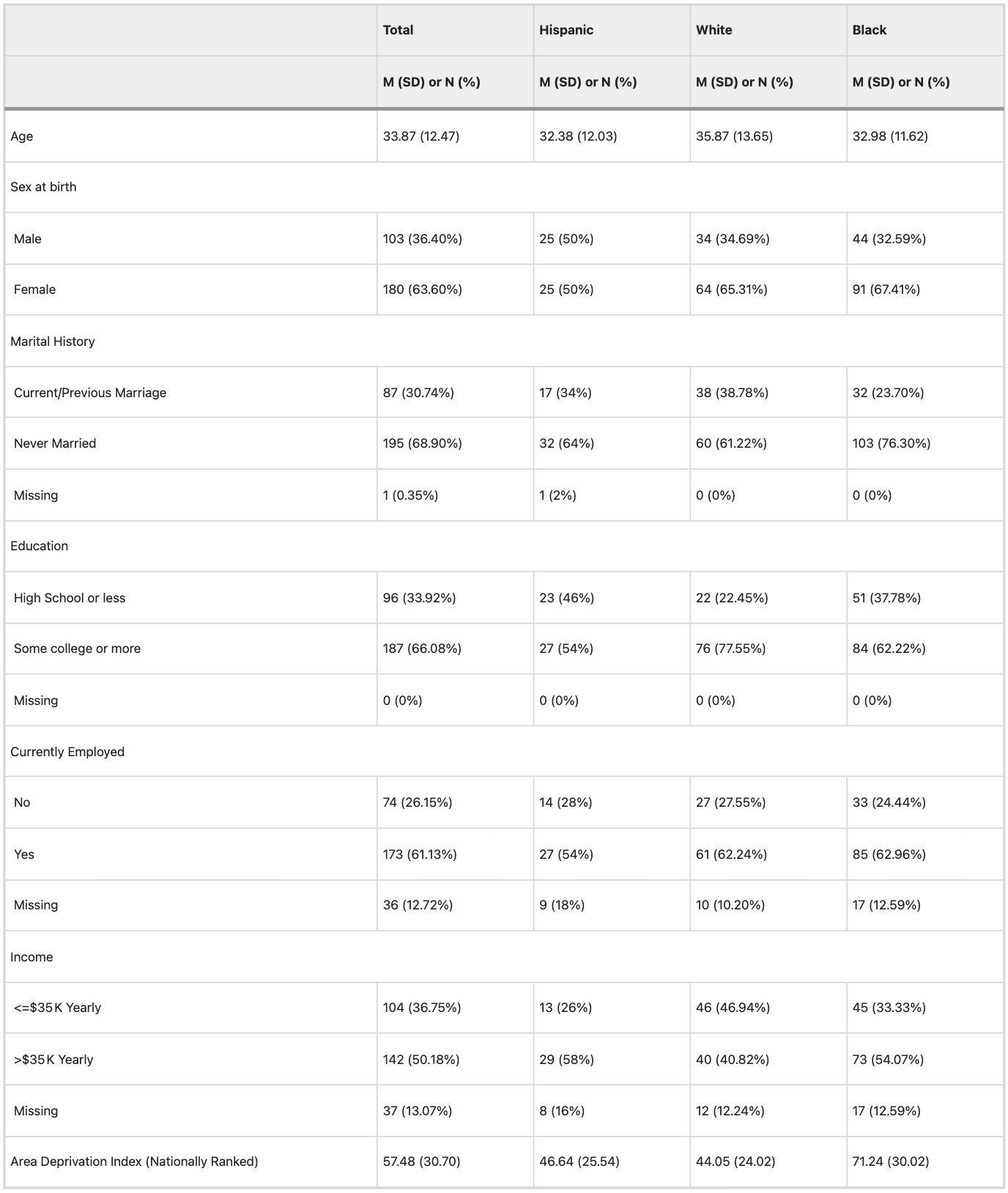
Data for the present analyses were obtained as part of the AURORA Study, a multisite longitudinal study of adverse neuropsychiatric sequelae. Details of the larger AURORA project are described elsewhere. Briefly, trauma-exposed participants were recruited from 22 Emergency Departments (EDs) from across the United States. Trauma was defined as a medical incident requiring admission to the ED, and participants who experienced events such as a motor vehicle collision, high fall (>10 feet), physical assault, sexual assault, or mass casualty incidents were included in the study. Other trauma exposures were also qualifying if: (a) the individual responded to a screener question that they experienced the exposure as involving actual or threatened serious injury, sexual violence, or death, either by direct exposure, witnessing, or learning about the trauma and (b) the research assistant agreed that the exposure was a plausible qualifying event. Trauma was a necessary inclusion criterion for the present study, and no participants without trauma were included. Data were collected for 436 participants recruited between 09/25/2017 and 07/31/2020 who had an MRI and physiological data collection approximately 2-weeks after trauma exposure. A subset of participants in the current report were also included in earlier MRI analyses from the AURORA study though the analyses here are distinct. The present analyses were focused on racial/ethnic differences in early amygdala reactivity/connectivity, and we thus excluded participants who did not have corresponding fMRI data (n = 55). Participants were also excluded listwise on the basis of motion or technical issues during task-fMRI (n = 74) or rs-fMRI (n = 59) (see below and supplement) leaving n = 295 participants with complete MRI data of acceptable quality. Participants self-reported their race/ethnicity and were coded into four categories of “Hispanic (“Hispanic”; n = 50)”, “non-Hispanic White (“White”; n = 98)”, “non-Hispanic Black (“Black”; n = 135)”, “non-Hispanic other-race (“Other”; n = 11)”, and one participant with no reported race/ethnicity. For the present analyses, we also excluded participants from the “other” or unreported racial category due to small sample size that may impact statistical analyses. In total, 283 participants were included in the analyses (Table 1). All participants gave written informed consent as approved by each study site’s Institutional Review Board.

Table 1 Demographic characteristics of the sample.

Table 1

Demographic and psychometric data collection

Initial participant demographic and psychometric data were collected after admission to the ED which included trauma exposure type, participant marital status, income, education level, and employment. Participants’ home address was geocoded to derive an area deprivation index (ADI) to reflect neighborhood disadvantage. Participants’ posttraumatic symptoms were assessed within the ED (i.e., a retrospective report in the past 30 days prior to trauma), 2-weeks, 8-weeks, 3-months, and 6-months after trauma exposure. In the present analyses, we focused on potential associations of 2-week fMRI measures with 3- and 6-month symptoms. The 3- and 6-month assessments queried participant symptoms in the past 30 days. PTSD symptoms were assessed using the PTSD Checklist for DSM-5 (PCL-5), a 20-item self-report questionnaire on trauma symptom expression and severity. Depression symptoms were assessed using the Patient-Reported Outcomes Measurement Information System (PROMIS) Depression instrument from the PROMIS short form 8b. T-scores were derived from total responses to eight items scored on a Likert scale from 1 (never) to 5 (always). Anxiety symptoms were assessed using four items from the PROMIS Anxiety Bank. Prior life trauma was assessed using the Life Events Checklist version 5. Participant’s prior trauma exposure was defined by (a) happened directly, (b) witnessed, (c) happened to someone close to them, or (d) exposed to details due to their occupation. Responses to all questions were summed to derive a prior trauma index.

Psychophysiological responses to threat

Psychophysiological data were collected during a Pavlovian fear conditioning procedure within a day of the MRI session and collected outside of the MRI scanner described in prior reports. Briefly, a shape on a computer screen (a blue square; CS+) was repeatedly paired with an aversive unconditioned stimulus (US) (140 psi airblast to the larynx, 250 ms duration). A different shape (a purple triangle; CS−) was never paired with the aversive stimulus. The paradigm included a 108 dB white noise startle probe that elicited the eyeblink startle response. The startle probe was presented during CS+ and CS− trials, and on its own (noise alone [NA] trials) to assess individual baseline startle response. Following habituation, acquisition consisted of three conditioning blocks with four trials of each type (NA, CS+ paired with US, CS-) in each block, for a total of 12 trials of each type. Ten minutes after acquisition, the extinction phase consisted of four blocks with four trials of each type (CS+, CS−, NA), wherein the airblast never occurred. There were a total of 16 trials of each type during extinction (20 min in duration). Given the focus of the present report on amygdala and threat reactivity, we focused on baseline startle response (EMG activity to the probe during noise alone), tonic skin conductance level (SCL), and fear-potentiated startle (FPS)/SCRs to the CS+ and CS− during the acquisition blocks. For statistical analyses, we excluded EMG and SCL/SCR data if scores were equal to or above 3 standard deviations from the sample mean (individually for each data-type/contrast).

Magnetic resonance imaging

Task-fMRI, rs-fMRI, and anatomical MRI data were collected across five sites with relatively harmonized acquisition parameters (Table S1). Results included in this manuscript come from preprocessing performed using FMRIPREP version stable 1.2.2 [1, 2, RRID:SCR_016216], a Nipype [3, 4, RRID:SCR_002502] based tool as in our prior reports. Further processing information is provided in the supplement.

Task-fMRI of amygdala reactivity

To index neural reactivity to threat, participants completed an emotional reactivity task designed to probe reactivity to social threat cues via passive view of fearful and neutral facial expressions. The task is described in prior work. Briefly, faces from the Ekman faces library were presented in a block design (15 fear blocks and 15 neutral blocks, 10 s rest in between) presented in a pseudorandom order. The order was counterbalanced across participants. SPM12 was used for the initial statistical models after using ICA-AROMA as part of the FMRIPREP pipeline. Emotion blocks were modeled with separate boxcar functions representing the onset and 8 s duration of each block, convolved with a canonical hemodynamic response function. Separate regressors for white matter, cerebrospinal fluid and global signal were included to account for motion/physiological noise. Amygdala reactivity from the 1st level contrasts of fearful – neutral face conditions was extracted from the left and right medial amygdala defined by the Brainnetome atlas [41] and used in statistical analyses (see statistical analysis section).

Resting-state amygdala connectivity

Following ICA-AROMA, the rs-fMRI data were further processed within the Analysis for Functional NeuroImages (AFNI) program 3dTproject to perform linear detrending, censoring of non-steady state volumes identified by FMRIPREP, bandpass filtering (0.01–0.1 Hz), and regression of white matter, corticospinal fluid, and global signal to account for potential physiological noise. The mean fMRI signal time-course was extracted separately from the left and right medial amygdala defined by the Brainnetome atlas [41] and Z-transformed Pearson correlation coefficients were calculated between each ROI and the rest of the brain (i.e., two voxel-wise connectivity maps for left/right amygdala per participant). Group-level statistical modeling was completed in AFNI using the separate voxelwise connectivity maps.

Statistical analyses

Statistical analyses were completed using IBM SPSS version 24, the JASP Statistical Package (https://jasp-stats.org/), and the Analysis of Functional NeuroImages (AFNI) software package. Demographic data such as grade-level, employment, marital status, and income were dummy-coded as per our prior analyses. Univariate ANOVAs assessed racial/ethnic differences in tonic SCLs and baseline startle responses. Post-hoc pairwise comparisons were completed for significant omnibus effects with adjustments to degrees of freedom for inequality of variance between groups were completed when a significant violation was detected (e.g., Levene’s test). Repeated-measures ANOVAs assessed racial/ethnic differences in amygdala activity for the fearful–neutral contrast. For ANOVA models, given the pronounced differences in prior trauma exposure (see results), we completed sensitivity analyses with prior trauma exposure as a covariate. For non-significant planned post-hoc comparisons, we ran confirmatory equivalence tests (described in the supplement). Voxelwise group-level models were completed using 3dMVM in AFNI that included a factor for racial/ethnic group. Due to collinearity between race/ethnicity and site/scanner (as well as missing racial/ethnic categories for some sites), we did not include a covariate of site/scanner in any analyses. For completeness, we completed an additional 3dMVM focusing on the effects of scanner to determine if there was overlap in the observed regions for our primary analysis. In addition, quality control metrics of the fMRI data by site and by racial group are provided in the supplement (see supplementary results; Figures S1 and S2). A gray matter mask that included subcortical areas and the cerebellum was applied to the data. Cluster-based methods for multiple comparison correction were applied to determine the voxel extent k needed at a cluster forming threshold of p = 0.005 to maintain α = 0.05. Specifically, 3dFWHMx was applied to the 1st-level contrasts of the preprocessed rs-fMRI data to derive the autocorrelation function parameters for 3dClustSim (10,000 iterations). The minimum k for analyses of the rs-fMRI data was 99 voxels. Given our strong a priori hypotheses about amygdala reactivity during faces task, we also extracted beta values for left and right medial amygdala from the Brainnetome atlas for statistical analysis in SPSS. We further completed univariate analyses of covariance (ANCOVA) to determine if racial/ethnic variability in amygdala connectivity patterns were related to differential outcomes in PTSD, depression, and anxiety symptoms at 3 or 6-months. ANCOVAs included between subject factors for racial/ethnic group and continuous covariates for posttraumatic assessment (one for each type and timepoint) to assess each effect for each connectivity pattern. We applied Benjamini-Hochberg false discovery rate corrections for each analysis within each posttraumatic assessment (i.e., correcting for 14 tests –7 connectivity patterns x 2 timepoints for PTSD, depression, and anxiety separately). Finally, to estimate the effect of racial/ethnic disparities on brain connectivity after accounting for demographic factors, we completed parallel mediation models to determine if demographic factors (marital status, income, education, employment, prior trauma, and area deprivation) mediated race-related differences in amygdala connectivity patterns.

Results

Demographic characteristics

Demographic data by racial/ethnic group are reported in Table 1. We observed significant differences in education level [χ2 = 9.90, p = 0.007], income [χ2 = 7.47, p = 0.023], and marital status [χ2 = 6.46, p = 0.040]. White participants tended to have more education, while Black participants were more often unmarried and—along with Hispanic participants—had lower income. No significant difference was observed in employment within the sample [χ2 = 0.59, p = 0.745]. A significant difference in the area deprivation index (ADI) was observed between the groups [F(2,280) = 31.73, p < 0.001]. Post-hoc comparisons showed no differences between Hispanic and White participants, but significantly greater ADI in Black compared to Hispanic [t(179) = 5.42, d = 0.91, p < 0.001] and White participants [t(221.58) = 8.06, d = 1.07, p < 0.001]. We further observed a significant effect of prior trauma [F(2,220) = 3.12, p = 0.046]. Post-hoc comparisons revealed White participants had greater prior trauma exposure than Black participants [t(182) = 2.47, d = 0.37, p = 0.015] consistent with prior research from the AURORA study [26]. Broad-class trauma exposures by group are presented in Table S2. Given the significant group differences in prior trauma exposure, we conducted sensitivity analyses for significant effects of racial/ethnic group.

Racial/ethnic differences in tonic physiological arousal but not reactivity during threat learning

A paired-samples t-test revealed significantly greater SCRs to the CS + than the CS- [t(134) = 2.67, d = 0.23, p = 0.009] during acquisition, confirming successful fear conditioning across the full sample. A one-way ANOVA revealed significant racial/ethnic differences in tonic SCLs [F(2,130) = 7.78, p < 0.001]. In sensitivity analyses that included a covariate for prior trauma exposure (i.e., ANCOVA), tonic SCL still differed by racial/ethnic group [F(2,109) = 5.90, p = 0.003]. Posthoc pairwise comparisons revealed Black participants showed significantly lower tonic SCL compared to White participants [t(55.76) = 3.36, d = 0.69, p = 0.001] (Figure S3). These effects survive a Bonferroni correction (criterial p = 0.05/3 = 0.016). No difference was observed between Hispanic and White, or Hispanic and Black, participants (all p > 0.05). Subsequently, a repeated measures ANOVA did not reveal a significant main effect of racial/ethnic group (p = 0.216) or racial/ethnic group by stimulus-type interaction (p = 0.820) on SCRs during acquisition. The main effect of stimulus type remained significant [F(1,132) = 4.93, p = 0.03]. Thus, racial and ethnic groups differed in baseline levels of peripheral arousal, but did not show differences in physiological reactivity during threat acquisition after trauma exposure.

We next investigated if tonic SCL was related to demographic factors (education, employment, marital status, income, prior trauma, and ADI). Tonic SCL and ADI were correlated at trend-level (r = −0.16, p = 0.076). Tonic SCL was not associated with other demographic variables. Given the difference in tonic SCL was driven by differences between White and Black participants, we focused a follow-up parallel mediation analysis on these groups. A parallel mediation analysis revealed significant total [Z-statc = −4.01, p < 0.001] and direct [Z-statc’ = −3.18, p = 0.001] effects of racial group, but there was not a significant indirect effect [Z-statab = −0.36, p = 0.719]. These data suggest the indexed structural inequities do not directly mediate the differences in tonic skin conductance between Black and White participants.

Racial/ethnic differences in baseline, but not fear-potentiated, startle during threat learning

A paired-samples t-test revealed significantly greater FPS response to the CS+ than the CS- [t(208) = 7.80, d = 0.54, p < 0.001] during acquisition. A one-way ANOVA revealed significant racial/ethnic differences in baseline startle responses [F(2,213) = 5.98, p = 0.003]. In sensitivity analyses that included a covariate for prior trauma exposure (i.e., ANCOVA), baseline startle responses still differed by racial/ethnic group F(2,166) = 4.05, p = 0.019. Post-hoc pairwise comparisons revealed Black participants showed significantly lower baseline startle compared to White participants [t(157.58) = 3.31, d = 0.50, p = 0.001] (Figure S3). These effects survive a Bonferroni correction (criterial p = 0.05/3 = 0.016). No difference was observed between Hispanic and White, or Hispanic and Black, participants (all p > 0.05). Subsequently, a repeated measures ANOVA did not reveal a significant main effect of racial/ethnic group (p = 0.732) or racial/ethnic group by stimulus-type interaction (p = 0.910) on FPS responses during acquisition. The main effect of stimulus type remained significant [F(1,206) = 51.55, p < 0.001]. These data further confirm race-related differences in general physiologic arousal, but not differences in threat reactivity.

We next investigated if baseline startle response was related to demographic factors (education, employment, marital status, income, prior trauma, and ADI). Baseline EMG and ADI were significantly correlated (r = −0.22, p = 0.001). Given the difference in baseline EMG was driven by differences between White and Black participants, we focused a follow-up parallel mediation analysis on these groups. A parallel mediation analysis revealed significant total [Z-statc = −3.38, p = 0.008] and a significant indirect effect [Z-statab = −1.98, p = 0.048], but not a significant direct [Z-statc’ = −1.75, p = 0.080] effects of racial group. These data suggest structural adversity mediates differences in baseline startle responses between Black and White trauma survivors.

Racial/ethnic groups do not differ in amygdala reactivity to fearful faces

A mixed measures ANOVA revealed no main effect of racial/ethnic group, and no racial/ethnic group by hemisphere interaction, on amygdala reactivity to threat (Fearful - Neutral faces; p > 0.05) (Figure S4). Exploratory post-hoc analyses revealed a difference in left amygdala reactivity between Hispanic and White participants (t(146) = 2.35, d = 0.41, p = 0.020, uncorrected) that did not survive multiple comparison correction. These data suggest racial/ethnic groups do not differ in amygdala reactivity to threat.

Racial/ethnic groups differ in basal amygdala to salience network connectivity patterns

General patterns of left and right amygdala connectivity are presented in the supplement (Figure S5). Racial/ethnic-related differences in amygdala connectivity during rs-fMRI are highlighted in Fig. 1. We observed significant differences in connectivity patterns with the left amygdala seed to regions such as cerebellum and dorsolateral PFC, as well as nodes in the canonical salience network, specifically the dorsal anterior cingulate cortex and insula (Table 2). We observed significant differences in connectivity between the right amygdala seed to the cerebellum. Sensitivity analyses including prior trauma exposure as a covariate did not show a significant effect for prior trauma exposure (all p > 0.05) and race/ethnicity remained significant (all p < 0.05) for the observed clusters. In general, Hispanic and Black participant groups showed higher resting-state connectivity between the amygdala seeds and these nodes than White participants (Table 3). Although left and right amygdala seeds both showed significant race-related connectivity patterns with cerebellum, a subsequent conjunction analysis did not meet a statistically significant cluster extent (k = 84). Finally, comparative models focused on effects of scanner did not reveal spatial overlap with the models focused on racial/ethnic group (Figure S6). These results highlight race-related differences in connectivity between the amygdala to major nodes of the salience network that does not appear to be driven by scanner effects.

Fig. 1: Lower amygdala to salience network connectivity in White, compared to Hispanic and Black, trauma survivors.

Fig 1

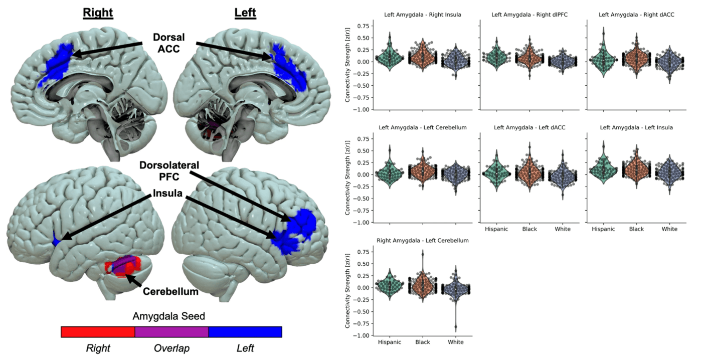
Several brain regions such as the dorsal anterior cingulate cortex (ACC), dorsolateral prefrontal cortex (PFC), insula, and cerebellum showed racial/ethnic differences in connectivity to both right (red) and left (blue) amygdala. Hispanic (green) and Black (orange) groups showed greater connectivity than White (purple) participants. Violin plots show distribution of participant connectivity strength (dots in overlaid swarm plot) for each group.

Table 2 Loci of racial/ethnic differences in amygdala connectivity.

Table 2

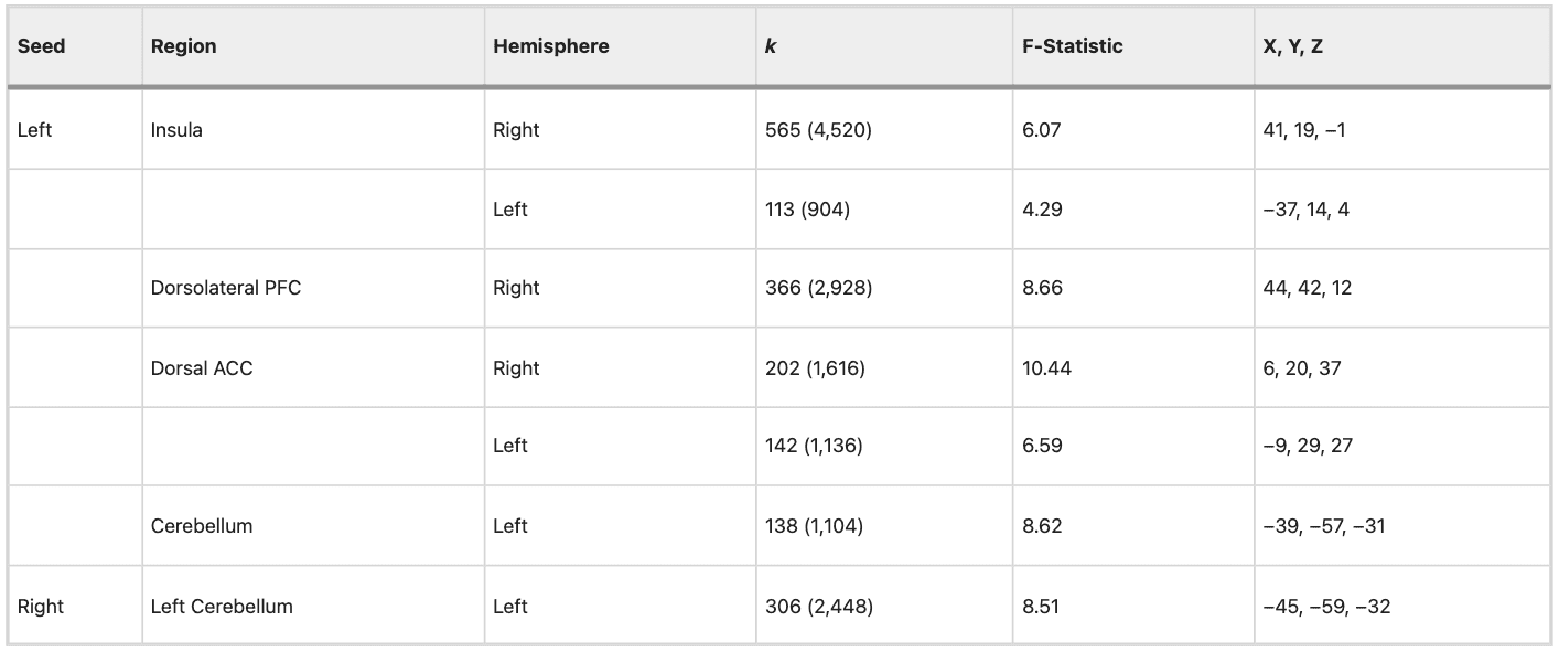
Coordinates are provided in Montreal Neurological Institute (MNI) standard space. F-statistic represents the F-value at the center of mass of the cluster. Cluster size (k) expressed as voxels (volume in mm3).

Table 3 Post-hoc tests of race-related differences in amygdala connectivity.

Table 3

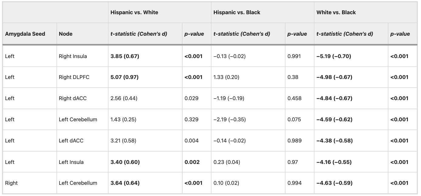
Bold values indicate post-hoc tests survive a Bonferroni correction (0.05 / 21 comparisons = 0.002) for multiple comparisons.

Next, we completed parallel mediation models to determine if accounting for indices of adversity-mediated race-related differences in amygdala connectivity patterns. Adversity metrics partially mediated the difference in amygdala-to-left insula connectivity between White and Black participants (Table S3). No other indirect effects were significant suggesting these metrics did not mediate race-related differences in amygdala connectivity patterns. Separate correlations and t-tests between the demographic variables and amygdala connectivity patterns are described in the supplement (Table S4).

Racial/ethnic differences in connectivity and posttraumatic outcomes

Racial/ethnic group by posttraumatic symptom interactions on amygdala connectivity patterns are summarized in Table S5. FDR correction using the Benjamini-Hochberg approach per posttraumatic cluster revealed racial/ethnic group moderated the relationship between connectivity of left amygdala with right DLPFC, right dACC, and left cerebellum, and PCL-5 scores at 3-months (Fig. 2). Specifically, greater connectivity between the amygdala and these regions was associated with lower PCL-5 scores for Hispanic individuals, but greater PCL-5 scores for Black individuals. White individuals showed no relationship between amygdala connectivity and PCL-5 scores. We then re-ran these analyses using residuals from models of demographic factors (i.e., prior trauma, ADI, income, education, marriage, and employment) on amygdala connectivity to determine if accounting for structural inequities affected the relationship. After accounting for structural inequities, only left amygdala to left dACC connectivity was differentially associated with 3-month PCL-5 scores between racial/ethnic groups [F(2,181) = 3.13, p = 0.046]. These data suggest neural patterns may predict future PTSD symptom severities differently for racial/ethnic groups and this variability is driven—in part—by structural inequities between groups.

Fig. 2: Amygdala connectivity shows differential associations with PTSD symptom development across different races/ethnicities.

Fig 2

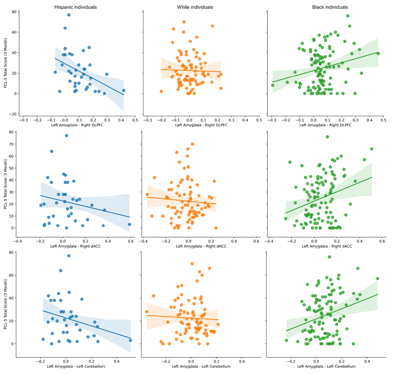
Racial/ethnic group moderated the relationship between amygdala connectivity to the right dorsolateral prefrontal cortex (DLPFC), right dorsal anterior cingulate cortex (dACC), and left cerebellum. Hispanic individuals (blue) showed negative relationships between connectivity and post-traumatic stress disorder (PTSD) symptoms at 3-months, White individuals (orange) showed an orthogonal relationship, and Black individuals (green) showed a positive relationship. Dots represent individual data points, and the solid lines represent the linear lines of best fit. Shaded areas represent the 95% confidence interval of the linear line of best fit.

Discussion

Despite well-documented racial inequities in societal risk and recovery factors for PTSD, limited research has investigated how these inequities may manifest in the neural markers of PTSD susceptibility. The current study used multisite rs-fMRI data from the AURORA study to identify race-related differences in amygdala functional dynamics after trauma and the moderating role of structural inequities. Black and Hispanic individuals displayed heightened connectivity between the amygdala and nodes of the salience network as well as dorsolateral PFC and cerebellum compared to White individuals. Further, Black participants showed lower tonic skin conductance levels (SCLs) and baseline startle responses compared to White participants. There were no racial or ethnic differences in amygdala, skin conductance, or startle reactivity to threat. Accounting for structural inequities attenuated baseline startle responses and the magnitude of racial/ethnic differences in amygdala connectivity. Importantly, these results demonstrate that lower socioeconomic position conveys higher resting amygdala connectivity to the salience network, and that the racial disparities in socioeconomic factors contribute to the appearance of race-related differences in neurophysiological tone. The present findings are critical for the development of generalizable neurobiological markers of susceptibility to trauma and stress-related disorders.

Neurophysiological differences were observed in tonic arousal between racial/ethnic groups, specifically with race-related variability in amygdala connectivity to the insula, dACC, dlPFC, and cerebellum. Of note, the insula and dACC are thought to be part of a salience network that directs attention towards biologically relevant stimuli. Given the role of the amygdala in threat learning and expression, increased amygdala-salience network connectivity may be thought to represent heightened emotional readiness for impending threat that potentiates physiological arousal. Prior research has observed greater amygdala-salience network connectivity in those with PTSD compared to those without, which may suggest that this connectivity pattern is indicative of emotion dysregulation. However, Black participants showed lower SCLs and baseline startle responses compared to White participants indicative of lower tonic physiological arousal. The lower physiological tone is more suggestive of desensitization to threat which is in line with previous neurophysiological research in Black individuals, which has found amygdala sensitization to threat cues as well as lower observed rates of internalizing disorders in Black individuals. In fact, prior work has found that increased connectivity between the salience network and other brain regions in those with a history of childhood maltreatment is related to increased psychological resiliency. It is noteworthy however that Hispanic participants were not significantly different from Black or White participants in analyses of SCLs and baseline startle responses. Prior work in non-psychiatric samples has found that Hispanic individuals may show blunted startle responses compared to non-Hispanic individuals. Despite the behavioral differences, Black and Hispanic individuals both showed heightened connectivity of the amygdala compared to White individuals which may suggest groups engage in similar adaptive neural strategies to mitigate the deleterious effects of race/ethnicity-related stressors. The current results may therefore suggest the neurophysiological profiles are indicative of differential emotion regulation approaches wherein Black and Hispanic groups utilize amygdala-salience network connectivity to promote regulated emotion at baseline.

Neuroimaging studies on the brain health consequences of racial discrimination lend some support to the hypothesis of baseline emotion regulation as a correlate of greater amygdala-salience network connectivity in Black individuals. Greater endorsement of discrimination is associated with greater amygdala to dACC and insula (i.e., salience network) connectivity in Black older adults. Further, a prior study found that trauma-exposed Black women with more experiences of racial discrimination had increased response in threat processing network regions that accompanied relatively better performance on an emotional stroop task that included threatening distractors. Relatively less work on the neurobiological consequences of discrimination and race-related stress has been completed in Hispanic individuals although exposed to race/ethnicity-related stressors such as discrimination. Racial discrimination is a component of multi-level racism that is often experienced by minoritized groups. Structural and systemic inequities in income, education, and other socioeconomic factors are considered components of structural racism. One speculative hypothesis then is that the present racial/ethnic differences in neural connectivity are a result of chronic, repeated racism-related stress throughout development. These findings may help to contextualize lower acute and chronic posttraumatic psychopathology symptoms after trauma. Taken together, individuals exposed to factors related to multi-level racism show greater amygdala-salience network connectivity that may allow for greater emotion-regulation during tasks that also contributes to general desensitization, including lower levels of resting peripheral arousal. However, it should also be noted that racial discrimination is associated with increased depressive symptoms and thus more research is needed at the intersection of racism, neurobiology, and psychiatry to fully understand neural associations of racial health disparities. Importantly, the race/ethnicity-related differences in neural connectivity patterns observed herein may also have implications for neuromodulatory-based treatments. For example, dlPFC to amygdala connectivity is a suggested prognostic marker of PTSD, and modulating this connection may be a mechanism for early evidence of transcranial magnetic stimulation (TMS) efficacy in PTSD. However, based on our results, disparate rates of exposure to early life stressors may affect generalizability of such neuromodulatory targets for different racial/ethnic groups particularly when considering additional socioeconomic barriers to treatment.

Of note, we observed no differences in brain or behavioral reactivity to threat after trauma exposure across the racial/ethnic groups. Amygdala reactivity before or in the early aftermath of trauma is predictive of later PTSD symptoms and was a feature of biotypes of posttraumatic sequalae in previous work from the AURORA study. Similarly, SCR and FPS appear to be reproducible physiological markers of PTSD susceptibility. These findings may suggest that measures of threat reactivity obtained relatively soon after trauma may be more generalizable markers of trauma outcomes. However, stimuli used to index amygdala reactivity to threat predominately consisted of white faces which may elicit differing responses from each racial/ethnic group due to in-group/out-group effects. Though similar results were observed using non-racial stimuli during Pavlovian conditioning in the present study, it is possible that a balanced mixed-race stimulus set may have led to different results and may be more ecologically valid. Additional research is needed using balanced stimulus sets to fully explore potential race-related differences in threat reactivity in the early aftermath of trauma.

Several limitations should be noted for the present investigation. First, our sample was limited to racial/ethnic groups of Hispanic, non-Hispanic White, and non-Hispanic Black. The present sampling did not allow for a more nuanced breakdown of racial or ethnic categories which may influence the current findings. Second, the present study only included participants who had experienced a DSM-5 criterion A traumatic event. It is technically and ecologically difficult to recruit previously trauma-unexposed individuals or recruit individuals right before trauma given both the preponderance of trauma in the US and lack of knowledge as to which individuals will soon experience trauma. However, the current and prior work suggests that pre-traumatic psychopathology symptoms vary between racial/ethnic groups, which may be related to pre-traumatic variability in amygdala connectivity. Thus, neuroimaging in the pre-traumatic period, perhaps in pre-post first-responder or military deployment studies, may be useful for understanding race-related differences in neural connectivity and psychiatry disorders. We also note that the socioeconomic factors assessed here may not fully capture the degree of structural inequities between racial/ethnic groups relevant for neuropsychiatric research. Exploratory analyses revealed that ADI—a neighborhood-level measure—was more consistently associated with connectivity patterns than individual socioeconomic measures (Table S3). Emergent research suggests that multidimensional indices of structural inequities, such as those that account for differential exposure to pollutants, may be particularly important for understanding health disparities. Further research is needed that combines granular assessments of structural inequities with neuroimaging in the early aftermath of trauma to understand racial/ethnic disparities in PTSD development. It is also prudent to note that the present analyses were completed as a secondary analysis within the AURORA dataset. Although the largest study of its kind, sampling was limited to five imaging sites which may limit generalizability to participants from other regions. Likewise, the parent study was not specifically designed to investigate other sources of race-related stress (e.g., racial discrimination). Further research including participants from other areas with more in-depth demography is needed to confirm and extend the present findings. Finally, physiological and rs-fMRI data were not collected concurrently in the present study. Continuous psychophysiological measurement during fMRI may allow for better identification of race-related brain-behavior differences important for understanding posttraumatic psychopathology.

In conclusion, the present study identified racial/ethnic variation in amygdala connectivity at rest and tonic physiological arousal during a threat conditioning task, however, no differences were observed between racial/ethnic groups in reactivity to threat. The racial/ethnic variability in amygdala connectivity was also related to expression of PTSD symptoms at 3-months and was partially attributable to the differences in the assessed socioeconomic factors. Our findings have important implications for the development of generalizable neuroimaging markers of posttraumatic dysfunction, and for the usage of neuromodulatory treatments in the aftermath of trauma. Full consideration of the ways in which systemic inequities may produce racial/ethnic variability in neural connectivity after traumatic stress will be necessary for equitable neuroscience-based treatment outcomes.

Open Article as PDF

Abstract

Considerable racial/ethnic disparities persist in exposure to life stressors and socioeconomic resources that can directly affect threat neurocircuitry, particularly the amygdala, that partially mediates susceptibility to adverse posttraumatic outcomes. Limited work to date, however, has investigated potential racial/ethnic variability in amygdala reactivity or connectivity that may in turn be related to outcomes such as post-traumatic stress disorder (PTSD). Participants from the AURORA study (n = 283), a multisite longitudinal study of trauma outcomes, completed functional magnetic resonance imaging and psychophysiology within approximately two-weeks of trauma exposure. Seed-based amygdala connectivity and amygdala reactivity during passive viewing of fearful and neutral faces were assessed during fMRI. Physiological activity was assessed during Pavlovian threat conditioning. Participants also reported the severity of posttraumatic symptoms 3 and 6 months after trauma. Black individuals showed lower baseline skin conductance levels and startle compared to White individuals, but no differences were observed in physiological reactions to threat. Further, Hispanic and Black participants showed greater amygdala connectivity to regions including the dorsolateral prefrontal cortex (PFC), dorsal anterior cingulate cortex, insula, and cerebellum compared to White participants. No differences were observed in amygdala reactivity to threat. Amygdala connectivity was associated with 3-month PTSD symptoms, but the associations differed by racial/ethnic group and were partly driven by group differences in structural inequities. The present findings suggest variability in tonic neurophysiological arousal in the early aftermath of trauma between racial/ethnic groups, driven by structural inequality, impacts neural processes that mediate susceptibility to later PTSD symptoms.

Introduction

Responses to stressful events can differ greatly, depending on an individual's past experiences and resources. Wealth and financial stability, for example, are known to protect against the lasting social, emotional, and financial burdens of trauma. In the United States, there are clear racial and ethnic differences in important protective factors like education, employment, and income. There has been limited research on how these differences might lead to race-related variations in responses to trauma and how they might affect brain processes involved in developing stress-related disorders. Understanding potential race-related differences in brain activity after trauma and how these relate to trauma outcomes is crucial for creating fair research and treatment plans.

Research into brain function consistently shows that areas involved in responding to threats, especially the amygdala, play a key role in vulnerability to negative outcomes like post-traumatic stress disorder (PTSD). The amygdala is essential for learning to respond to threats and directly controls physical reactions like skin conductance (sweating). Both amygdala activity and skin conductance responses to threats change in individuals diagnosed with PTSD. High amygdala activity in response to threats and strong skin conductance responses soon after trauma are linked to more severe PTSD symptoms later on. Recent studies show that changes in the amygdala and prefrontal cortex (PFC) activity, as well as how they connect, are associated with later PTSD symptoms, possibly indicating less control over amygdala activity. Therefore, amygdala function and related physical responses are considered potential brain markers for trauma-related mental health issues.

Despite the potential for a brain-based marker of PTSD vulnerability involving the amygdala, very little research has explored race/ethnicity-related differences in these findings, or the role of social inequalities. Minority groups are more likely to have experienced adverse events throughout their lives, which are known to affect amygdala function. Previous studies have shown lower skin conductance and startle responses in Black individuals, both in general populations and in those with PTSD. However, this earlier work did not fully consider how structural inequalities might contribute to these race-related differences in physical responses. Recent evidence suggests that unequal exposure to negative life experiences across development leads to lower amygdala activity and skin conductance responses to threats in Black individuals compared to White individuals. Additionally, previous work found that greater neighborhood disadvantage is linked to stronger connections between the amygdala and the inferior parietal lobule. This existing research suggests that racially/ethnically minority individuals might develop adaptive ways to regulate amygdala activity (e.g., emotional blunting) to cope with higher levels of life stress. These race-related structural inequalities may partly explain recently observed race-related differences in post-traumatic symptoms soon after trauma. However, there has been no prior research directly investigating racial/ethnic differences in the connections within the brain's threat network immediately after trauma, or the potential contributions of structural inequalities.

The current multi-site study examined potential racial/ethnic differences in brain and physiological responses and connectivity that might be related to post-traumatic dysfunction. This was done through an exploratory re-analysis of the AURORA study data. The study measured physical expressions of emotional responses to threat using skin conductance and startle responses during learned threat situations. It also looked at amygdala activity in response to social threats (viewing fearful and neutral faces) and brain connectivity during rest. Researchers hypothesized that racial/ethnic differences would be observed in physical arousal and amygdala activity during threat, with participants from minority groups showing lower threat reactivity compared to White participants. Differences were also expected in amygdala connectivity patterns between racial/ethnic groups. Furthermore, it was predicted that racial/ethnic variations in amygdala connectivity would be linked to later reported post-traumatic dysfunction at 3 and 6 months after the trauma. Finally, the study investigated whether observed race-related brain and physiological differences could be explained by racial inequalities in socioeconomic factors (e.g., neighborhood deprivation or income). The findings of this study highlight important race-related variations in brain circuits linked to PTSD development and have significant implications for using brain targets in predicting and treating trauma and stress-related disorders.

Methods and materials

Data for the current analysis came from the AURORA Study, a large, multi-site study tracking mental health outcomes after trauma. Participants were recruited from 22 emergency departments across the United States after experiencing a traumatic event. Data were collected from 436 participants between 2017 and 2020 who had MRI and physiological data collected approximately two weeks after their trauma exposure. To focus on racial/ethnic differences in early amygdala activity and connectivity, participants without fMRI data or with poor data quality due to motion or technical issues were excluded, leaving 283 participants for the final analysis. Participants self-reported their race/ethnicity and were categorized as Hispanic, non-Hispanic White, or non-Hispanic Black for this study.

Initial demographic information, including trauma type, marital status, income, education level, and employment, was collected soon after emergency department admission. Participants' home addresses were used to calculate an area deprivation index (ADI), reflecting neighborhood disadvantage. Post-traumatic symptoms were assessed at various times, with the current analysis focusing on associations between two-week fMRI measures and symptoms at 3 and 6 months post-trauma. PTSD symptoms were measured using the PCL-5 questionnaire, depression symptoms with the PROMIS Depression instrument, and anxiety symptoms with the PROMIS Anxiety Bank. Previous life trauma was assessed using the Life Events Checklist version 5, with responses summed to create a prior trauma index.

Psychophysiological data were collected during a fear conditioning procedure, outside the MRI scanner, around the same time as the MRI session. In this procedure, one shape (CS+) was repeatedly paired with an unpleasant airblast (US), while another shape (CS-) was never paired. A startle probe was used to measure eyeblink startle responses. The study focused on baseline startle response, tonic skin conductance level (SCL), and fear-potentiated startle (FPS)/skin conductance responses (SCRs) to the CS+ and CS- during the learning phase. Data considered outliers were excluded from statistical analysis.

Brain imaging data (task-fMRI, resting-state fMRI, and anatomical MRI) were collected across five sites using similar equipment settings. To measure brain activity related to threat, participants completed a task involving passive viewing of fearful and neutral facial expressions. Brain responses, specifically amygdala activity, were measured from these images. For resting-state fMRI, the mean brain signal over time was extracted from the left and right medial amygdala, and connections between these areas and the rest of the brain were analyzed.

Statistical analyses used various software packages to examine the collected data. Differences in demographics, physiological responses, and brain activity/connectivity across racial/ethnic groups were assessed. When significant differences were found, further comparisons were made between specific groups. Given observed differences in prior trauma exposure among groups, sensitivity analyses included prior trauma as a factor. Brain imaging data underwent specific processing and correction methods to ensure reliable results. Additionally, analyses explored whether racial/ethnic differences in amygdala connectivity were linked to different outcomes in PTSD, depression, and anxiety symptoms at 3 or 6 months. Finally, parallel mediation models were used to determine if socioeconomic factors like marital status, income, education, employment, prior trauma, and area deprivation explained any race-related differences in amygdala connectivity patterns.

Results

Differences were observed in several demographic characteristics among racial/ethnic groups. White participants generally had more education, while Black participants were more often unmarried and, along with Hispanic participants, had lower incomes. Significant differences were also found in the area deprivation index (ADI), indicating greater neighborhood disadvantage for Black participants compared to Hispanic and White participants. There was also a significant difference in prior trauma exposure, with White participants reporting more prior trauma than Black participants. Given these differences, additional analyses were performed to account for prior trauma exposure.

Regarding physiological responses, the study confirmed that participants successfully learned to associate the CS+ with fear. Significant racial/ethnic differences were found in general physiological arousal. Black participants showed lower baseline skin conductance levels and lower baseline startle responses compared to White participants, even after accounting for prior trauma. These differences suggest race-related variations in general physiological arousal, but not in how groups reacted to learned threats. Further analysis indicated that neighborhood disadvantage partially explained the differences in baseline startle responses between Black and White trauma survivors. However, other socioeconomic factors did not directly explain differences in skin conductance.

When examining amygdala activity in response to fearful faces, no significant differences were found among racial/ethnic groups. This suggests that these groups did not differ in their direct brain reactivity to social threat cues.

However, racial/ethnic groups did differ in how the amygdala connected with other brain regions during rest. Black and Hispanic participants showed stronger resting-state connections between the amygdala and regions part of the salience network (involved in detecting important stimuli), as well as the dorsolateral prefrontal cortex and cerebellum, compared to White participants. These differences remained significant even after accounting for prior trauma exposure and were not attributed to differences in MRI scanner effects. Generally, Hispanic and Black groups exhibited greater connectivity in these brain areas than White participants. Further analysis revealed that certain adversity metrics partially explained the differences in amygdala-to-left insula connectivity between White and Black participants, but not other connectivity patterns.

Finally, the study found that racial/ethnic groups showed different relationships between amygdala connectivity and future PTSD symptoms. Specifically, greater connectivity between the left amygdala and certain regions (right dorsolateral PFC, right dorsal anterior cingulate cortex, and left cerebellum) was associated with lower PTSD symptoms at 3 months for Hispanic individuals, but with greater PTSD symptoms for Black individuals. White individuals showed no such relationship. After accounting for socioeconomic factors, the differential association between left amygdala to left dorsal anterior cingulate cortex connectivity and 3-month PTSD symptoms across racial/ethnic groups remained. These results suggest that brain activity patterns may predict future PTSD symptom severity differently across racial/ethnic groups, and that structural inequalities partly contribute to this variability.

Discussion

This study identified racial/ethnic differences in amygdala connectivity during rest and in general physiological arousal during a threat conditioning task; however, no differences were observed in how racial/ethnic groups reacted to direct threats. Black and Hispanic individuals displayed heightened connectivity between the amygdala and brain regions of the salience network, as well as the dorsolateral PFC and cerebellum, compared to White individuals. Additionally, Black participants showed lower general arousal, indicated by lower skin conductance levels and baseline startle responses, compared to White participants. These findings suggest that a lower socioeconomic position is linked to higher resting amygdala connectivity to the salience network, and that racial inequalities in socioeconomic factors contribute to observed race-related differences in general brain and physiological tone. These results are vital for developing reliable brain markers of vulnerability to trauma and stress-related disorders that apply to all populations.

The observed neurophysiological differences in general arousal and amygdala connectivity are noteworthy. Increased amygdala-salience network connectivity might suggest a heightened emotional readiness for threat, potentially boosting physiological arousal. However, Black participants showed lower physiological arousal, which could indicate a desensitization to threat, aligning with previous research on Black individuals. This desensitization might also be an adaptive response to chronic stress, consistent with findings that increased connectivity in the salience network for individuals with a history of childhood maltreatment is linked to greater psychological resilience. While Hispanic participants did not significantly differ from Black or White participants in arousal measures, both Black and Hispanic individuals showed increased amygdala connectivity compared to White individuals. This suggests that both groups might use similar adaptive brain strategies to cope with the harmful effects of race/ethnicity-related stressors. These neurophysiological profiles could represent different emotion regulation approaches, where Black and Hispanic groups use amygdala-salience network connectivity to maintain regulated emotions at baseline.

Studies on the brain effects of racial discrimination support the idea that increased amygdala-salience network connectivity in Black individuals may reflect baseline emotion regulation. Greater experiences of discrimination are linked to stronger amygdala-to-dorsal anterior cingulate cortex and insula connectivity in Black older adults. One possible explanation for the current racial/ethnic differences in brain connectivity is chronic, repeated stress from racism throughout development. These findings could help explain why some minority groups may experience lower acute and chronic post-traumatic mental health symptoms after trauma. Overall, individuals exposed to multi-level racism may show greater amygdala-salience network connectivity, which could enable better emotion regulation during tasks and contribute to a general desensitization, including lower resting physiological arousal. However, it is also known that racial discrimination is associated with increased depressive symptoms, highlighting the need for more research to fully understand the complex links between racism, brain biology, and mental health. These findings also have implications for brain-based treatments, as racial disparities in socioeconomic factors might affect how well such treatments work for different racial/ethnic groups.

It is important to note that the study found no differences in brain or behavioral reactivity to threats across racial/ethnic groups after trauma. Amygdala reactivity soon after trauma predicts later PTSD symptoms and is considered a reliable physiological marker of PTSD vulnerability. This might suggest that measures of threat reactivity taken relatively soon after trauma could be more universally applicable markers of trauma outcomes. However, the stimuli used to assess amygdala reactivity (predominantly White faces) might have elicited different responses based on racial/ethnic identity, which could have influenced the results. Future research using diverse stimulus sets is needed to fully explore potential race-related differences in threat reactivity.

Several limitations should be considered. The study sample was limited to Hispanic, non-Hispanic White, and non-Hispanic Black groups, which restricts a more detailed understanding of other racial or ethnic categories. The study also only included individuals who had experienced trauma, making it difficult to assess pre-trauma brain activity. Additionally, the socioeconomic factors examined might not fully capture the extent of structural inequalities. For instance, neighborhood-level measures like ADI showed more consistent links with brain connectivity than individual socioeconomic measures. Future research needs to combine detailed assessments of structural inequities with brain imaging soon after trauma to better understand racial/ethnic disparities in PTSD development. Lastly, physiological and resting-state fMRI data were not collected simultaneously, which might limit the ability to identify certain brain-behavior differences.

In conclusion, this study identified racial/ethnic variations in resting amygdala connectivity and general physiological arousal, but not in reactivity to threats, following trauma. The racial/ethnic differences in amygdala connectivity were also related to PTSD symptom severity at 3 months and were partly explained by socioeconomic factors. These findings have significant implications for developing generalizable brain imaging markers of post-traumatic dysfunction and for using neuromodulatory treatments after trauma. It is essential to fully consider how systemic inequities may lead to racial/ethnic variations in brain connectivity after traumatic stress to ensure equitable outcomes in neuroscience-based treatments.

Open Article as PDF

Abstract

Considerable racial/ethnic disparities persist in exposure to life stressors and socioeconomic resources that can directly affect threat neurocircuitry, particularly the amygdala, that partially mediates susceptibility to adverse posttraumatic outcomes. Limited work to date, however, has investigated potential racial/ethnic variability in amygdala reactivity or connectivity that may in turn be related to outcomes such as post-traumatic stress disorder (PTSD). Participants from the AURORA study (n = 283), a multisite longitudinal study of trauma outcomes, completed functional magnetic resonance imaging and psychophysiology within approximately two-weeks of trauma exposure. Seed-based amygdala connectivity and amygdala reactivity during passive viewing of fearful and neutral faces were assessed during fMRI. Physiological activity was assessed during Pavlovian threat conditioning. Participants also reported the severity of posttraumatic symptoms 3 and 6 months after trauma. Black individuals showed lower baseline skin conductance levels and startle compared to White individuals, but no differences were observed in physiological reactions to threat. Further, Hispanic and Black participants showed greater amygdala connectivity to regions including the dorsolateral prefrontal cortex (PFC), dorsal anterior cingulate cortex, insula, and cerebellum compared to White participants. No differences were observed in amygdala reactivity to threat. Amygdala connectivity was associated with 3-month PTSD symptoms, but the associations differed by racial/ethnic group and were partly driven by group differences in structural inequities. The present findings suggest variability in tonic neurophysiological arousal in the early aftermath of trauma between racial/ethnic groups, driven by structural inequality, impacts neural processes that mediate susceptibility to later PTSD symptoms.

Introduction

Responses to severe stress, or trauma, differ based on a person's past experiences and available resources. For instance, wealth and financial stability are known factors that can help lessen the lasting social, emotional, and financial challenges of trauma. In the United States, there are clear racial and ethnic differences in how these protective socioeconomic factors, like education, jobs, and income, are distributed. Little research has explored how these visible differences might lead to race-related variations in how people react to trauma, or how they might interact with brain mechanisms involved in developing trauma and stress-related conditions. Understanding potential race-related differences in brain and body function after trauma, and how they relate to outcomes, is crucial for creating fair research and treatment methods.

Studies of the brain have consistently shown that brain circuits involved in responding to threats, especially a part called the amygdala, are very important in determining who might develop problems like posttraumatic stress disorder (PTSD) after trauma. The amygdala is vital for learning how to react to threats and directly controls the body's skin conductance response (SCR) to danger. In people with PTSD, both how the amygdala reacts and how the skin conductance responds to threats are different. Being overly active in the amygdala and having strong skin conductance responses soon after a trauma are linked to more severe PTSD symptoms later on. Recent research also shows that differences in activity and connections between the amygdala and the prefrontal cortex (PFC) are linked to later PTSD symptoms, possibly because of less control over amygdala reactions. This suggests that amygdala function and related body responses could be brain markers for mental health issues related to trauma.

Even though amygdala activity could be a brain marker for who is likely to get PTSD, very little research has explored how race or ethnicity, and the effects of social inequality, might change these findings. Racial/ethnic minority groups often experience more negative events throughout their lives, which are known to affect how the amygdala works. Earlier studies have shown that Black individuals, both with and without PTSD, tend to have lower skin conductance and startle responses. However, these studies did not fully consider how structural inequalities might lead to these race-related differences in body responses. Newer evidence suggests that different exposures to negative life experiences over time contribute to both lower amygdala activity and skin conductance responses to threats in Black individuals compared to White individuals. Other research found that more disadvantaged neighborhoods are linked to stronger connections between the amygdala and another brain area. This current research suggests that racial/ethnic minority individuals might develop adaptive brain responses, like emotional blunting, to cope with more life stress. Race-related structural inequalities might partly explain the race-related differences in posttraumatic symptoms seen soon after trauma. However, no previous study has directly looked at racial/ethnic differences in how threat-related brain circuits connect after trauma, or how structural inequalities might play a role.

This multi-site study examined potential racial/ethnic differences in brain and body responses and connections that might relate to problems after trauma. This was done through a new analysis of data from the AURORA study. Researchers measured the body's physical response to emotional threat using skin conductance and startle responses while participants learned about conditioned threats. They also looked at amygdala activity when viewing social threats (fearful and neutral faces) and brain connections during rest. It was predicted that racial/ethnic differences would appear in physiological arousal and amygdala activity during threat, with racial/ethnic minority groups showing less threat reactivity compared to White participants. Differences in amygdala connectivity patterns between racial/ethnic groups were also expected. Additionally, it was thought that these variations in amygdala connectivity would be linked to problems reported 3 and 6 months after the trauma. Finally, the study investigated whether racial differences in socioeconomic factors, like neighborhood deprivation or income, explained any observed race-related brain and body differences. The results of this study point to important race-related variations in brain circuits involved in PTSD development, with significant implications for using brain targets to predict and treat trauma and stress-related disorders.

Methods and materials

This study used data from the AURORA Study, a large, ongoing study that collects information from many sites about mental health problems after trauma. Participants were recruited from 22 emergency departments across the United States. Trauma was defined as a medical incident requiring an emergency department visit, such as car crashes, falls, or assaults. Only participants who had experienced trauma were included. Data were collected from 436 participants who underwent MRI and physiological data collection about two weeks after their trauma. Some participants were excluded due to incomplete MRI data or technical issues, resulting in 283 participants for the final analyses. Participants reported their race/ethnicity, and were categorized as Hispanic, non-Hispanic White, or non-Hispanic Black. All participants gave written informed consent.

Initial data collected in the emergency department included participant demographics like marital status, income, education level, and employment. The home address of each participant was used to calculate an Area Deprivation Index (ADI), which shows neighborhood disadvantage. Posttraumatic symptoms were assessed multiple times, starting from a retrospective report of the past 30 days before trauma, and then at 2 weeks, 8 weeks, 3 months, and 6 months after the trauma. This study specifically focused on how measures taken at two weeks related to symptoms at 3 and 6 months. PTSD symptoms were measured using the PCL-5 questionnaire, depression symptoms with the PROMIS Depression instrument, and anxiety symptoms with the PROMIS Anxiety Bank. Prior life trauma was assessed using the Life Events Checklist version 5.

Psychophysiological data were collected shortly after the MRI session, outside the scanner. This involved a fear conditioning task where participants saw shapes, with one shape (CS+) paired with an unpleasant airblast stimulus and another (CS-) not. Measurements included skin conductance response (SCR) and startle response, which involved a loud noise probe. The study focused on baseline startle response, overall skin conductance level (SCL), and conditioned fear responses (FPS/SCR) during the learning phase. Magnetic resonance imaging (MRI) data, including task-fMRI, resting-state fMRI, and anatomical MRI, were collected across five sites using standardized settings. Task-fMRI measured amygdala activity in response to social threats, specifically when participants passively viewed fearful and neutral facial expressions. Resting-state fMRI measured how different parts of the brain, particularly the amygdala, were connected when participants were at rest.

Statistical analyses were performed using various software packages. Differences between racial/ethnic groups in overall skin conductance levels, baseline startle responses, and amygdala activity were assessed using statistical tests like ANOVAs. For significant overall findings, further tests compared specific groups. Given known differences in prior trauma exposure among groups, sensitivity analyses were conducted by including prior trauma as a factor to see if the main findings still held. Voxel-wise models were used for analyzing brain connectivity, applying methods to correct for multiple comparisons. Researchers also conducted analyses to see if racial/ethnic differences in amygdala connectivity patterns were linked to different PTSD, depression, and anxiety symptoms at 3 and 6 months. Finally, parallel mediation models were used to explore if demographic factors, such as marital status, income, education, employment, prior trauma, and neighborhood disadvantage, explained the observed race-related differences in amygdala connectivity.

Results

The demographic data showed significant differences among racial/ethnic groups. White participants generally had more education, while Black participants were more often unmarried and, along with Hispanic participants, reported lower income. No significant differences were found in employment. There was a significant difference in the Area Deprivation Index (ADI), with Black participants living in significantly more disadvantaged neighborhoods compared to both Hispanic and White participants. The study also found a significant difference in prior trauma exposure, with White participants reporting more past traumas than Black participants. Because of these differences in prior trauma exposure, additional analyses were done to check if these factors affected the main findings.

The fear conditioning task was successful for all participants, showing that the threatening stimulus created a response. However, significant racial/ethnic differences were found in overall skin conductance levels (SCLs). Black participants showed significantly lower SCLs compared to White participants, and this difference remained even when accounting for prior trauma exposure. No differences in SCL were found between Hispanic and White or Hispanic and Black participants. Importantly, while groups differed in baseline physiological arousal, they did not show differences in how their skin conductance reacted to threat during the learning phase. When examining if SCL related to demographic factors, a slight trend showed a connection between SCL and neighborhood disadvantage (ADI), but other demographic variables were not linked. A follow-up analysis focusing on Black and White participants suggested that the measured structural inequalities did not directly explain the differences in tonic skin conductance.

Similarly, significant racial/ethnic differences were observed in baseline startle responses. Black participants had notably lower baseline startle responses compared to White participants, a difference that persisted even after accounting for prior trauma exposure. No differences were found between Hispanic and White or Hispanic and Black participants. Like skin conductance, there were no differences across groups in how much the startle response increased when exposed to threats. Baseline startle response was significantly linked to neighborhood disadvantage (ADI). Further analysis indicated that structural adversity, as measured by these factors, helped explain the differences in baseline startle responses between Black and White trauma survivors.

In contrast to physiological arousal, no significant differences were found in amygdala activity when racial/ethnic groups viewed fearful versus neutral faces, suggesting similar brain reactivity to social threats. However, significant racial/ethnic differences were found in resting-state amygdala connectivity. The study observed different connectivity patterns between the left amygdala and areas like the cerebellum, dorsolateral prefrontal cortex (PFC), and key parts of the salience network (dorsal anterior cingulate cortex and insula). Differences were also seen in connections between the right amygdala and the cerebellum. Generally, Hispanic and Black participants showed stronger resting-state connections between the amygdala and these brain regions compared to White participants. These connectivity differences remained significant even when accounting for prior trauma exposure, and they did not appear to be caused by differences in MRI scanners across sites.

Further analyses explored whether measures of adversity explained the race-related differences in amygdala connectivity. Adversity factors partly explained the difference in connectivity between the amygdala and the left insula in White and Black participants, but did not mediate other observed connectivity differences. The study also found that racial/ethnic group influenced the relationship between amygdala connectivity and PTSD symptom scores (PCL-5) at 3 months. For Hispanic individuals, stronger connectivity between the amygdala and certain brain areas (right DLPFC, right dACC, left cerebellum) was linked to lower PTSD scores. However, for Black individuals, stronger connectivity in these same areas was linked to higher PTSD scores. White individuals showed no such link. When structural inequalities were taken into account, only the connection between the left amygdala and the left dACC remained differently associated with 3-month PTSD scores across racial/ethnic groups. These findings suggest that brain connection patterns might predict future PTSD symptom severity differently for various racial/ethnic groups, and structural inequalities partly explain these variations.

Discussion

While racial inequalities in factors affecting PTSD risk and recovery are well-known, little research has examined how these inequalities appear in brain markers related to PTSD. This study used multi-site MRI data from the AURORA study to find race-related differences in amygdala function after trauma and to understand how structural inequalities play a role. Black and Hispanic individuals showed stronger connections between the amygdala and parts of the salience network, as well as the dorsolateral prefrontal cortex and cerebellum, compared to White individuals. Additionally, Black participants had lower overall skin conductance levels and baseline startle responses than White participants. However, no racial or ethnic differences were found in how the amygdala, skin conductance, or startle responses reacted to threat. When structural inequalities were considered, they reduced the differences in baseline startle responses and the strength of race-related amygdala connectivity. These results highlight that a lower socioeconomic position is linked to stronger resting amygdala connectivity to the salience network, and that racial differences in socioeconomic factors contribute to observed race-related differences in basic brain and body responses. These findings are vital for creating brain markers that can be generally applied to predict who might develop trauma and stress-related disorders.

Differences in basic brain and body arousal were seen among racial/ethnic groups, particularly in how the amygdala connected to the insula, dorsal anterior cingulate cortex (dACC), dorsolateral prefrontal cortex (dlPFC), and cerebellum. The insula and dACC are part of the salience network, which focuses attention on important stimuli. Because the amygdala is involved in learning about and expressing threats, stronger amygdala-salience network connectivity might suggest a heightened emotional readiness for danger, which could increase physiological arousal. Previous research has linked stronger amygdala-salience network connectivity to PTSD, possibly indicating difficulty regulating emotions. Yet, Black participants showed lower skin conductance levels and baseline startle responses compared to White participants, suggesting lower overall physiological arousal. This lower arousal might point to desensitization to threat, aligning with past research showing both amygdala sensitization to threat cues and lower rates of internalizing disorders in Black individuals. Some studies even link increased salience network connectivity to greater psychological resilience in those who experienced childhood maltreatment. Interestingly, Hispanic participants did not significantly differ from Black or White participants in skin conductance or startle responses. Despite these behavioral differences, both Black and Hispanic individuals had stronger amygdala connectivity than White individuals. This could mean these groups use similar adaptive brain strategies to reduce the harmful effects of stress related to their race or ethnicity.

Brain imaging studies on how racial discrimination affects brain health support the idea that stronger amygdala-salience network connectivity in Black individuals might reflect a way to regulate emotions at baseline. For Black older adults, reporting more discrimination is linked to stronger connections between the amygdala and the dACC and insula. Another study found that Black women who experienced trauma and more racial discrimination showed increased activity in brain regions that process threats, alongside better performance on an emotional task with distracting threats. Less research has explored the brain consequences of discrimination and race-related stress in Hispanic individuals, even though they also face such stressors. Racial discrimination is one part of broader racism often experienced by minority groups. Differences in income, education, and other socioeconomic factors are considered parts of structural racism. One idea is that the racial/ethnic differences in brain connectivity seen in this study result from ongoing, repeated stress related to racism throughout a person's life. These findings might help explain why some groups show lower acute and chronic mental health problems after trauma. Overall, individuals exposed to various forms of racism appear to have stronger amygdala-salience network connectivity, which could allow for better emotion regulation during tasks and contribute to a general desensitization, including lower basic physiological arousal. However, racial discrimination is also linked to increased depressive symptoms, so more research is needed to fully understand the connections between racism, brain biology, and mental health disparities. The race-related differences in brain connectivity found here could also affect brain stimulation treatments. For example, the connection between the dlPFC and amygdala is a potential predictor for PTSD, and changing this connection might be how treatments like transcranial magnetic stimulation (TMS) work for PTSD. But based on these results, different levels of exposure to early life stressors might mean these brain targets work differently for various racial/ethnic groups, especially when considering other socioeconomic obstacles to treatment.

The study observed no differences across racial/ethnic groups in how the brain or body reacted to threat after trauma. Amygdala reactivity and physiological markers like skin conductance response (SCR) and fear-potentiated startle (FPS) are known to predict later PTSD symptoms. This suggests that these immediate post-trauma threat reactivity measures might be more widely applicable markers of trauma outcomes. However, the stimuli used for amygdala reactivity mainly showed White faces, which might cause different responses from various racial/ethnic groups due to "in-group/out-group" effects. Although the study used non-racial stimuli for physiological conditioning with similar results, using a balanced set of faces from different races might have led to different findings and been more relevant to real-world situations. More research with diverse stimuli is needed to fully explore race-related differences in threat reactivity soon after trauma. This study has several limitations. The sample included only Hispanic, non-Hispanic White, and non-Hispanic Black groups, limiting a more detailed analysis of other racial or ethnic categories. Also, the study only included individuals who had experienced a specific type of traumatic event. It is challenging to recruit individuals without prior trauma or right before a trauma occurs, given how common trauma is and the unpredictability of when it will happen. However, prior work suggests that mental health symptoms before trauma vary by racial/ethnic group, potentially linking to pre-trauma amygdala connectivity differences. Therefore, brain imaging before trauma, possibly in studies of first responders or military deployments, could help understand race-related differences in brain connections and mental health conditions.

The socioeconomic factors measured in this study might not fully capture the extent of structural inequalities relevant to brain and mental health research. For example, neighborhood-level disadvantage (ADI) was more consistently linked to brain connectivity patterns than individual socioeconomic measures. Newer research suggests that comprehensive measures of structural inequalities, including exposure to environmental pollutants, are especially important for understanding health disparities. More research is needed that combines detailed assessments of structural inequalities with brain imaging soon after trauma to understand racial/ethnic disparities in PTSD development. It is also important to note that this study involved re-analyzing existing data from the AURORA dataset. Despite being the largest study of its kind, data came from only five imaging sites, which might limit how broadly the findings apply to people from other areas. The original study was also not designed to specifically investigate other sources of race-related stress, such as racial discrimination. Future research should include participants from more diverse areas and gather more in-depth demographic information to confirm and expand these findings. Finally, physiological and resting-state fMRI data were not collected at the same time in this study. Taking continuous physiological measurements during fMRI could help better identify race-related differences in brain and behavior, which is important for understanding mental health problems after trauma. In summary, this study found racial/ethnic differences in amygdala connectivity at rest and in basic physiological arousal during a threat conditioning task. However, no differences were seen in how racial/ethnic groups reacted to threats. The racial/ethnic variability in amygdala connectivity was also linked to PTSD symptoms at 3 months, and these differences were partly explained by variations in socioeconomic factors. These findings have significant implications for developing brain imaging markers that can be generally applied to understand problems after trauma, and for using brain stimulation treatments. Fully considering how systemic inequalities might create racial/ethnic differences in brain connectivity after traumatic stress will be essential for ensuring fair and effective neuroscience-based treatment outcomes.

Open Article as PDF

Abstract

Considerable racial/ethnic disparities persist in exposure to life stressors and socioeconomic resources that can directly affect threat neurocircuitry, particularly the amygdala, that partially mediates susceptibility to adverse posttraumatic outcomes. Limited work to date, however, has investigated potential racial/ethnic variability in amygdala reactivity or connectivity that may in turn be related to outcomes such as post-traumatic stress disorder (PTSD). Participants from the AURORA study (n = 283), a multisite longitudinal study of trauma outcomes, completed functional magnetic resonance imaging and psychophysiology within approximately two-weeks of trauma exposure. Seed-based amygdala connectivity and amygdala reactivity during passive viewing of fearful and neutral faces were assessed during fMRI. Physiological activity was assessed during Pavlovian threat conditioning. Participants also reported the severity of posttraumatic symptoms 3 and 6 months after trauma. Black individuals showed lower baseline skin conductance levels and startle compared to White individuals, but no differences were observed in physiological reactions to threat. Further, Hispanic and Black participants showed greater amygdala connectivity to regions including the dorsolateral prefrontal cortex (PFC), dorsal anterior cingulate cortex, insula, and cerebellum compared to White participants. No differences were observed in amygdala reactivity to threat. Amygdala connectivity was associated with 3-month PTSD symptoms, but the associations differed by racial/ethnic group and were partly driven by group differences in structural inequities. The present findings suggest variability in tonic neurophysiological arousal in the early aftermath of trauma between racial/ethnic groups, driven by structural inequality, impacts neural processes that mediate susceptibility to later PTSD symptoms.

Introduction

Responses to traumatic stress vary based on the challenges individuals face before a traumatic event. Things like wealth and economic resources are known to protect people from the long-term social, emotional, and financial burdens of trauma. In the United States, there are clear racial and ethnic differences in who has access to these protective socioeconomic factors, including education, jobs, and income. Limited research has explored how these differences might show up as race-related variations in how people respond to trauma and how these responses might connect with brain mechanisms related to stress disorders. Understanding potential race-related differences in brain activity after trauma and how these relate to trauma outcomes is important for creating fair research and clinical treatments.

Brain research consistently shows that the brain's threat system, especially a part called the amygdala, plays a big role in whether someone develops serious problems after trauma, such as post-traumatic stress disorder (PTSD). The amygdala is vital for learning how to react to threats, and it directly influences the skin conductance response (SCR) to danger. Both amygdala activity and SCR to threat change in people diagnosed with PTSD. Specifically, an overactive amygdala and strong SCRs soon after trauma are linked to more severe PTSD symptoms later. Newer studies show that changes in the amygdala and prefrontal cortex (PFC) activity, and how these areas connect, are linked to later PTSD symptoms, possibly reflecting less control over amygdala responses. This suggests that amygdala function and related body responses could be brain markers for mental health issues related to trauma.

Despite the amygdala potentially being a key brain marker for PTSD risk, very little research has looked at racial/ethnic differences in these findings, or the role social inequalities play. Minority groups are more likely to have experienced adverse events throughout life, which are known to affect amygdala function. Previous studies have shown lower SCRs and startle responses in Black individuals, both in general populations and those with PTSD. However, this past work did not fully consider how structural inequalities might contribute to race-related differences in body responses. Recent evidence suggests that different experiences of negative life events lead to lower amygdala activity and SCRs to threat in Black individuals compared to White individuals. Also, greater neighborhood disadvantage has been linked to stronger connections between the amygdala and another brain area called the inferior parietal lobule. This suggests that racially/ethnically minoritized individuals might develop adaptive ways to regulate their emotions (like emotional blunting) to cope with more life stress. These race-related structural inequalities may partly explain recently observed race-related differences in PTSD symptoms soon after trauma. However, no prior study has directly examined racial/ethnic differences in the connections within the brain's threat system shortly after trauma, or how structural inequalities might contribute.

This multi-site study explored potential racial/ethnic differences in brain and body responses and connections that might relate to post-traumatic problems. This was done through a secondary analysis of the AURORA study data. Researchers measured skin conductance and startle responses (physical signs of emotional response) during a threat conditioning task. They also looked at amygdala activity when people viewed social threats (fearful and neutral faces) and how the amygdala connected with other brain regions during rest. The study expected to see racial/ethnic differences in physical arousal and amygdala activity during threat, with participants from racially/ethnically minoritized groups showing lower threat responses than White participants. They also anticipated differences in amygdala connectivity patterns between groups. Additionally, they thought that these racial/ethnic differences in amygdala connectivity would be linked to later reported post-traumatic problems 3 and 6 months after the trauma. Finally, the study assessed if racial inequities in socioeconomic factors (like neighborhood disadvantage or income) could explain the observed race-related brain differences. The findings highlight important race-related variations in brain circuits linked to PTSD development and have significant implications for using brain targets for predicting and treating trauma and stress-related disorders.

Methods and Materials

Data for these analyses came from the AURORA Study, a large-scale study tracking negative mental health outcomes after trauma. Trauma-exposed participants were recruited from 22 emergency departments across the United States. Trauma was defined as a medical incident requiring an emergency department visit, such as a car crash, severe fall, physical or sexual assault, or mass casualty events. Data were collected for 436 participants who had MRI and physiological data collection about two weeks after their trauma. The current report included a subset of these participants. For these analyses, participants were excluded if they lacked MRI data or if their MRI data had too much motion or technical issues, leaving 295 participants with usable MRI data. Participants reported their race/ethnicity, categorized as Hispanic, non-Hispanic White, non-Hispanic Black, or non-Hispanic other-race. Participants from the "other" category or with unreported race/ethnicity were excluded due to small numbers. In total, 283 participants were included in the analyses. All participants gave written informed consent.

Demographic and Symptom Data Collection

Initial demographic and psychological data were collected after emergency department admission, including trauma type, marital status, income, education level, and employment. Participants' home addresses were used to calculate an "area deprivation index" (ADI) to measure neighborhood disadvantage. Post-traumatic symptoms were assessed in the emergency department (reporting symptoms from the past 30 days before trauma), then 2 weeks, 8 weeks, 3 months, and 6 months after trauma. For this study, researchers focused on how the 2-week brain measures related to symptoms at 3 and 6 months. PTSD symptoms were measured using the PTSD Checklist for DSM-5 (PCL-5). Depression and anxiety symptoms were measured using parts of the PROMIS instruments. Prior life trauma was assessed using the Life Events Checklist version 5, and responses were summed to create a prior trauma index.

Physiological Responses to Threat

Physiological data were collected during a fear conditioning experiment, completed outside the MRI scanner, around the same day as the MRI session. In this experiment, a blue square (CS+) was repeatedly paired with an unpleasant airblast to the throat (unconditioned stimulus, US). A purple triangle (CS−) was never paired with the airblast. The experiment also included a loud white noise startle probe to measure the eyeblink startle response, presented during CS+ and CS− trials, and alone (noise alone, NA) to establish a baseline. The study focused on baseline startle response (EMG activity during noise alone), general skin conductance level (SCL), and fear-potentiated startle (FPS)/SCRs to the CS+ and CS− during the learning blocks.

Brain Imaging Data Collection and Analysis

Task-fMRI, resting-state fMRI (rs-fMRI), and anatomical MRI data were collected across five sites using similar settings. For task-fMRI, participants completed an emotional reactivity task designed to measure brain responses to social threat cues by passively viewing fearful and neutral facial expressions. Amygdala activity (fearful minus neutral faces) was extracted from specific regions of the left and right amygdala for statistical analysis. For rs-fMRI, brain activity was measured during rest. The average fMRI signal over time was extracted separately from the left and right amygdala, and statistical connections between each amygdala region and the rest of the brain were calculated.

Statistical Analyses

Statistical analyses used IBM SPSS, JASP, and AFNI software. Demographic data like education, employment, marital status, and income were coded for analysis. Univariate ANOVAs examined racial/ethnic differences in general SCLs and baseline startle responses. Post-hoc comparisons were made for significant findings. Repeated-measures ANOVAs assessed racial/ethnic differences in amygdala activity. Given significant differences in prior trauma exposure, sensitivity analyses included prior trauma as a covariate. Voxel-wise group-level models in AFNI looked for racial/ethnic differences in amygdala connectivity across the brain. Due to the relationship between race/ethnicity and study site/scanner, site was not included as a covariate. Further analyses checked for scanner effects. Analyses also determined if racial/ethnic differences in amygdala connectivity patterns were related to differences in PTSD, depression, and anxiety symptoms at 3 or 6 months. Finally, parallel mediation models were used to see if demographic factors (marital status, income, education, employment, prior trauma, and area deprivation) explained race-related differences in amygdala connectivity.

Results

Demographic Characteristics

The study found significant racial/ethnic differences in education level, income, and marital status. White participants generally had more education, while Black participants were more often unmarried and, along with Hispanic participants, had lower incomes. No significant differences were found in employment. There was a significant difference in the area deprivation index (ADI), with Black participants living in areas with significantly higher ADI compared to both Hispanic and White participants. White participants reported more prior trauma exposure than Black participants.

Physiological Arousal During Threat Learning

Participants successfully learned the fear association, showing significantly greater skin conductance responses (SCRs) to the threat signal (CS+) than to the safe signal (CS-). Black participants showed significantly lower general skin conductance levels (tonic SCLs) and lower baseline startle responses compared to White participants, even after accounting for prior trauma. No significant differences were found in how skin conductance or startle responses changed specifically in response to threat signals between racial/ethnic groups during learning. These findings suggest racial/ethnic groups differed in their general physiological arousal, but not in their specific physiological reactions to threats. The difference in baseline startle responses between Black and White participants was partially explained by structural adversity (e.g., area deprivation), but structural inequities did not directly explain differences in tonic SCL.

Amygdala Activity and Connectivity

The study found no significant racial/ethnic differences in how the amygdala reacted to fearful faces, either overall or between brain hemispheres. However, significant racial/ethnic differences were observed in how the amygdala connected with other brain regions while at rest. Hispanic and Black participants generally showed stronger resting-state connectivity between the amygdala and regions like the cerebellum, dorsolateral prefrontal cortex, and key parts of the salience network (specifically the dorsal anterior cingulate cortex and insula), compared to White participants. These connectivity differences were not significantly affected by prior trauma or scanner differences. These results highlight race-related differences in connections between the amygdala and major parts of the salience network.

Connectivity and Long-Term Outcomes

Racial/ethnic group influenced the relationship between amygdala connectivity patterns and 3-month PTSD symptoms. Specifically, stronger connectivity between the left amygdala and certain brain regions (right dorsolateral prefrontal cortex, right dorsal anterior cingulate cortex, and left cerebellum) was linked to lower PTSD symptoms for Hispanic individuals. In contrast, for Black individuals, stronger connectivity in these same areas was linked to higher PTSD symptoms. White individuals showed no clear relationship between amygdala connectivity and PTSD symptoms. After accounting for structural inequities, only the link between left amygdala to left dorsal anterior cingulate cortex connectivity and 3-month PTSD symptoms remained significantly different across racial/ethnic groups. This suggests that certain brain patterns may predict future PTSD symptom severity differently for various racial/ethnic groups, and these differences are partly driven by structural inequalities between groups.

Discussion

Despite clear racial inequalities in societal factors that influence PTSD risk and recovery, little research has explored how these differences manifest in brain markers of PTSD risk. This study, using multisite resting-state fMRI data, identified race-related differences in amygdala functional dynamics after trauma and highlighted the role of structural inequities. Black and Hispanic individuals showed stronger connections between the amygdala and areas like the salience network, dorsolateral prefrontal cortex, and cerebellum compared to White individuals. Additionally, Black participants had lower general skin conductance levels and baseline startle responses than White participants. However, no racial or ethnic differences were found in how the amygdala, skin conductance, or startle responses reacted specifically to a threat. Accounting for structural inequities reduced the baseline startle responses and lessened the size of the race-related differences in amygdala connectivity. These findings demonstrate that a lower socioeconomic position is linked to higher resting amygdala connectivity to the salience network, and that racial differences in socioeconomic factors contribute to the appearance of race-related differences in physiological state. These findings are crucial for developing brain markers of trauma-related disorders that are applicable to everyone.

Differences in general arousal between racial/ethnic groups were observed, particularly in amygdala connectivity to the insula, dorsal anterior cingulate cortex, dorsolateral prefrontal cortex, and cerebellum. The insula and dorsal anterior cingulate cortex are thought to be part of a "salience network" that directs attention to important stimuli. Given the amygdala's role in threat learning, increased amygdala-salience network connectivity might suggest a heightened emotional readiness for threats. However, Black participants showed lower skin conductance levels and baseline startle responses, indicating lower general physiological arousal. This lower physiological state suggests a desensitization to threat, which aligns with previous research on Black individuals that found amygdala sensitization to threat cues alongside lower observed rates of internalizing disorders. Interestingly, Hispanic participants did not significantly differ from Black or White participants in arousal levels. Despite these behavioral differences, both Black and Hispanic individuals showed stronger amygdala connectivity compared to White individuals, which might suggest these groups use similar adaptive brain strategies to lessen the harmful effects of race/ethnicity-related stressors. The current results may therefore suggest that these brain and body profiles point to different emotion regulation approaches, where Black and Hispanic groups use amygdala-salience network connectivity to promote regulated emotions at baseline.

Brain imaging studies on the health impacts of racial discrimination support the idea that greater amygdala-salience network connectivity in Black individuals may reflect baseline emotion regulation. Higher reported discrimination is linked to stronger amygdala-dorsal anterior cingulate cortex and insula (salience network) connectivity in older Black adults. Also, a previous study found that trauma-exposed Black women with more experiences of racial discrimination showed increased activity in threat processing networks, which was accompanied by relatively better performance on an emotional task involving threatening distractions. Less research has been done on the brain consequences of discrimination and race-related stress in Hispanic individuals, despite their exposure to such stressors. Racial discrimination is a part of multi-level racism often experienced by minority groups. Structural inequalities in income, education, and other socioeconomic factors are components of structural racism. One possible explanation is that the current racial/ethnic differences in brain connectivity are a result of chronic, repeated stress related to racism throughout development. These findings might help explain why some groups show lower acute and chronic mental health symptoms after trauma. Overall, individuals exposed to factors related to multi-level racism show greater amygdala-salience network connectivity, which may allow for better emotion regulation during tasks and contribute to general desensitization, including lower levels of general physiological arousal. It is important to note that racial discrimination is also linked to increased depressive symptoms, so more research is needed to fully understand the connections between racism, brain biology, and mental health disparities. These race/ethnicity-related differences in brain connectivity patterns also have implications for brain stimulation treatments.

Notably, no differences were observed in brain or behavioral responses to threat itself after trauma across racial/ethnic groups. Amygdala activity either before or soon after trauma is known to predict later PTSD symptoms and was a feature of different types of post-traumatic problems in earlier work from the AURORA study. Similarly, skin conductance response and fear-potentiated startle appear to be reliable physical markers of PTSD risk. These findings might suggest that measures of threat reactivity obtained relatively soon after trauma could be more broadly applicable markers of trauma outcomes. However, the stimuli used to measure amygdala reactivity to threat mostly consisted of White faces, which might elicit different responses from each racial/ethnic group due to in-group/out-group effects. Although non-racial stimuli were used during the physiological conditioning task, a balanced set of mixed-race stimuli might have led to different results and could be more realistic. More research using diverse stimulus sets is needed to fully explore potential race-related differences in threat reactivity soon after trauma.

Several limitations should be noted. First, the sample was limited to Hispanic, non-Hispanic White, and non-Hispanic Black racial/ethnic groups, which limits a more detailed understanding of other categories. Second, this study only included participants who had experienced a traumatic event. It is difficult to recruit people who have not experienced trauma or to recruit people just before a trauma, given how common trauma is in the U.S. and the unpredictability of who will experience it. However, current and past work suggests that pre-trauma mental health symptoms vary between racial/ethnic groups, which might relate to pre-trauma differences in amygdala connectivity. Therefore, brain imaging before trauma, perhaps in studies of first responders or military personnel before deployment, could help understand race-related differences in brain connectivity and mental health disorders. Also, the socioeconomic factors assessed here might not fully capture the extent of structural inequalities between racial/ethnic groups relevant for brain research. Initial analyses suggested that the area deprivation index (a neighborhood-level measure) was more consistently linked to connectivity patterns than individual socioeconomic measures. Emerging research indicates that multi-faceted measures of structural inequalities, such as those that consider exposure to pollutants, may be especially important for understanding health disparities. More research is needed that combines detailed assessments of structural inequalities with brain imaging soon after trauma to understand racial/ethnic disparities in PTSD development. It is also important to note that these analyses were a secondary part of the AURORA dataset. Although the largest study of its kind, sampling was limited to five imaging sites, which may affect how broadly the findings can be applied to people from other regions. Similarly, the main study was not specifically designed to investigate other sources of race-related stress, such as racial discrimination. Future research should include participants from more diverse areas with more in-depth demographic information to confirm and expand these findings. Finally, physiological and resting-state fMRI data were not collected at the exact same time in this study. Continuous physiological measurement during fMRI could better identify race-related brain-behavior differences important for understanding post-traumatic mental health.

In conclusion, this study identified racial/ethnic variation in amygdala connectivity at rest and general physiological arousal during a threat conditioning task. However, no differences were observed between racial/ethnic groups in their responses to threat itself. The racial/ethnic variability in amygdala connectivity was also linked to the presence of PTSD symptoms at 3 months and was partly explained by differences in the socioeconomic factors examined. Our findings have important implications for developing generalizable brain imaging markers of post-traumatic problems and for using brain stimulation treatments after trauma. Fully considering how systemic inequalities may lead to racial/ethnic differences in brain connectivity after traumatic stress will be necessary for equitable brain science-based treatment outcomes.

Open Article as PDF

Abstract

Considerable racial/ethnic disparities persist in exposure to life stressors and socioeconomic resources that can directly affect threat neurocircuitry, particularly the amygdala, that partially mediates susceptibility to adverse posttraumatic outcomes. Limited work to date, however, has investigated potential racial/ethnic variability in amygdala reactivity or connectivity that may in turn be related to outcomes such as post-traumatic stress disorder (PTSD). Participants from the AURORA study (n = 283), a multisite longitudinal study of trauma outcomes, completed functional magnetic resonance imaging and psychophysiology within approximately two-weeks of trauma exposure. Seed-based amygdala connectivity and amygdala reactivity during passive viewing of fearful and neutral faces were assessed during fMRI. Physiological activity was assessed during Pavlovian threat conditioning. Participants also reported the severity of posttraumatic symptoms 3 and 6 months after trauma. Black individuals showed lower baseline skin conductance levels and startle compared to White individuals, but no differences were observed in physiological reactions to threat. Further, Hispanic and Black participants showed greater amygdala connectivity to regions including the dorsolateral prefrontal cortex (PFC), dorsal anterior cingulate cortex, insula, and cerebellum compared to White participants. No differences were observed in amygdala reactivity to threat. Amygdala connectivity was associated with 3-month PTSD symptoms, but the associations differed by racial/ethnic group and were partly driven by group differences in structural inequities. The present findings suggest variability in tonic neurophysiological arousal in the early aftermath of trauma between racial/ethnic groups, driven by structural inequality, impacts neural processes that mediate susceptibility to later PTSD symptoms.

Introduction

When people go through a very stressful event, how they react can depend on what they've already been through. Things like having money or other resources can help people deal with stress better in the long run. In the United States, Black and Hispanic people often have fewer of these helpful resources. This study looked at how these differences might affect how people's brains and bodies react after a stressful event.

A part of the brain called the amygdala is very important for how people react to danger and how they develop problems like PTSD (Post-Traumatic Stress Disorder). When someone has PTSD, their amygdala might act differently. This study explores if the amygdala's activity and related body responses can show who might get PTSD after a bad event.

There hasn't been much research on how a person's race or background, and their access to resources, affects these brain reactions. People from minority groups often face more difficult events in life, which can change how their amygdala works. Past studies have shown some differences in how Black individuals react physically to stress. This study aims to understand these differences better, especially how life disadvantages might play a role.

This study, called AURORA, looked at possible differences in brain and body responses among different racial and ethnic groups after a stressful event. Researchers thought that minority groups might show less reaction to threats and have different brain connections. They also wanted to see if differences in money or neighborhood played a part. The goal is to find better ways to help people recover from stress, fairly for everyone.

Methods and materials

This study used information from the AURORA Study, which collected data from many hospitals across the United States. It included people who had just been through a serious event, like a car crash or an assault, and went to an emergency room. Researchers collected brain scans (MRI) and body response data about two weeks after the event. They focused on 283 people who identified as Hispanic, White, or Black. Everyone in the study agreed to take part.

Researchers gathered information about the people in the study. This included details like their income, education, job, and marital status, as well as how disadvantaged their neighborhood was. They also asked about any other difficult events people had experienced in their lives before the study. Symptoms of PTSD, depression, and anxiety were measured at 2 weeks, 3 months, and 6 months after the stressful event.

To see how the body reacts to threats, people did a test outside the MRI scanner. They saw shapes, where one shape was sometimes followed by a puff of air to the throat, while another was not. Researchers measured how much people sweated (skin conductance) and how much they jumped or blinked (startle response) when they saw these shapes. This helped them understand how people learned to react to threats.

Brain scans were done using MRI machines to look at brain activity. For one task, participants looked at pictures of fearful and neutral faces, and researchers measured how a brain part called the amygdala reacted. For another part, they looked at how the amygdala connected with other parts of the brain when people were simply resting in the scanner. Special computer programs were used to prepare and analyze these brain scan images.

Researchers used advanced computer programs to analyze all the data. They looked for differences between racial and ethnic groups in body responses and brain activity. They also checked to see if things like income, education, neighborhood disadvantage, or past difficult experiences could help explain any race-related differences in brain connections and how these related to future PTSD, depression, and anxiety symptoms.

Results

The study found clear differences between racial and ethnic groups in their life situations. White participants generally had more education, while Black and Hispanic participants had lower incomes. Black participants were more often unmarried and lived in more disadvantaged neighborhoods. Also, White participants reported having experienced more difficult life events in the past compared to Black participants.

When researchers measured how the body reacted to stress, they found differences in general arousal. Black participants showed less sweating and a weaker baseline startle response compared to White participants. However, all groups reacted similarly when faced with a new threat. This suggests that while there were differences in how generally "ready" the body was, the actual threat response itself was similar across groups. The study also found that neighborhood disadvantage was linked to baseline startle responses.

Interestingly, the amygdala, the brain's "danger alarm," did not react differently to fearful faces across the groups. However, when people were just resting, there were differences in how the amygdala connected with other parts of the brain. Black and Hispanic participants showed stronger connections between the amygdala and parts of the brain involved in paying attention to important things, as well as parts of the prefrontal cortex and cerebellum, compared to White participants. These brain connection differences were not due to the MRI machines used.

The study also looked at how these brain connections related to PTSD symptoms three months later. For Hispanic individuals, stronger amygdala connections to certain brain areas were linked to fewer PTSD symptoms. For Black individuals, stronger connections were linked to more PTSD symptoms. For White individuals, there was no clear link. These findings suggest that a person's life disadvantages played a part in these race-related brain differences and how they relate to future PTSD symptoms.

Discussion

This study found differences among racial and ethnic groups in how the amygdala (a brain part linked to fear) connects with other brain areas when a person is at rest. It also found differences in how generally "ready" the body was for stress (tonic arousal). Black participants, for example, showed lower levels of general body arousal than White participants. However, all groups reacted similarly to actual threats. These differences seem to be partly explained by existing disadvantages in society. These findings are important for finding brain markers for PTSD and for creating fair treatments.

Black and Hispanic individuals showed stronger connections between the amygdala and areas of the brain involved in noticing important things. This might mean they are more emotionally "ready" for stress. But Black participants also showed lower general body arousal, which could mean their bodies have become less sensitive to stress over time, possibly as a way to cope with many difficult life events. This suggests different ways people regulate their emotions at a basic level, possibly as an adaptive strategy to handle ongoing stress related to race and ethnicity.

These brain differences might come from the ongoing stress of racism and discrimination that minority groups often face. For example, previous studies have shown that discrimination is linked to stronger amygdala connections in Black adults. These brain patterns could be a way the brain adapts to constant stress, which might help explain why some groups show lower rates of certain mental health issues after trauma. This also means that future brain-based treatments for PTSD need to consider these differences to work well for everyone.

The study did not find differences in how the brain or body reacted to new threats right after a stressful event. This might mean that these types of reactions are more general ways to predict how someone will recover from trauma, regardless of their background. However, it's worth noting that the pictures of faces used as threats were mostly White, which might affect how different groups reacted. More research with diverse faces is needed to fully understand any differences in threat reactions.

This study had some limits. It only looked at Hispanic, White, and Black individuals, and only those who had experienced a serious trauma. Also, the study might not have fully captured all the societal disadvantages that exist between groups. Despite these limits, the results clearly show that differences in brain connections and body arousal exist between racial and ethnic groups after trauma. These differences are linked to future PTSD symptoms and are partly due to societal disadvantages. Understanding these factors is key to developing fair and effective treatments for all people.

Open Article as PDF

Footnotes and Citation

Cite

Harnett, N. G., Fani, N., Carter, S., Sanchez, L. D., Rowland, G. E., Davie, W. M., ... & Ressler, K. J. (2023). Structural inequities contribute to racial/ethnic differences in neurophysiological tone, but not threat reactivity, after trauma exposure. Molecular psychiatry, 28(7), 2975-2984.

    Highlights