Structural and functional brain recovery in individuals with substance use disorders during abstinence: A review of longitudinal neuroimaging studies
Muhammad A. Parvaz
Rachel A. Rabin
Faith Adams
Rita Z. Goldstein
SimpleOriginal

Summary

Longitudinal neuroimaging shows partial structural, functional, and neurochemical brain recovery during abstinence from substances. Frontal cortex, insula, hippocampus, and cerebellum show early repair, supporting relapse prevention.

2022

Structural and functional brain recovery in individuals with substance use disorders during abstinence: A review of longitudinal neuroimaging studies

Keywords Abstinence; Addiction; Alcohol; Cocaine; Longitudinal; Neuroimaging; Recovery; Substance use disorders; SUD

Abstract

Background: Neuroimaging studies reveal structural and functional including neurochemical brain abnormalities in individuals with substance use disorders compared to healthy controls. However, whether and to what extent such dysfunction is reversible with abstinence remains unclear, and a review of studies with longitudinal within-subject designs is lacking. We performed a systematic review of longitudinal neuroimaging studies to explore putative brain changes associated with abstinence in treatment-seeking individuals with substance use disorders. Methods: Following PRISMA guidelines, we examined articles published up to May 2021 that employed a neuroimaging technique and assessed neurobiological recovery in treatment-seeking participants at a minimum of two time-points separated by a period of abstinence (longer than 24 h apart) or significant reduction in drug use. Results: Forty-five studies met inclusion criteria. Encouragingly, in this limited but growing literature, the majority of studies demonstrated at least partial neurobiological recovery with abstinence. Structural recovery appeared to occur predominantly in frontal cortical regions, the insula, hippocampus, and cerebellum. Functional and neurochemical recovery was similarly observed in prefrontal cortical regions but also in subcortical structures. The onset of structural recovery appears to precede neurochemical recovery, which begins soon after cessation (particularly for alcohol); functional recovery may require longer periods of abstinence. Conclusions: The literature is still growing and more studies are warranted to better understand abstinence-mediated neural recovery in individuals with substance use disorders. Elucidating the temporal dynamics between neuronal recovery and abstinence will enable evidence-based planning for more effective and targeted treatment of substance use disorders, potentially pre-empting relapse.

1. INTRODUCTION

Substance use disorders (SUD) are chronically relapsing disorders. They are characterized by compulsive drug-seeking and drug-taking behaviors despite a decrease in the pleasure derived from the drug and harmful or even catastrophic consequences. Impairments in response inhibition and salience attribution, functions of the prefrontal cortex, are hypothesized to contribute to the cycle of addiction (Goldstein and Volkow, 2002, 2011). Indeed, neuroimaging studies provide reliable evidence for structural and functional including neurochemical abnormalities in the prefrontal cortex and numerous other cortical and subcortical brain regions with chronic exposure to substances of abuse, irrespective of the specific drug consumed (Chang et al., 2007; Ende et al., 2013; Ersche et al., 2013; Fritz et al., 2014; Luijten et al., 2017; Moselhy et al., 2001; Sullivan and Pfefferbaum, 2005). Importantly, these alterations often parallel changes in cognitive functioning (Chanraud et al., 2007; Moreno-Lopez et al., 2012), affective symptoms (London et al., 2004), and treatment outcomes (Rando et al., 2011) implying a clinically significant impact.

However, whether these neurobiological changes associated with long-term substance use are permanent or recover with abstinence has been a matter of debate. Commonly, studies investigating the potential for human brain recovery with abstinence compare data from a group of current substance users with those from abstainers (i.e., former users) as well as to those from healthy volunteers. Although necessary (to explore such between-group effects), these cross-sectional studies are often confounded by between-subject variability (e.g., comorbid drug use or mood symptoms) and preclude the possibility of teasing apart pre-existing vulnerabilities (e.g., traits and/or environmental insults associated with greater likelihood of drug consumption) from the effects of chronic drug use. Moreover, cross-sectional studies have a propensity for both Type I (reporting false positives) and Type II errors (driven by lack of power/statistical sensitivity to detect subtle changes) due to small sample sizes. In contrast, designs that use longitudinal data can control for within-subject variability (each subject serves as their own control while their brain outcomes are measured repeatedly over time) and provide greater statistical power. These designs allow for a more direct examination of change while accounting for (at least some of) the extraneous variables that may contribute to results.

Although changes in brain structure and function including neurochemistry during abstinence have previously been reviewed (see Charlet et al., 2018; Moeller and Paulus, 2018 for recent reviews), the current manuscript uniquely focuses on longitudinal neuroimaging studies that examined within-subjectbrain changes between early and protracted abstinence in treatment-seeking humans with SUDs. Appropriate within-subject modeling of brain changes with abstinence is critical for a better understanding of the neurobiological trajectory that may help identify mechanisms associated with sustained long-term abstinence or with the propensity for relapse. Importantly, these results may also help to identify vital individualized predictors of, and targets for, timely prevention and treatment to enhance recovery and reduce the chronically relapsing nature of drug addiction.

2. METHODS

We performed a systematic literature search to identify neuroimaging studies investigating the effects of alcohol/drug abstinence on brain structure, function, and neurochemistry. First, we searched Pubmed using a search term that included (“magnetic resonance imaging” OR “MRI” OR “functional magnetic resonance imaging” OR “fMRI” OR “diffusion tensor imaging” OR “DTI” OR “positron emission tomography” OR “PET” OR “electroencephalography” OR “EEG” OR “magnetic resonance spectroscopy” OR “MRS” OR “single photon emission computed tomography” OR “SPECT”). These terms were combined with a term related to SUDs (“substance use disorder” OR “addiction” OR “dependence” OR “drug abuse” OR “alcohol” OR “cocaine” OR “crack” OR “speed” OR “methamphetamine” OR “amphetamine” OR “opioids” OR “heroin” OR “hallucinogens” OR “MDMA” OR “ecstasy” OR “mushrooms” OR “ketamine” OR “sedative” OR “tobacco” OR “nicotine” OR “cannabis” OR “marijuana”) as well as with a term referring to abstinence or treatment (“abstinence” OR “cessation” OR “treatment” OR “recovery”; note we did not use the term “relapse” because our focus was on the effects of recovery based on abstinence or significant reduction in drug use). The initial search was limited to full text articles and studies published in English, in a peer-reviewed journal in any year and were assessed using Endnote X9 following the PRISMA guidelines.

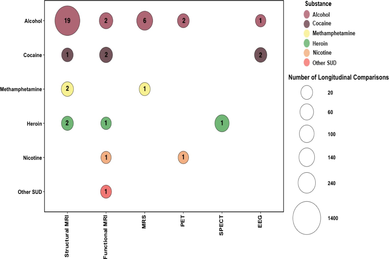
This initial database search yielded 7,749 records, and included studies published up to May 2021. Titles and abstracts from all articles identified through the search were screened. Articles were excluded for being non-relevant (i.e., not related to SUD), case studies, reviews (i.e., literature or systematic), clinical trials (since they examine the impact of a treatment intervention and not of abstinence-mediated recovery, which is the focus of this review), meta-analyses and/or non-human research. A total of 106 articles remained and were assessed more closely for eligibility. Full text articles and studies adhering to the following criteria were included; (1) employed a neuroimaging technique; (2) assessed participants at a minimum of two time-points with an inter-scan duration of greater than 24 hours, separated by a period of abstinence or significant reduction in drug use; (3) SUD sample at the first follow-up was at least n=10; and (4) participants were defined as seeking treatment for SUD. Forty-five studies met eligibility criteria (Figure 1). We summarized the imaging modalities, brain regions, abstinence period prior to any of the scans, statistical analysis thresholds, and within-subject changes in structural, functional, and neurochemical outcomes (emerging during the defined abstinence period) in Table 1.

Fig. 1. PRISMA flowchart detailing the procedure used to for article inclusion.
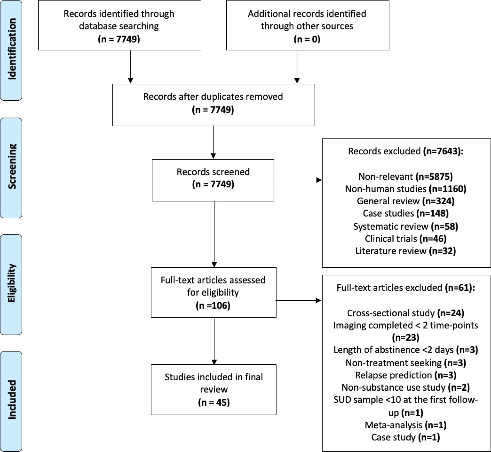
Table 1 - Summary of Brain Recovery in in Human Substance-Using Samples. Studies are sorted by imaging measure, drug type and by the increasing duration of abstinence (at the final neuroimaging scan).

3. STRUCTURAL STUDIES

Abnormalities in brain structure are well documented in individuals with SUD (Mackey et al., 2019). Magnetic resonance imaging (MRI) allows for the detection of subtle variations in the volume and shape of cortical and subcortical regions as well as cortical thickness, area and folding patterns (Ashburner and Friston, 2000). Voxel-based morphometry is a technique that segments brain images into gray matter (GM) volume, white matter (WM) volume, and cerebrospinal fluid to index neuroanatomical abnormalities (Ashburner and Friston, 2000). Although both GM and WM can be assessed using this method, changes in WM integrity are evaluated more accurately using imaging techniques, such as diffusion tensor imaging (DTI), which provides more subtle information about tissue microstructure and organization (Jones et al., 2013; Whitwell, 2009). Common measures computed from DTI are fractional anisotropy, an indicator of WM track myelination (Nucifora et al., 2007), radial diffusivity, a marker of myelin integrity (Harsan et al., 2006), mean diffusivity, a marker of the magnitude of (isotropic) water diffusivity, and axial diffusivity, a marker of the magnitude of diffusion parallel to the fiber tracts.

3.1. Alcohol

3.1.1. Gray Matter

Structural changes have reliably been reported in individuals with alcohol use disorders (AUD) who have achieved abstinence. Using a regions of interest (ROI) approach in 49 alcohol-dependent patients, van Eijk et al., (2013) revealed GM volume increases in several brain regions including the cingulate gyrus, insula, temporal gyrus, precuneus, parietal lobule, and cerebellum following 2 weeks of abstinence (van Eijk et al., 2013). These results were partially supported by a more recent study that used a whole-brain approach to investigate GM volume recovery in 62 individuals with AUD (Bach et al., 2020). Here, GM volume increased in the middle and inferior frontal gyri, middle cingulate gyrus, insula, supramarginal gyrus, and precuneus from approximately 12 days of abstinence to 27 days. Cortical thickness measures show a similar pattern (Wang et al., 2016; Bach et al., 2020). Specifically, in the first 2 weeks of abstinence in 49 alcohol-dependent individuals increased cortical thickness was observed in the medial orbitofrontal cortex (OFC), middle frontal, superior frontal, rostral anterior cingulate cortex (ACC), cuneus, and inferior parietal and lateral occipital regions; greater cortical thickness recovery in sulci compared to gyri, particularly for frontal regions, suggested that sulci may be more sensitive than gyri to excessive alcohol consumption and abstinence-induced recovery (Wang et al., 2016). The Bach et al., (2020) study similarly reported increases primarily in frontal regions encompassing the superior frontal cortices, lateral OFC, and rostral middle frontal cortex, and in the insula and lateral occipital cortex in 62 alcohol-dependent individuals. In general, in these studies, GM (volume and/or thickness) increased in brain regions that showed reductions when individuals with AUD were compared to healthy controls.

When studies investigated abstinence durations of a least 4 weeks, increases in frontal regions became more apparent. For example, increased frontal GM volume (but not parietal, temporal or occipital) was found in 42 individuals with AUD following 4 weeks of abstinence (Durazzo et al., 2017). A study from the same group showed that differences between individuals with AUD and controls in GM volume in frontal regions, such as the OFC (but not the rostral anterior cingulate cortex or insula) observed at one week of abstinence dissipated following 4 weeks of abstinence; yet within-subject frontal changes did not achieve significance when examined longitudinally (Durazzo and Meyerhoff, 2020). Interestingly, following 5 weeks of abstinence, Mon and colleagues (2013) reported that in 41 alcohol-dependent individuals, GM volume increases in the frontal lobe were genotype-specific for a polymorphism (Val66Met) in the neurotrophin, brain-derived neurotrophic factor (BDNF) (Mon et al., 2013). Given that BDNF supports the survival of neurons and promotes neurogenesis in the brain (Lu and Chow, 1999), it has clear relevance to GM volume recovery. Specifically, relative to Val/Val carriers, Met carriers have a significant reduction (~30%) in BDNF secretion, which may compromise their potential for GM volume recovery (Chen et al., 2008). Indeed, while some GM changes were observed with abstinence in alcohol-dependent individuals who were Val/Met carriers (n=15) (increased GM volume in the cerebellum exclusively in this group and increases in the temporal lobe in both genotype groups), only those who were Val/Val carriers (n=26) had increased frontal GM volume following abstinence (Mon et al., 2013). There were also GM volume increases in the parietal lobe and the thalamus, and decreased GM volume in the caudate, again exclusive to Val/Val carriers. Of note, collapsed across genotype group, increases in frontal and parietal GM volume were related to improvement in working memory, with the latter also associated with improvement in processing speed, and increases in all cortical and subcortical GM were related to improvement in visuospatial learning (Mon et al., 2013).

Significant recovery in frontal GM volume, including in the OFC, was observed in investigations following 4 and 7 months of abstinence. Demirakca et al., (2011) showed increases in GM volume in the OFC, as well as the cingulate gyrus, and insula after 4 months of abstinence in 14 alcohol-dependent individuals (Demirakca et al., 2011), while Cardenas et al., (2007) demonstrated increased GM volume in the OFC and parietal lobes following 7 months of abstinence in 17 alcohol-dependent individuals (Cardenas et al., 2007).

Many studies have adopted an ROI approach to specifically examine the hippocampus, a structure that is highly susceptible to neuronal injury (Geddes et al., 2003) but also possesses high potential for neuronal plasticity and regeneration (Leuner and Gould, 2010). Studies have reported increased hippocampal GM volume following 2 weeks (Kuhn et al., 2014), 4 weeks (Gazdzinski et al., 2008), and 7.5 months of abstinence (Zou et al., 2018). Interestingly, similarly to Mon et al., (2013), Hoefer et al., (2014)reported a trend towards increased hippocampal volume following 7 months of abstinence in Val/Val but not Met/Val carriers or controls (Hoefer et al., 2014). Differences from healthy control participants even after this abstinence duration (reduced hippocampal volumes in the AUD) suggest only partial recovery of the hippocampus (Hoefer et al., 2014; Zou et al., 2018). Encouragingly, better visuospatial processing (Hoefer et al., 2014) and visual short- and long-term memory (Gazdzinski et al., 2008) were observed even with only partial hippocampal recovery. In contrast, other studies have reported no change in hippocampal volume early in abstinence [2 weeks (van Eijk et al., 2013; Wang et al., 2016)] or with longer periods of abstinence [4 months (Demirakca et al., 2011)]. Similarly no significant changes in GM volume were found in several other subcortical regions examined as ROIs, such as the amygdala (Demirakca et al., 2011; Wang et al., 2016; Zou et al., 2018) and putamen/lenticular nucleus (Durazzo et al., 2015; Mon et al., 2013; Wang et al., 2016) following abstinence; results observed in the caudate were mixed (Durazzo et al., 2015; Mon et al., 2013; Wang et al., 2016).

Investigations that include more than two time-points provide a window into the trajectory/slope of brain volume recovery over abstinence. Overall, results in individuals with AUD suggest that over protracted abstinence (~ 1 year), the majority of GM volume recovery occurs within the first month of abstinence (Durazzo et al., 2015; Gazdzinski et al., 2005; Pfefferbaum et al., 1995). A large cohort scanned over three time-points (n=82 at 1 week and 1 month; and n=36 at 7.5 months) demonstrated increased GM volumes in the frontal, parietal and occipital lobes, thalamus, caudate, and cerebellum over 7.5 months of abstinence with greater increases occurring between 1 week and 1 month of abstinence compared to 1 month and 7.5 months of abstinence (specifically for frontal and parietal lobes, thalamus, and cerebellum) (Durazzo et al., 2015). With the exception of GM volume in the thalamus, these changes over the 7.5 month of abstinence were associated with better processing speed, a relationship that was only evident in non-smoking individuals (n=18) and did not extend to smokers with AUD (n=18) (Durazzo et al., 2015). Interestingly, a follow-up study in a similar cohort and similar time points (n=65 at 1 week; n=82 at 1 month; and n=36 at 7.5 months; 23 participants had no/unusable data from 1 week of abstinence and thus were first assessed at 1 month of abstinence) revealed that the rate of recovery may be region-specific (Zou et al., 2018). Specifically, whereas the dorsolateral prefrontal cortex (DLPFC), OFC, and insula showed greater increases in GM volume within the first month of abstinence relative to 1 week and 7.5 months, the ACC increased only between 1 month and 7.5 months of abstinence. Together, these studies indicate that GM volume recovery follows a nonlinear trajectory (i.e., steeper slope earlier in recovery) that may vary region by region (Durazzo et al., 2015; Zou et al., 2018).

3.1.2. White Matter

Studies using voxel-based morphometry to index morphological abnormalities in WM have reported mixed results, perhaps because of this technique’s limited sensitivity to quantifying WM volume. Mon et al., (2013) observed increased WM volume in the frontal lobes (and trends for the parietal and temporal lobes) in Val/Met carriers of the BDNF gene following 5 weeks of abstinence. Given the GM findings and BDNF’s contributions as described above, this unexpected finding remains to be replicated in larger sample sizes. Following 4 months of abstinence, two studies reported increased total WM volume (Demirakca et al., 2011; Shear et al., 1994), while following 7.5 months of abstinence, another study demonstrated increased WM volume specifically in the parietal, temporal, and occipital lobes, with the predominance of recovery occurring between 1 and 7.5 months (Durazzo et al., 2015). In contrast, other studies have reported no changes in regional WM volume following 7 months of abstinence (Cardenas et al., 2007) or in total WM volume following 2 weeks (van Eijk et al., 2013), 4 weeks (Durazzo et al., 2017) and 12 months of abstinence (Pfefferbaum et al., 1995).

Using diffusion tensor imaging, the pattern of results is more consistent. Following one month of abstinence, Gazdzinski et al., (2010) observed increased fractional anisotropy in the frontal, temporal, parietal, and occipital lobes and a decrease in mean diffusivity in the temporal lobe in non-smoking in alcohol-dependent individuals (n=10), an effect not present in alcohol-dependent smokers (n=11) (Gazdzinski et al., 2010), suggesting that smoking may selectively affect WM microstructure and recovery in alcohol-dependent individuals. Increased fractional anisotropy and reduced radial diffusivity were seen in the genu and body of the corpus callosum during the first year of abstinence in 15 alcohol-dependent individuals (Alhassoon et al., 2012). Although not directly correlated with the diffusion metrics, significant improvements in working memory at follow-up were noted in these subjects. Increased fractional anisotropy in multiple brain areas (20 out of 27 ROIs assessed, including the corpus collosum, the fornix, and corona radiata) was also observed over a follow-up period of up to 8 years in individuals with alcohol dependence (Pfefferbaum et al., 2014).

3.2. Stimulants and Opioids

A study from our group investigated regional GM volume recovery in 19 abstinent individuals with cocaine use disorder who either achieved abstinence or significantly reduced their cocaine use from baseline (≥ 3 weeks after last drug use) to 6 month follow-up (Parvaz et al., 2016a). Using a whole-brain approach, we demonstrated that GM volume increased in the ventromedial PFC, OFC and inferior frontal gyrus with the latter increases associated with improvements in cognitive flexibility and decision-making measured by the Wisconsin Card Sorting Test and Iowa Gambling Task, respectively (Parvaz et al., 2016a). In another whole-brain study examining individuals with methamphetamine use (n=29), cerebellar GM volume increased, but the cingulate gyrus GM volumes decreased, from 6 months to 12 months of abstinence (Ruan et al., 2018). Over a similar timeframe, Zhuang et al., (2016) showed that compared with baseline (at a 6 months abstinence), individuals with methamphetamine use had continued fractional anisotropy reductions in the internal capsule and superior corona radiata at 13 months of abstinence (Zhuang et al., 2016).

To date only two studies used a longitudinal design to investigate structural recovery in individuals with an opioid use disorder. Employing a whole-brain approach, Wang et al., (2012b) examined the effects of one month of abstinence (compared to a 3 day abstinent scan) in 20 treatment-seeking males with heroin use disorder. While there were no significant longitudinal improvements, the superior frontal gyrus GM abnormalities (as compared to healthy controls) that were documented after 3 days of abstinence were not detectable following one month of abstinence (abnormalities in the other cortical regions, including the middle frontal gyrus, persisted) (Wang, X. et al., 2012). Similarly, although white matter showed no within-subject longitudinal changes with abstinence, abnormalities in fractional anisotropy in the frontal gyrus and cingulate gyrus that were evident after 3 days of abstinence were no longer detectable following one month of abstinence (Wang, X. et al., 2013).

3.3. Interim Summary

Among individuals with AUD, GM volume recovery following abstinence was predominantly assessed via an ROI approach with findings generally indicating increased GM volume in cortical regions spanning the frontal (Cardenas et al., 2007; Demirakca et al., 2011; Durazzo et al., 2017; Durazzo et al., 2015; Gazdzinski et al., 2005; Mon et al., 2013; Pfefferbaum et al., 1995; van Eijk et al., 2013; Zou et al., 2018) and temporal, parietal, and occipital lobes (Cardenas et al., 2007; Durazzo et al., 2015; Mon et al., 2013; van Eijk et al., 2013) as well as the insula (Bach et al., 2020; Demirakca et al., 2011; van Eijk et al., 2013; Zou et al., 2018). Increases were also noted in the hippocampus (Gazdzinski et al., 2005; Gazdzinski et al., 2008; Hoefer et al., 2014; Kuhn et al., 2014; Zou et al., 2018), thalamus (Durazzo et al., 2015; Mon et al., 2013), and cerebellum (Durazzo et al., 2015; Gazdzinski et al., 2005; Mon et al., 2013; van Eijk et al., 2013) but not in the caudate [where mixed results were reported (Durazzo et al., 2015; Mon et al., 2013; Wang et al., 2016)] and lentiform nucleus/putamen (Durazzo et al., 2015; Mon et al., 2013; Wang et al., 2016). Encouragingly, GM recovery occurred as early as 2 weeks post-cessation in select regions (Kuhn et al., 2014; van Eijk et al., 2013) and multi-time point studies suggest that the majority of GM recovery occurs within the first month of abstinence (Durazzo et al., 2015; Gazdzinski et al., 2005; Pfefferbaum et al., 1995; Zou et al., 2018). These changes are associated with improved cognitive function and may be more discernable in certain subgroups of individuals (e.g., with select genes and/or non-smokers).

Given the paucity of studies investigating WM, the general pattern of its recovery remains unclear. While studies using non-DTI methods report both regional and global increases (Demirakca et al., 2011; Durazzo et al., 2015; Mon et al., 2013; Shear et al., 1994), as well as some mixed findings, DTI studies more consistently point to increased WM integrity (as measured by fractional anisotropy) of the corpus collosum following abstinence (Alhassoon et al., 2012; Pfefferbaum et al., 2014).

The majority of the above reviewed studies have been conducted in individuals with AUD following abstinence. Only a handful of studies have examined structural recovery in individuals with substance use disorders other than alcohol including stimulant and opioid use disorder, and no study has investigated structural recovery in treatment-seeking marijuana users. Future research that addresses structural changes associated with these substances following abstinence is clearly warranted.

4. FUNCTIONAL STUDIES

Human brain function is commonly assessed using imaging modalities such as functional MRI (fMRI) and psychophysiological tools such as electroencephalography (EEG). Functional MRI measures local changes in cerebral blood flow and brain metabolism using the blood oxygen level-dependent (BOLD) signal, which is an indirect measure of neural activity that relies on a cascade of physiological events linking neural activity in specific brain regions to the MRI signal. Electroencephalography assesses electrical signals with high temporal resolution, allowing to track human brain function in almost real time, although it is limited by poor spatial resolution. These techniques provide an evaluation of how the brain works dynamically, its physiology, regional connectivity and functional architecture either during rest or in response to specific stimuli. Accordingly, these tools can index neural changes and reorganization that are associated with cessation of or reduction in substance use in individuals with SUD.

4.1. Alcohol

In contrast to the numerous structural neuroimaging studies, to date there have only been a few neuroimaging studies that examined longitudinal changes in brain function during abstinence in AUD. Mon et al. (2009) used arterial spin labeling to examine longitudinal cerebral blood flow changes after 1 month of sobriety relative to baseline (one week of abstinence) in individuals with AUD (n=41) as compared to light social drinkers (n=13). Similarly to the GM volume results reported above, at baseline and compared to the light drinkers, individuals with AUD showed lower cerebral blood flow in frontal and parietal GM. Longitudinally, although there were no significant changes with abstinence in the entire sample of individuals with AUD, recovery (i.e., increase to the level of light social drinkers) in frontal and parietal GM cerebral blood flow was observed only in non-smoking AUD (n=19) but not in the smokers (n=22) (Mon et al., 2009). A subsequent study from the same group divided the 41 individuals with AUD to those who remained abstinent (n=19) vs. those who relapsed (n=22) at 12-months follow-up; here again, although there were no longitudinal changes across the entire group, recovery in cerebral blood flow was observed in those who maintained abstinence but not in those who relapsed (Durazzo et al., 2010). Taken together, these two studies suggest that longitudinal recovery between 1 week and 1 month of abstinence in cerebral blood flow can be observed in non-smoking AUD and/or those who can maintain abstinence.

Further evidence of functional recovery in abstaining alcohol users (n=15) is provided by an EEG study that reported recovery of sleep evoked potentials, recorded from frontal electrodes, after a longer-term (>12 months) abstinence (Colrain et al., 2012). These sleep evoked potentials (mainly the K-complex comprised of the N550 and P900 amplitudes), previously reported to be reduced in individuals with AUD as compared to healthy controls by the same group (Colrain et al., 2009), reflect the functional integrity of the underlying cortex (Colrain, 2005; Tononi and Cirelli, 2006), also representing memory consolidation (Poe et al., 2010).

4.2. Nicotine and Cocaine

In a longitudinal fMRI cue-reactivity study, Janes et al, (2009) reported an increase in fMRI BOLD activity in prefrontal, temporal and parietal regions in response to smoking-related relative to neutral pictures in 13 tobacco-dependent individuals from a pre-cessation baseline to about 1–2 months of abstinence (52 ± 11 days). These results suggest that reactivity to substance-related cues increased during the early phase of abstinence, which is consistent with the trajectory of incubation of cue-reactivity (or craving) as has previously been shown in individuals with other types of SUD using self-reported (Bedi et al., 2011; Li et al., 2015; Wang, G. et al., 2013; Wang, G.B. et al., 2012) and EEG correlates of (Parvaz et al., 2016b) cue-induced craving.

Two fMRI studies investigated longitudinal changes as a function of abstinence duration in individuals with cocaine use disorders, both reporting improved activation in the midbrain and the thalamus. In the first study we used a monetarily rewarded drug Stroop task and showed decreased fMRI BOLD activation (overall task versus baseline) in the midbrain of 15 treatment-seeking cocaine-addicted individuals compared to 13 non-addicted healthy controls at baseline (after detoxification; ≥ 3 weeks after last drug use). After about 6 months of mostly abstinence/substantially reduced drug use, the fMRI BOLD signal in cocaine-addicted individuals was comparable to that in non-addicted healthy controls at baseline. We interpreted these results to suggest a restoration of dopaminergic activity, supported by correlations with reduced drug-seeking behavior in these subjects (Moeller et al., 2012). The later study by Balodis et al. (2016) used the Monetary Incentive Delay task (Knutson et al., 2000) in a larger sample of cocaine-addicted individuals (n=29) and non-addicted healthy controls (n=28), to show similar increases from approximately 2 to 5 months of abstinence in the midbrain and the thalamus, and in the posterior cingulate cortex and the precuneus. Notably, the increase in midbrain activity correlated positively with abstinence duration at follow-up (Balodis et al., 2016). Taken together these results suggest that recovery in the midbrain and thalamus in response to salient reward-relevant tasks is associated with better clinical outcomes (i.e., reduced drug seeking and longer abstinence duration).

Using EEG, our group focused on the late positive potential (an event-related potential observed typically at centroparietal recording sites and indicative of bottom-up attentional change) to report that motivated attention to pleasant cues, which was lower at baseline (after detoxification; ≥ 3 weeks after last drug use) in 19 treatment-seeking cocaine-addicted individuals compared to healthy controls, increased with six months of significantly reduced cocaine use (Parvaz et al., 2017). This increase in reactivity to pleasant cues correlated with longer duration of abstinence at baseline and with decreased craving at follow-up. Nevertheless, reactivity to pleasant cues in the cocaine-addicted individuals at follow-up was still lower than that in healthy controls at baseline, suggesting only a partial recovery with 6 months of significant reduction of cocaine use. Notably, motivated attention to drug-related cues, which was increased in the cocaine-addicted individuals at baseline as compared to the healthy controls, did not change at follow-up, highlighting the protracted nature of the disproportionate attention attributed to drug-related cues in drug addiction (Parvaz et al., 2017). Similarly, a previous study investigating the EEG power in delta band frequency [that reflects frontal cortical regulation of behavioral impulses or concentration/attention allocation to extraneous cues (Fernandez et al., 1995)] also did not show abstinence-related recovery in 17 cocaine addicted individuals from 5 – 10 days to 1 or 6 months of abstinence (Alper et al., 1998). Taken together, these results suggest that while motivated attention to non-drug-related reinforcers may partially recover with 6 months of abstinence or significant reduction in cocaine use, the processes that underlie heightened attention allocation to drug-related cues and maladaptive impulse control may persist. The plasticity in reactivity to salient (including drug) reinforcers could therefore serve as an important target for long-term interventions.

4.3. Heroin and Other SUD

In a resting-state fMRI study, Wang et. al., (2011) showed higher BOLD fMRI signal in the OFC and lower activity in the cerebellar tonsil in 15 individuals with heroin use disorder at 3 days of abstinence compared to 16 non-addicted healthy controls; the activity in the cerebellar tonsil continued to decline as assessed after 1 month of abstinence when activity of frontopolar and subgenual ACC regions was also decreased (Wang et al., 2011). Although, no longitudinal changes were observed in the OFC, the absence of significant cross-sectional differences with the non-addicted healthy controls at 1-month suggests that the OFC activity may have recovered during the first month of abstinence.

In an fMRI study that employed the Balloon Analogue Risk task, 21 treatment-seeking individuals with SUD (12 AUD, four polysubstance dependence, two opioid dependence and one each sedative/hypnotic/anxiolytic dependence, cannabis dependence, and amphetamine dependence) were scanned first at 1 – 4 weeks (baseline) and then at 3 months of abstinence (follow-up) (Forster et al., 2016). Compared to baseline, at follow-up there were increased activations in the dorsal premotor cortex to decision events (when participants are deciding whether or not to inflate the balloon) and in the caudal anterior and posterior cingulate cortices for the success feedback (when participants are informed whether they succeeded and the balloon did not explode on that trial), while the inferior frontal gyrus and the caudal anterior cingulate cortex showed decreased activations to the failure feedback (when the balloon exploded) (Forster et al., 2016). Together, findings were interpreted to reflect an increased surprise signal to unexpected outcomes (i.e., high risk successes and low risk balloon explosions) in recovery, suggestive of the formation of stronger expectancies (Alexander and Brown, 2010, 2014). This study is unique in combining treatment-seeking individuals across different alcohol/drug classes potentially enhancing generalizability of results if replicated in additional samples of individuals with SUDs.

4.4. Interim Summary

There is a paucity of neuroimaging studies examining changes in brain function with abstinence in individuals with SUD. Studies in AUD point to a general recovery in frontal brain regions, showing increased cerebral blood flow in non-smoking and/or abstinent AUD (Durazzo et al., 2010; Mon et al., 2009) and increased amplitude of the EEG-derived auditory sleep evoked potentials (Colrain et al., 2012). In nicotine users, increased reactivity to smoking-related cues during the first 2 months of abstinence (Janes et al., 2009) supports the notion of incubation of cue-reactivity/craving during earlier phases of abstinence (Li et al., 2016). Reports of abstinence-mediated recovery in cocaine use disorder paint a more complex picture with a pattern of recovery that may depend on context. Within the first six months of abstinence, midbrain and thalamic responses to salient stimuli (including money) recover (Balodis et al., 2016; Moeller et al., 2012) with a similar, albeit partial, recovery in reactivity to pleasant cues as documented using an EEG-derived marker of bottom-up/automatic processing (Parvaz et al., 2017). In contrast, heightened reactivity to drug-related cues (Parvaz et al., 2017) or dysregulated attention allocation to extraneous cues (Alper et al., 1998) may be more protracted. In heroin users, however, some evidence for recovery in the OFC resting activity within the first month of abstinence (Wang et al., 2011) warrants replication and further validation. Overall, these studies point to both cortical and subcortical functional recovery during the first year of abstinence in alcohol and cocaine use disorders. More studies are needed to explore functional (cognitive and emotional) recovery in drug addiction (across all drugs of abuse and alcohol) and the effects of context and time in this non-linear multi-layered process. A drug related context may be a crucial variable predisposing addicted individuals to relapse especially at specific times in abstinence as potentially amenable for timely interception.

5. NEUROCHEMICAL STUDIES

In humans, chemical and molecular integrity of brain cells and tissue are quantified using either nuclear imaging techniques such as Positron Emission Tomography (PET) and Single-Photon Emission Computed Tomography (SPECT) or Magnetic Resonance Spectroscopy (MRS). The two nuclear imaging modalities provide a quantitative assessment of regional distribution and kinetics of chemical compounds labeled with short-lived positron (in PET) or gamma (in SPECT) emitting isotopes in the living body. The molecules labeled with these isotopes bind to specific proteins (i.e., receptors and transporters) and can be measured in the tissues of interest as a function over time; PET is also used to assess the cerebral metabolic rate of glucose utilization as well as regional cerebral blood flow. Magnetic Resonance Spectroscopy is also a molecular neuroimaging technique, but does not use ionizing radiation of PET and SPECT, instead leveraging magnetic fields to localize a specific volume of tissue for spectral analysis. Each frequency in the spectrum corresponds to a metabolite nucleus, and the amplitude represents its concentration within the volume. This technique is typically used to study neuronal integrity, most reliably quantifiable via N-acetylaspartate [NAA; a surrogate marker for neuronal density and integrity (Licata and Renshaw, 2010)], choline-containing compounds [Cho; a marker of cell membrane integrity, increased in diseases with increased membrane turnover or axonal injury (Lin et al., 2012)], Creatine [Cr; a metabolite that provides a measure of cellular energy storage (Rackayova et al., 2017)], and myo-inositol [mI; a marker of glial cell density and therefore a measure of inflammation (Brand et al., 1993)], together providing a snapshot of the chemical environment of the selected brain region.

5.1. Alcohol

Most longitudinal studies employed MRS in AUD, showing partial recovery within the first three months of abstinence (Bartsch et al., 2007; Durazzo et al., 2006; Mon et al., 2012; Parks et al., 2002) of the deficits documented in this population within one month of abstinence as compared to light drinkers, encompassing reduced NAA in the ACC (Mon et al., 2012), medial temporal lobe (Gazdzinski et al., 2008), the cerebellum (Ende et al., 2005; Parks et al., 2002) and parietal (Durazzo et al., 2006) and frontal WM (Ende et al., 2005), as well as reduced Cr in the cerebellum and frontal WM (Ende et al., 2005). The partial nature of the recovery is evident in both a differential pattern of results for the different neurochemicals within different ROIs and their different recovery trajectories whereby, for example, cerebellar NAA levels increased from 3 weeks to 3 months of abstinence (n=11), whereas cerebellar Cho levels as well as frontal NAA remained below that of controls during this time frame (Parks et al., 2002). Adding to the evidence for partial neurochemical recovery, Ende et al. (2005)showed increased Cho levels in frontal WM, cerebellar cortex and vermis in AUD individuals following 3-months of complete abstinence (n=14), but reported no change in NAA levels over 3 (n=14) or 6 months (n=11) of abstinence (Ende et al., 2005). These measures of neurochemical recovery correlated with both cognitive function and brain structure. For example, increased levels of fronto-mesial NAA and cerebellar Cho from baseline (<1 week abstinence) to 6 – 7 weeks of abstinence in 15 individuals with AUD were associated with global volumetric brain gain as well as improved attention (Bartsch et al., 2007). In another study, Mon et al (2012) observed lower concentrations of glutamate and glutamate + glutamine in the ACC in treatment-seekers with AUD (n=20) at approximately nine days of abstinence, which normalized (compared to healthy controls, n=16) over four weeks of sustained abstinence but were not associated with cognitive improvement (Mon et al., 2012).

Similarly to the above reviewed structural and functional studies, the lack of consistency of results between these studies may reflect diverse subject characteristics including comorbid cigarette smoking. For example, Durazzo et al. (2006) reported significant increases in NAA (frontal GM and WM), Cho (frontal and parietal GM & WM, and occipital WM), and mI and Cr (frontal WM) concentrations in 25 individuals with AUD following approximately one-month of abstinence. However, when further stratified based on cigarette smoking status, the results diverged between non-smokers (n=11) and smokers (n=14) such that the former drove most of these changes with the latter showing increased NAA and Cho only in frontal GM and a decrease in NAA in parietal and occipital WM, suggesting that cigarette smoking may adversely affect metabolite recovery in AUD (Durazzo et al., 2006). A later study from the same group reported a similar trend for a partial recovery in Cho and NAA in non-smokers but not in smokers (Gazdzinski et al., 2008). Taken together, studies using MRS present an encouraging, albeit not entirely consistent, pattern of results with respect to changes in Cho and NAA recovery with abstinence in AUD. Low sample sizes may be another source contributing to this discrepancy.

In a PET study Ceccarini et al (2014) reported a global deficiency (−16.1%) of the endocannabinoid signaling pathways, especially in the availability of the type 1 cannabinoid receptor (CB1R), in individuals with AUD (n=26) as compared to healthy controls (n=17). Such blunted CB1R availability in the cerebellum and parieto-occipital cortex, the ventral striatum and the mesotemporal lobe did not recover after one month of abstinence (−17.0%), highlighting persistent deficits in the endocannabinoid signaling pathways, at least within the first month of abstinence in AUD (Ceccarini et al., 2014). In a recent PET study, the same group showed reduced corticolimbic metabotropic glutamate receptor 5 (mGluR5) availability in AUD at baseline (<2 weeks post-detoxification), which recovered up to the levels observed in healthy controls across 2 and 6 months of abstinence in most cortical and subcortical regions, except for the hippocampus, nucleus accumbens and thalamus. Interestingly, lower striatopallidal mGluR5 availability at baseline was associated with higher propensity to relapse at 6 months and its longitudinal normalization was associated with lower craving. Together, these results suggest that, unlike deficits in CB1R, those in mGluR5 availability normalize with abstinence in AUD (Ceccarini et al., 2014; Ceccarini et al., 2020), and such normalization, especially in the mGluR5 availability, is associated with decreased craving in this population (Ceccarini et al., 2020).

5.2. Nicotine, Methamphetamine and Heroin

Using 6-[18F]fluoro-L-DOPA (FDOPA)-PET, Rademacher et al (2016) compared presynaptic dopamine function between 15 nonsmokers and 30 nicotine-dependent smokers studied before and after 3 months of abstinence. Results revealed a 15% to 20% lower capacity of dopamine synthesis in the dorsal and ventral regions of the caudate nuclei of sated smokers as compared to non-smokers, which normalized during three months of abstinence to the level of non-smoking controls. Interestingly, this time course is consistent with earlier research suggesting that the cholinergic system takes approximately three months to normalize in abstinent tobacco smokers (Cosgrove et al., 2009).

Using MRS, Burger et al. (2018) showed lower NAA and NAA with n-acetyl-aspartyl-glutamate concentration in the DLPFC and lower Cho concentration in frontal WM in 31 individuals with a methamphetamine use disorder compared to 22 non-addicted controls. In contrast to the results of partial NAA recovery with abstinence in the AUD population, a longitudinal examination (n=22) from acute (up to 2 weeks) to short-term (up to 6 weeks) abstinence revealed further reduction in NAA and NAA with n-acetyl-aspartyl-glutamate concentrations in the ACC and frontal WM. Over time, there were also decreased levels of myo-inositol in the left frontal WM, while an increase in myo-inositol was seen in the ACC (Burger et al., 2018).

Treatment-seeking heroin users (n=55), randomly assigned to receive either Placebo or Jitai (a traditional Chinese medicine that has been approved by the China Food and Drug Administration for treatment of opioid addiction), were scanned with SPECT using [99mTc]TRODAT-1 to examine longitudinal changes in dopamine transporter concentration from baseline (almost 20 days abstinent), to three, six, and 12 months of abstinence (Xu et al., 2015). At baseline, compared to healthy controls (n=20; scanned once), the heroin addicted individuals showed lower dopamine transporter concentrations in the striatum (by 30%). Longitudinal analyses showed that the individuals assigned to the Jitai group had a steady increase in the dopamine transporter concentrations, while in the placebo group results were mixed, such that there was an increase from baseline to three months, a slight decrease from three to six months and then an increase from six to 12 months follow-up. Importantly, both groups showed a longitudinal increase in dopamine transporter concentrations from baseline to 12 months follow-up (by 20%) (Xu et al., 2015).

5.3. Interim Summary

Neurochemical techniques, especially MRS in alcohol use, have been predominantly used to quantify molecular recovery as a function of abstinence in individuals with SUD. Most studies in AUD have shown consistent increases in NAA concentration within the first 3 months of abstinence (Bartsch et al., 2007; Durazzo et al., 2006; Parks et al., 2002). The frontal cortex and the cerebellum have been the most studied ROIs, whereas some studies have also examined the parietal cortex and the medial temporal lobe. Early recovery (within one month of abstinence) shows increased NAA in the frontal cortex and the medial temporal lobe and increased Cho in frontal, temporal, parietal and occipital lobes, driven by non-smoking individuals with AUD (Durazzo et al., 2006; Gazdzinski et al., 2008). During more protracted abstinence (2 to 6 months) studies show conflicting results. For example, whereas some show increase in the cerebellar Cho (Bartsch et al., 2007; Ende et al., 2005), others do not (Parks et al., 2002), and whereas some show no change in frontal NAA (Ende et al., 2005; Parks et al., 2002), others do (Bartsch et al., 2007). Thus, in AUD, variability of results was observed based on length and status of abstinence, the metabolite and ROI under investigation, and cigarette smoking. In methamphetamine use disorder there was no recovery as a function of abstinence, with results instead suggesting continued reduction in NAA in the ACC and frontal WM up to 5 weeks of abstinence (Burger et al., 2018).

Nuclear imaging results suggest a dopaminergic recovery with abstinence. For example, a PET study in nicotine users showed increased dopamine synthesis in the dorsal and ventral caudate with over 5 weeks of abstinence (Rademacher et al., 2016). A study in heroin users showed recovery in dopamine transporter concentration in the striatum after 6 – 12 months of abstinence (Xu et al., 2015). In AUD, although mGluR5 availability showed overall recovery in both cortical and subcortical regions during the first 6 months of abstinence (Ceccarini et al., 2020), CB1R availability did not recover, at least within the first month of abstinence (Ceccarini et al., 2014).

6. DISCUSSION

Longitudinal studies that assess within-subject changes in brain morphology and function including neurochemistry following sustained abstinence are optimal for identifying the potential for and the trajectory of neural recovery in individuals with SUD. Overall, the reviewed research suggests that neural deficits dissipate following a period of sustained abstinence from substance use in individuals with SUD. In each of the three reviewed sections, sustained abstinence was predominantly associated with (at least partial) recovery, such that over time deficits in select regions appeared to normalize, implying that these abnormalities are likely consequences of substance consumption rather than premorbid or risk factors for SUD. Importantly, these neural substrates may serve as potential biomarkers that can be targeted for treatment of SUDs.

Structural recovery occurred predominantly in frontal cortical regions, the insula, hippocampus, and cerebellum. In addition to prefrontal cortical regions, functional recovery was also observed in subcortical structures (midbrain, striatum, thalamus). While reversal of neural damage was evident across studies and modalities used, numerous instances of regional specificity and variability/inconsistencies in time-course and pattern of these changes were noted. These discrepancies may reflect between-study differences in the use of ROI versus whole-brain approaches (with the former increasing the susceptibility to selection biases), inter-scan intervals and clinical characteristics (e.g., concurrent substance use, severity of SUD), calling for more and larger studies of this type. A question to explore is whether specific brain regions may be faster or more amenable to recovery, particularly the frontal cortex, while other regions may show a slower trajectory or be more impervious to change.

For structural studies, where recovery was primarily indexed as GM volume increases in individuals with AUD, greater changes occurred relatively early in the course of abstinence (i.e., within the first month of cessation), while relatively less change occurred with longer abstinence [i.e., post 6 months (Durazzo et al., 2015; Pfefferbaum et al., 1995; Zou et al., 2018)] indicating that GM structural recovery may follow a nonlinear trajectory (Gazdzinski et al., 2005). More longitudinal multi-interval studies that assess WM integrity are needed to determine the trajectory of WM recovery, particularly studies that employ DTI, rather than voxel-based morphometry, to more sensitively index changes in WM microstructure and organization following abstinence.

Similarly, early recovery was observed in neurometabolite levels in individuals with AUD (Ende et al., 2005; Parks et al., 2002). However, such early recovery was not observed in CB1R availability (Ceccarini et al., 2014) and the recovery in mGluR5 availability was most evident several months post-cessation (Ceccarini et al., 2020), suggesting a heterogeneous functional molecular recovery profile. An exception was noted for individuals with a methamphetamine use disorder, where studies generally demonstrated that neural abnormalities may worsen with abstinence, even up to a year (Burger et al., 2018; Ruan et al., 2018; Zhuang et al., 2016), raising the possibility that neurotoxic effects are not only associated with current methamphetamine use, but also with withdrawal from the drug, and/or with premorbid factors. Similar maladaptive longitudinal change was seen in chronic cigarette smokers who showed increased reactivity to smoking related cues during the course of early abstinence (Janes et al., 2009). Such an increase in (or incubation of) drug cue-reactivity during the initial phase of abstinence has been consistently seen in animal models of addiction, and is now being observed in human studies as well (Li et al., 2016). Overall, evidence suggests that recovery is not a uniform process but instead may occur along a non-linear trajectory (i.e., different phases of recovery). This non-linear trajectory mirrors findings from a mega-analysis that demonstrated the absence of substance-specific linear effects on brain volume where both the impact of the drug and GM recovery may be more complex than can be elucidated by a simple linear analysis (Mackey et al., 2019). While speculative, it is plausible that functional recovery may be dependent, to some degree, on initial structural recovery, which may explain its delayed onset. More specifically, because GM volume reductions in specific regions may be related to alteration in functional response (Fu et al., 2008; Yuan et al., 2010), improvement in brain function may be subsequent to neural repair of structural networks (Crews et al., 2005). However, it is also possible that structural imaging is simply more sensitive and hence can detect effects earlier compared to functional imaging (Johansen-Berg, 2012).

It is important to note that the dynamic brain changes observed within the first few days following cessation may be associated, in part, with withdrawal and compensatory actions in response to drug removal [e.g. (Wang, X. et al., 2012)]. Thus, any damage associated with excessive drug use needs to be teased apart from that associated with the sub-acute, residual, effects of the drug (Fernandez-Serrano et al., 2011). It has been suggested that a minimum of two weeks of abstinence must be maintained to make this distinction (Schulte et al., 2014), which indeed has been undertaken by all but three studies reviewed here [(Kuhn et al., 2014; van Eijk et al., 2013; Wang et al., 2016)]. Considering the other end of the time spectrum, only one study (Pfefferbaum et al., 2014) assessed an abstinence period greater than 13 months, prohibiting the examination of the long-term trajectory of recovery, which clearly awaits future studies. Studies examining relationships between protracted abstinence length and neural outcomes suggest that recovery is ongoing and not the result of a single neural process (He et al., 2018; O’Neill et al., 2001). Future studies employing multiple assessments at different time-points and combining different imaging modalities over extended abstinence periods (>13-months), are warranted to accurately capture the precise recovery time-course associated with protracted abstinence in individuals with SUD.

A goal of this body of neuroimaging research is to leverage these data to improve prognostic outcomes in individual with SUDs, thus linking neural recovery to clinical improvements is fundamental. Findings from the reviewed studies demonstrate that structural (Durazzo et al., 2006; Gazdzinski et al., 2008; Hoefer et al., 2014; Mon et al., 2013; Parvaz et al., 2016a) and functional (DeVito et al., 2012; Forster et al., 2016) including neurochemical (Bartsch et al., 2007; Durazzo et al., 2006) improvements correlate with enhanced cognitive performance, indicating putative neurobiological substrates for cognitive recovery following abstinence in individuals with SUD. While not assessed in every study or across every substance, these findings are encouraging given that cognitive dysfunction is a vulnerability factor for relapse (Stevens et al., 2014) and better cognitive function is associated with achieving more favorable treatment outcomes in recovering individuals (Sofuoglu et al., 2013). Further, while not assessed in any of the reviewed studies, predictors of relapse beyond cognition, such as craving, have been associated with changes in GM volume (Makris et al., 2004). Thus, the full breadth of improved clinical outcomes and their relation to neural regeneration over abstinence remains to be discerned. Lastly, demonstrating that better brain health is achievable with abstinence may serve as a powerful motivational tool to encourage cessation and inspire treatment engagement among individuals with SUD and treatment providers.

The prevalence of cigarette smoking among individuals with SUD is high (John et al., 2003; Weinberger et al., 2018; Weinberger and Sofuoglu, 2009; Zale et al., 2015) and evidence suggests that cigarette smoking negatively affects neural recovery. Compared to non-smokers, heavy cigarette smokers have lower GM volume (Brody et al., 2004; Gallinat et al., 2006), lower global cerebral blood flow (Domino et al., 2004), and altered neurochemistry (Ashok et al., 2019; Moffett et al., 2007). Functional MRI studies suggest that nicotine may modulate task-induced BOLD responses and have performance enhancing properties (Hahn et al., 2009). Collectively, this evidence underscores the importance to control for the confounding effects of cigarette smoking on results. While some of the studies in individuals with AUD prospectively parsed smokers from non-smokers to determine if recovery differed as a function of cigarette smoking [e.g., (Durazzo et al., 2006; Durazzo et al., 2015; Gazdzinski et al., 2008; Mon et al., 2009)], this was not done for any other SUD. Some studies did, however, include baseline cigarette smoking as a covariate in longitudinal analyses when differences between the control and SUD group emerged (Mon et al., 2013). Given that it is common for individuals in treatment for SUD to also quit smoking cigarettes (Orleans and Hutchinson, 1993), future studies may consider modeling the trajectory of cigarette consumption over SUD abstinence to appropriately disassociate the effects of the primary substance from cigarette use on the changes in neural outcomes.

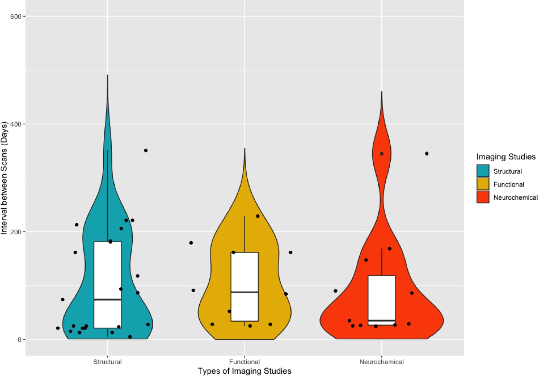
Several limitations of this review are worth noting. First, this review included studies that employed behavioral and/or other interventions [e.g., cognitive behavioral therapy (Balodis et al., 2016; DeVito et al., 2012) and traditional Chinese medicine (Xu et al., 2015)] to facilitate abstinence. These treatments may have induced positive neural or neurochemical changes over abstinence (DeVito et al., 2017; Seminowicz et al., 2013). Disassociating the intervention effects from those related directly to abstinence is yet to be achieved. Second, a caveat of this research is that treatment-seekers in general, or those who successfully achieve abstinence, might possess less neurobiological vulnerability and be more apt to neural recovery compared to those who are unable to quit or maintain abstinence (e.g., non-treatment-seekers and/or relapsers) (Martinez et al., 2011; Wang, G.J. et al., 2012). Third, methodologies varied across studies (e.g., sample characteristics, abstinence length (see Figure 3 for the distribution of inter-scan interval in structural, functional, and neurochemical studies), treatment administered, imaging techniques, specific ROIs examined, statistical thresholds) making direct between-study comparisons difficult. In addition, while within-study paradigms offer benefits over cross-sectional designs, these studies must still ensure that adequate control groups are enrolled and assessed at equivalent time-points to the SUD group and the influence of potential confounders are controlled for in the analyses (e.g., age, comorbid substance use, baseline SUD severity). Fourth, our inferences are based on a limited number of studies, and most studies had limited sample sizes, primarily due to high attrition rates. High attrition rate has been a major limitation in conducting longitudinal studies in human drug addiction, partially because of the highly mobile, unstable, and transient lifestyles of many study participants (BootsMiller et al., 1998). Lastly, there is a predominance of studies examining recovery in treatment-seeking individuals with AUD, while recovery in other SUD (e.g., opiates and cannabis) has been generally overlooked, representing a critical gap in this area of research as seen in Figure 2. Nevertheless, the literature is growing, and we anticipate that future studies, with larger sample sizes, and longer follow-up periods will help clarify these issues.

Figure 2: Summary of the number of longitudinal comparisons (for participants with Substance Use Disorder) in the collected studies for each imaging modality and substance.Figure 3: Distribution of inter-scan interval in structural, functional, and neurochemical studies.

The use of imaging techniques in addiction research has increased substantially in the last decade and many of these studies have been instrumental in providing evidence that structural and functional including neurochemical deficits may recover with even short periods of abstinence. Beyond providing hope for individuals with SUD and encouraging them to seek treatment, and providing evidence-based treatment, characterizing these neurobiological processes may help to identify novel biomarkers that can be targeted for SUD timely intervention. Capturing the trajectory of neural changes over abstinence (or even using a harm-reduction approach) may help establish a neuroscience-informed framework for developing pharmacological, psychotherapeutic, and/or neuromodulatory interventions that can mimic and/or enhance the brain’s ability to repair itself, restoring cognitive function, and contributing to positive long-term treatment outcomes in individuals with SUD.

Highlights.

  • Structural recovery occurred predominantly in frontal cortical regions, the insula, hippocampus, and cerebellum.

  • In addition to prefrontal cortical regions, functional recovery was also observed in subcortical structures (midbrain, striatum, thalamus).

  • A question to explore is whether specific brain regions may be more amenable to recovery, particularly the frontal cortex, while other regions may be more impervious to change.

  • Characterizing these neurobiological processes may help to identify novel biomarkers that can be targeted for timely intervention for substance use disorders.

  • Results provide hope for treatment-seeking individuals with substance use disorders and encourage them to seek treatment.

Open Article as PDF

Abstract

Background: Neuroimaging studies reveal structural and functional including neurochemical brain abnormalities in individuals with substance use disorders compared to healthy controls. However, whether and to what extent such dysfunction is reversible with abstinence remains unclear, and a review of studies with longitudinal within-subject designs is lacking. We performed a systematic review of longitudinal neuroimaging studies to explore putative brain changes associated with abstinence in treatment-seeking individuals with substance use disorders. Methods: Following PRISMA guidelines, we examined articles published up to May 2021 that employed a neuroimaging technique and assessed neurobiological recovery in treatment-seeking participants at a minimum of two time-points separated by a period of abstinence (longer than 24 h apart) or significant reduction in drug use. Results: Forty-five studies met inclusion criteria. Encouragingly, in this limited but growing literature, the majority of studies demonstrated at least partial neurobiological recovery with abstinence. Structural recovery appeared to occur predominantly in frontal cortical regions, the insula, hippocampus, and cerebellum. Functional and neurochemical recovery was similarly observed in prefrontal cortical regions but also in subcortical structures. The onset of structural recovery appears to precede neurochemical recovery, which begins soon after cessation (particularly for alcohol); functional recovery may require longer periods of abstinence. Conclusions: The literature is still growing and more studies are warranted to better understand abstinence-mediated neural recovery in individuals with substance use disorders. Elucidating the temporal dynamics between neuronal recovery and abstinence will enable evidence-based planning for more effective and targeted treatment of substance use disorders, potentially pre-empting relapse.

INTRODUCTION

Substance use disorders (SUDs) are chronic conditions marked by compulsive drug seeking and use, even when pleasure from the drug decreases and harmful outcomes occur. Brain areas responsible for impulse control and recognizing important signals are thought to play a role in the cycle of addiction. Brain imaging studies consistently show changes in brain structure, function, and chemistry in various brain regions after long-term substance exposure, regardless of the specific drug used. Importantly, these brain changes often align with shifts in thinking abilities, mood symptoms, and treatment success, indicating a significant impact on recovery.

However, whether these brain changes linked to long-term substance use are permanent or recover with abstinence has been debated. Studies often compare current substance users with former users and healthy individuals. While these comparisons are necessary, such studies, which look at different groups at one point in time, can be unclear due to differences between individuals, such as other drug use or mood issues. They also make it hard to distinguish changes caused by chronic drug use from pre-existing brain characteristics or vulnerabilities. Furthermore, these single-time-point studies often have small sample sizes, which increases the risk of reporting false findings or missing real, subtle changes. In contrast, studies that track the same individuals over time can account for individual differences and offer more statistical power. These designs allow for a more direct look at how changes unfold while controlling for other variables that might influence results.

This document specifically reviews longitudinal brain imaging studies that tracked brain changes within the same individuals between early and extended periods of abstinence in people seeking treatment for SUDs. Carefully tracking within-subject brain changes during abstinence is crucial for understanding how the brain recovers over time. This understanding can help identify brain processes linked to maintaining long-term abstinence or to the risk of relapse. Furthermore, these findings may help identify important personalized indicators and targets for early prevention and treatment to improve recovery and reduce the chronic relapsing nature of drug addiction.

METHODS

A careful search of scientific literature was conducted to identify brain imaging studies that looked at how stopping alcohol or drug use affects brain structure, function, and chemistry. The search used terms for various imaging techniques, such as MRI, fMRI, PET, EEG, MRS, and SPECT. These were combined with terms related to substance use disorders (SUDs), including specific drugs, and terms referring to abstinence or treatment. The initial search was limited to full-text, English-language articles published in peer-reviewed journals up to May 2021 and followed standard guidelines for scientific reviews.

This initial search found 7,749 articles. The titles and summaries of all found articles were reviewed. Articles were excluded if they were not related to SUDs, were single case studies, reviews, clinical trials (as these focus on treatment effects rather than recovery due to abstinence), meta-analyses, or non-human research. After this screening, 106 articles remained and were assessed in more detail. Studies were included if they: (1) used a brain imaging technique; (2) assessed participants at least two times, with more than 24 hours between scans, and a period of abstinence or much less drug use between these scans; (3) had at least 10 participants with SUD at the first follow-up; and (4) involved participants seeking treatment for SUD. Forty-five studies met these criteria. Information on imaging methods, brain regions, abstinence duration before scans, statistical analysis, and within-subject changes in structural, functional, and chemical brain outcomes were summarized.

STRUCTURAL STUDIES

Changes in brain structure are well known in individuals with substance use disorders (SUDs). Magnetic resonance imaging (MRI) can detect subtle changes in the volume and shape of brain regions, including gray matter (GM) and white matter (WM). Techniques like voxel-based morphometry measure GM and WM volume, while diffusion tensor imaging (DTI) provides more detailed information about WM organization and integrity, using measures like fractional anisotropy (indicating myelination) and various diffusivities (reflecting tissue microstructure).

For individuals with alcohol use disorders (AUD), structural changes in gray matter have been reliably observed during abstinence. Studies show increases in GM volume and/or thickness in various brain regions, particularly in frontal areas, the insula, hippocampus, temporal, parietal, and occipital lobes, as well as the thalamus and cerebellum. These increases often begin as early as two weeks into abstinence, with most significant recovery occurring within the first month. Such brain recovery is frequently linked to improvements in thinking abilities, and factors like an individual's genetic makeup and smoking status can influence the extent of recovery. The trajectory of GM recovery often appears to be non-linear, with more rapid changes occurring in the initial phases of abstinence. White matter studies, especially those using DTI, more consistently indicate increased WM integrity in regions like the corpus callosum during abstinence, although findings using other methods are mixed.

Fewer longitudinal studies have investigated structural recovery in individuals with stimulant or opioid use disorders. For cocaine use disorder, some studies have reported increased GM volume in areas of the prefrontal cortex, which was associated with better cognitive function and decision-making. In methamphetamine use disorder, findings are mixed, with some studies showing increases in cerebellar GM volume but decreases in cingulate gyrus GM, and continued reductions in white matter integrity even with abstinence. For heroin use disorder, some initial GM abnormalities in the superior frontal gyrus appeared to normalize within a month of abstinence, but no significant within-subject changes in white matter were consistently observed.

Overall, brain structure recovery, primarily involving increases in gray matter volume, is seen in individuals with AUD, especially in frontal cortical regions, the insula, hippocampus, and cerebellum. This recovery can begin early in abstinence and often shows a non-linear pattern, with significant changes occurring within the first month. For white matter, DTI studies generally suggest improved integrity. Research on structural recovery in other SUDs, such as stimulant and opioid use disorders, is more limited and shows varied results, indicating a need for more studies across different substance types.

FUNCTIONAL STUDIES

Human brain function is often assessed using imaging methods like functional MRI (fMRI) and electroencephalography (EEG). Functional MRI measures changes in blood flow and metabolism in the brain, providing an indirect measure of brain activity. EEG measures electrical signals, offering high temporal detail to track brain function almost instantly, though it has less precise spatial information. These techniques allow for an evaluation of how the brain works dynamically, its physiology, connections, and functional organization, either at rest or in response to specific stimuli. They can track brain changes and reorganization associated with stopping or reducing substance use in individuals with SUD.

There have been fewer brain imaging studies examining longitudinal changes in brain function during abstinence in alcohol use disorder (AUD). Studies point to a general recovery in frontal brain regions, showing increased cerebral blood flow in non-smoking individuals with AUD or those who maintain abstinence. Additionally, long-term abstinence has been linked to the recovery of sleep evoked potentials, which are measured by EEG from frontal electrodes and reflect the functional health of the underlying brain areas and memory consolidation.

Studies on nicotine users suggest increased brain activity in response to smoking-related cues during the early phase of abstinence, supporting the idea of heightened craving. For cocaine use disorder, fMRI studies report improved activation in the midbrain and thalamus in response to salient rewards (like money) following abstinence. These improvements are associated with better clinical outcomes, such as reduced drug-seeking behavior and longer abstinence. While reactivity to pleasant cues may show partial recovery, heightened attention to drug-related cues and issues with impulse control may persist.

In heroin use disorder, there is some evidence for recovery in the resting activity of the orbitofrontal cortex within the first month of abstinence. A study involving individuals with various SUDs showed that during abstinence, there were increased brain activations related to decision-making and positive feedback, along with decreased activations for negative feedback. Overall, these studies indicate functional recovery in both cortical and subcortical regions during the first year of abstinence for alcohol and cocaine use disorders. More research is needed to explore functional recovery across all types of substance use disorders and to understand how context and time influence this complex, non-linear process.

NEUROCHEMICAL STUDIES

The chemical and molecular health of brain cells and tissue in humans can be measured using nuclear imaging techniques like Positron Emission Tomography (PET) and Single-Photon Emission Computed Tomography (SPECT), or with Magnetic Resonance Spectroscopy (MRS). Nuclear imaging methods provide information on the distribution of chemical compounds that bind to specific brain proteins, or they can measure brain metabolism and blood flow. MRS is a non-ionizing technique that uses magnetic fields to study metabolites, such as N-acetylaspartate (NAA), a marker for neuronal health; choline-containing compounds (Cho), indicating cell membrane integrity; creatine (Cr), related to cellular energy; and myo-inositol (mI), a marker for glial cell density and inflammation. These measurements offer insight into the chemical environment of specific brain regions.

Most longitudinal MRS studies in alcohol use disorder (AUD) show partial neurochemical recovery within the first three months of abstinence. Deficits in compounds like NAA and Cr in various brain regions (including the anterior cingulate cortex, medial temporal lobe, cerebellum, and frontal/parietal white matter) tend to increase, indicating some recovery, though they may not fully return to normal levels. Changes in Cho levels show mixed results. This recovery can vary depending on the brain region, duration of abstinence, and factors such as cigarette smoking. PET studies in AUD have shown that while deficits in endocannabinoid signaling pathways (CB1R) did not recover within one month of abstinence, deficits in metabotropic glutamate receptor 5 (mGluR5) availability generally normalized over two to six months, with this normalization linked to decreased craving.

For nicotine use, a PET study found that the brain's capacity for dopamine synthesis in the caudate nucleus, initially lower in smokers, normalized after three months of abstinence. In contrast, MRS studies in methamphetamine use disorder showed a further reduction in NAA concentrations in certain frontal brain areas during abstinence, suggesting ongoing neuronal damage rather than recovery. For heroin use disorder, a study observed a recovery in dopamine transporter concentrations in the striatum after six to twelve months of abstinence.

Neurochemical techniques, particularly MRS in alcohol use, have been primarily used to measure molecular recovery during abstinence in individuals with SUD. Most AUD studies consistently show increases in NAA concentration within the first three months of abstinence, mainly in the frontal cortex and cerebellum, with smoking potentially affecting these outcomes. In methamphetamine use disorder, some neurochemical abnormalities may worsen rather than improve with abstinence. However, nuclear imaging results suggest a recovery in dopamine systems for nicotine and heroin users. The recovery profile can be complex, with some neurochemical pathways normalizing while others do not.

DISCUSSION

Longitudinal studies that assess brain changes within the same individuals during sustained abstinence are crucial for understanding the potential and path of neural recovery in those with substance use disorders (SUDs). Overall, the reviewed research indicates that brain deficits linked to SUDs diminish after a period of abstinence. This suggests that these abnormalities are likely a result of substance use rather than pre-existing conditions, making these neural markers potential targets for SUD treatment.

Structural recovery, involving increases in gray matter volume, was predominantly observed in frontal cortical regions, the insula, hippocampus, and cerebellum. This recovery often begins early in abstinence, with the most significant changes occurring within the first month, following a non-linear pattern. White matter integrity also showed improvements. Functional recovery was seen in prefrontal brain regions and subcortical structures like the midbrain, striatum, and thalamus. Neurochemical recovery, while common, displayed varied patterns, with some metabolites recovering early, while others, like certain receptors, showed delayed or no recovery. This variability suggests that recovery is not uniform and may differ by brain region, type of deficit, and substance.

Inconsistencies across studies reflect methodological differences, such as the use of region-of-interest versus whole-brain approaches, varying scan intervals, and diverse participant characteristics including co-occurring substance use or SUD severity. Early brain changes immediately after stopping drug use may be partly due to withdrawal effects; therefore, studies typically require at least two weeks of abstinence to better distinguish drug-induced damage from acute withdrawal. Cigarette smoking is also a significant factor, often negatively affecting neural recovery in alcohol use disorder and likely impacting other SUDs. Furthermore, some studies included behavioral or other treatments that could have influenced neural changes, making it difficult to isolate the effects purely attributable to abstinence.

A significant limitation is the scarcity of studies with very long abstinence periods (over 13 months), which prevents a full understanding of the long-term trajectory of recovery. Most research has focused on alcohol use disorder, leaving a critical gap in understanding recovery for other SUDs like opioid and cannabis use disorders. Small sample sizes and high participant dropout rates also pose challenges. It is also possible that treatment-seeking individuals, or those who successfully achieve abstinence, may have been less severely vulnerable to begin with. Future research should include larger samples, multiple assessments over extended periods, and combine different imaging techniques to more accurately map the complex, non-linear path of brain recovery. Critically, linking these neural recoveries directly to improvements in clinical outcomes, such as cognitive function and reduced craving, is essential for developing neuroscience-informed treatments and for providing compelling motivation for individuals to seek and maintain abstinence.

Highlights

  • Brain structure recovery, mainly in areas like the frontal cortex, insula, hippocampus, and cerebellum, was observed.

  • Functional recovery was seen in prefrontal brain regions and deeper brain areas such as the midbrain, striatum, and thalamus.

  • Research should explore if certain brain regions, especially the frontal cortex, recover more easily than others.

  • Understanding these brain changes can help find new markers for early substance use disorder interventions.

  • These findings offer hope for individuals seeking treatment for substance use disorders and encourage treatment engagement.

Open Article as PDF

Abstract

Background: Neuroimaging studies reveal structural and functional including neurochemical brain abnormalities in individuals with substance use disorders compared to healthy controls. However, whether and to what extent such dysfunction is reversible with abstinence remains unclear, and a review of studies with longitudinal within-subject designs is lacking. We performed a systematic review of longitudinal neuroimaging studies to explore putative brain changes associated with abstinence in treatment-seeking individuals with substance use disorders. Methods: Following PRISMA guidelines, we examined articles published up to May 2021 that employed a neuroimaging technique and assessed neurobiological recovery in treatment-seeking participants at a minimum of two time-points separated by a period of abstinence (longer than 24 h apart) or significant reduction in drug use. Results: Forty-five studies met inclusion criteria. Encouragingly, in this limited but growing literature, the majority of studies demonstrated at least partial neurobiological recovery with abstinence. Structural recovery appeared to occur predominantly in frontal cortical regions, the insula, hippocampus, and cerebellum. Functional and neurochemical recovery was similarly observed in prefrontal cortical regions but also in subcortical structures. The onset of structural recovery appears to precede neurochemical recovery, which begins soon after cessation (particularly for alcohol); functional recovery may require longer periods of abstinence. Conclusions: The literature is still growing and more studies are warranted to better understand abstinence-mediated neural recovery in individuals with substance use disorders. Elucidating the temporal dynamics between neuronal recovery and abstinence will enable evidence-based planning for more effective and targeted treatment of substance use disorders, potentially pre-empting relapse.

INTRODUCTION

Substance use disorders (SUD) are chronic conditions marked by a persistent urge to seek and use drugs, even when the pleasure from them decreases and negative consequences arise. Problems with controlling impulses and recognizing important signals, which are functions of the brain's prefrontal cortex, are believed to play a role in the cycle of addiction. Studies using brain imaging consistently show changes in the structure, function, and chemistry of the prefrontal cortex and other brain areas after long-term exposure to addictive substances. These changes often align with difficulties in thinking, emotional problems, and how well people respond to treatment, suggesting a significant impact on health.

A key question is whether these brain changes caused by long-term substance use are permanent or can improve with abstinence. Many studies compare current users to those who have stopped using and to healthy individuals. However, these types of studies, which look at different groups at one point in time, can be complicated by individual differences, such as other drug use or mood issues. They also make it hard to tell if brain changes existed before drug use or are a result of chronic use. These studies can also be prone to errors due to small participant numbers.

In contrast, studies that track individuals over time (longitudinal studies) are better. They allow each person to serve as their own control, with brain changes measured repeatedly. This approach provides stronger statistical evidence and helps account for other factors that might influence the results. This document specifically examines longitudinal brain imaging studies that have looked at brain changes in treatment-seeking individuals with SUDs from early abstinence to longer periods of sobriety. Understanding how the brain changes over time is crucial for identifying mechanisms linked to sustained abstinence or the risk of relapse. These findings could also help pinpoint important individual markers and targets for prevention and treatment, aiming to improve recovery and reduce the chronic nature of addiction.

METHODS

A comprehensive search of medical literature was conducted to identify brain imaging studies exploring the effects of alcohol or drug abstinence on brain structure, function, and chemistry. The search combined terms related to various brain imaging techniques with terms for substance use disorders and words related to abstinence or treatment. The initial search yielded numerous records, which were then screened based on specific criteria.

Articles were excluded if they were not relevant to SUD, were case studies, reviews, clinical trials (as the focus was on abstinence-mediated recovery, not treatment intervention effects), meta-analyses, or non-human research. Full-text articles were then reviewed against stricter eligibility criteria. Studies had to use a brain imaging technique, assess participants at at least two points in time with more than 24 hours between scans, include a period of abstinence or significant reduction in drug use, have a minimum of 10 SUD participants at the first follow-up, and involve individuals seeking treatment for SUD. Forty-five studies met these criteria for inclusion. The studies' imaging methods, brain regions, abstinence periods, and observed changes in structural, functional, and neurochemical outcomes were summarized.

STRUCTURAL STUDIES

Individuals with substance use disorders often show abnormalities in brain structure. Magnetic resonance imaging (MRI) can detect subtle differences in the size and shape of brain regions, as well as the thickness and folding patterns of the brain's outer layer (cortex). Techniques like voxel-based morphometry measure volumes of gray matter (GM) and white matter (WM), which are key components of the brain. While voxel-based morphometry can assess both, white matter integrity is better evaluated using diffusion tensor imaging (DTI), which provides detailed information about nerve fiber organization.

For individuals with alcohol use disorders (AUD) who achieve abstinence, changes in brain structure have been consistently reported. Studies have shown increases in gray matter volume in several brain areas, including parts of the frontal lobe, insula, and cerebellum, often within weeks of abstinence. Cortical thickness, another measure of gray matter, also shows similar improvements. Generally, gray matter volume and/or thickness increase in areas that were reduced when individuals with AUD were compared to healthy individuals. Over longer periods of abstinence, such as four weeks or more, increases in frontal gray matter become more noticeable. Genetic factors, such as variations in the BDNF gene, can influence the extent of gray matter recovery, with some individuals showing more significant improvements in frontal lobe gray matter. These gray matter changes have been linked to better thinking abilities, such as improved working memory and processing speed. The hippocampus, a brain structure important for memory, also shows increased gray matter volume after various abstinence durations, though complete recovery to the level of healthy individuals is not always observed.

When examining white matter, studies using voxel-based morphometry have shown mixed results, possibly due to this technique's limited sensitivity for white matter. However, studies using diffusion tensor imaging (DTI) generally show more consistent patterns. Increased white matter integrity, an indicator of healthy nerve fibers, has been observed in various brain regions, including the corpus callosum (a major connection between the two brain hemispheres), after one month of abstinence and even over several years. This improvement in white matter integrity has been associated with better working memory.

In individuals with stimulant use disorders, some studies have shown increased gray matter volume in the ventromedial prefrontal cortex and orbitofrontal cortex, which was linked to improvements in cognitive flexibility and decision-making. However, in methamphetamine users, some studies reported decreases in other brain regions over time or continued reductions in white matter integrity even with abstinence. For opioid use disorder, limited longitudinal studies suggest that some gray matter and white matter abnormalities may become undetectable after one month of abstinence, though consistent within-subject recovery is not always significant. Overall, while some structural recovery is seen, especially in alcohol use disorder, more research is needed for other substance use disorders.

FUNCTIONAL STUDIES

Human brain function is often assessed using functional MRI (fMRI), which measures blood flow changes as an indirect sign of brain activity, and electroencephalography (EEG), which measures electrical signals with high temporal precision. These techniques can reveal how the brain works, its connectivity, and its functional organization, both at rest and in response to specific stimuli. They help track neural changes and reorganization linked to stopping or reducing substance use.

For individuals with alcohol use disorder, functional brain recovery has been observed. Studies show increased cerebral blood flow in frontal and parietal brain regions in non-smoking individuals with AUD or those who maintain abstinence, indicating a return to healthier brain function. EEG studies have also reported recovery of sleep-related brain potentials, reflecting improved cortical integrity. In tobacco-dependent individuals, fMRI studies show an increase in brain activity in response to smoking-related cues during early abstinence, consistent with a temporary increase in craving.

In cocaine use disorder, fMRI studies indicate improved activation in the midbrain and thalamus in response to rewarding tasks after several months of abstinence. These improvements are linked to reduced drug-seeking behavior and longer periods of abstinence. EEG studies show that the brain's response to pleasant cues, which was initially lower in cocaine-addicted individuals, increased with abstinence, although it did not fully return to normal levels. However, the heightened attention to drug-related cues did not change, suggesting that some dysfunctional processes may persist. For heroin use disorder, a study suggested recovery in orbitofrontal cortex resting activity within the first month of abstinence, though this needs further confirmation. A study across various SUDs found increased brain activation related to decision-making and positive feedback, and decreased activation to negative feedback, implying an improved ability to learn from outcomes during recovery. Overall, these studies point to both cortical and subcortical functional recovery during the first year of abstinence in alcohol and cocaine use disorders, with varied patterns depending on the substance and context.

NEUROCHEMICAL STUDIES

The chemical and molecular health of brain cells and tissues can be measured using nuclear imaging techniques like Positron Emission Tomography (PET) and Single-Photon Emission Computed Tomography (SPECT), or Magnetic Resonance Spectroscopy (MRS). PET and SPECT assess the distribution and activity of specific chemical compounds in the brain, while MRS measures the concentration of various metabolites, providing insights into neuronal health, cell membrane integrity, energy storage, and inflammation.

Most longitudinal studies on alcohol use disorder have used MRS, showing partial recovery of deficits in brain chemicals within the first three months of abstinence. For example, levels of N-acetylaspartate (NAA), a marker for neuronal integrity, increased in certain brain areas, although not all deficits fully recovered. Similarly, choline (Cho), a marker of cell membrane integrity, and other chemicals also showed recovery in some regions. These neurochemical improvements often correlated with better cognitive function and brain structure. However, findings can vary depending on the specific brain region, the duration of abstinence, and whether individuals also smoke cigarettes, as smoking appears to negatively affect metabolite recovery. PET studies in AUD have shown mixed results for specific receptors: endocannabinoid receptor availability did not recover within the first month of abstinence, while metabotropic glutamate receptor 5 (mGluR5) availability did normalize over several months, with this normalization linked to decreased craving.

In nicotine users, PET studies have shown that the capacity for dopamine synthesis in the striatum, which was lower in smokers, normalized after three months of abstinence. For methamphetamine use disorder, MRS studies indicated a continued reduction in NAA concentrations during early abstinence, suggesting that neural abnormalities might worsen or persist even after stopping drug use. In heroin users, SPECT studies revealed a recovery in dopamine transporter concentrations in the striatum after 6 to 12 months of abstinence. Overall, neurochemical techniques provide encouraging, though sometimes inconsistent, evidence of molecular recovery with abstinence, often related to dopaminergic systems.

DISCUSSION

Longitudinal studies, which track changes within the same individual over time, are crucial for understanding the potential for and path of brain recovery in individuals with substance use disorders. Research indicates that brain deficits often improve or normalize after a period of sustained abstinence. This suggests that these abnormalities are likely a result of substance use rather than pre-existing vulnerabilities. These neural changes could serve as potential targets for SUD treatments.

Structural recovery, particularly increases in gray matter volume, primarily occurs in frontal brain regions, the insula, hippocampus, and cerebellum. Functional recovery is also observed in cortical and subcortical areas like the midbrain and striatum. While neural repair is evident, the timing and pattern of these changes vary. It is not yet clear whether some brain regions recover faster or more completely than others. For structural changes, particularly in alcohol use disorder, the majority of gray matter recovery seems to happen relatively early, within the first month of abstinence, following a non-linear path. More longitudinal studies using advanced imaging techniques are needed to fully understand white matter recovery.

Similarly, neurochemical levels often show early recovery in alcohol use disorder. However, other neurochemical systems may recover at different rates or not at all within short abstinence periods. For methamphetamine use disorder, some studies even suggest that brain abnormalities may worsen with abstinence, raising questions about withdrawal effects or pre-existing factors. The concept of "incubation of cue-reactivity," where craving for drugs might intensify in early abstinence, is also observed in some cases. Overall, recovery is not a simple, uniform process but follows a complex, non-linear trajectory with different phases.

It is important to consider that brain changes observed in the very early days of abstinence might partly be due to withdrawal symptoms or the brain's immediate response to drug removal. Studies typically require at least two weeks of abstinence to distinguish drug-induced damage from acute withdrawal effects. Most reviewed studies adhere to this. However, very few studies track recovery beyond 13 months, making it difficult to understand the long-term trajectory of brain recovery. Future research with multiple assessments over extended periods and combining different imaging methods is needed to accurately map the full recovery timeline.

A key goal of this research is to improve outcomes for individuals with SUDs. Findings show that improvements in brain structure, function, and chemistry correlate with better cognitive performance, highlighting potential biological foundations for cognitive recovery. Since cognitive difficulties are a known risk factor for relapse, and better cognitive function is linked to more successful treatment outcomes, these findings are encouraging. Demonstrating that brain health can improve with abstinence provides a powerful motivational tool for individuals with SUD and encourages greater engagement in treatment.

The high prevalence of cigarette smoking among individuals with SUD is a significant factor, as smoking can negatively impact neural recovery. Some studies have begun to analyze smokers and non-smokers separately in AUD to see if recovery differs, but this has not been widely done for other SUDs. Future research should consider how concurrent cigarette use influences brain changes during abstinence from other substances. This review also has limitations, including the inclusion of studies that used behavioral or other interventions (making it hard to isolate the effects of abstinence alone), the possibility that treatment-seekers may have a greater capacity for recovery, and variations in methodologies across studies. Additionally, sample sizes are often small due to high participant attrition, and most research focuses on alcohol use disorder, leaving a critical gap in understanding recovery for other SUDs like opioid and cannabis use. Despite these limitations, the growing body of research offers hope and evidence that even short periods of abstinence can lead to brain repair, providing a foundation for developing neuroscience-informed treatments to enhance recovery and improve long-term outcomes.

HIGHLIGHTS

  • Structural recovery was primarily observed in frontal cortical regions, the insula, hippocampus, and cerebellum.

  • Functional recovery was noted in prefrontal cortical regions as well as subcortical structures such as the midbrain, striatum, and thalamus.

  • Research should continue to explore whether specific brain regions recover faster or are more resilient to change compared to others, particularly in the frontal cortex.

  • Identifying these neurobiological processes can help pinpoint new biomarkers for targeted and timely interventions in substance use disorders.

  • The findings offer encouragement to individuals seeking treatment for substance use disorders, motivating them to pursue and engage in recovery efforts.

Open Article as PDF

Abstract

Background: Neuroimaging studies reveal structural and functional including neurochemical brain abnormalities in individuals with substance use disorders compared to healthy controls. However, whether and to what extent such dysfunction is reversible with abstinence remains unclear, and a review of studies with longitudinal within-subject designs is lacking. We performed a systematic review of longitudinal neuroimaging studies to explore putative brain changes associated with abstinence in treatment-seeking individuals with substance use disorders. Methods: Following PRISMA guidelines, we examined articles published up to May 2021 that employed a neuroimaging technique and assessed neurobiological recovery in treatment-seeking participants at a minimum of two time-points separated by a period of abstinence (longer than 24 h apart) or significant reduction in drug use. Results: Forty-five studies met inclusion criteria. Encouragingly, in this limited but growing literature, the majority of studies demonstrated at least partial neurobiological recovery with abstinence. Structural recovery appeared to occur predominantly in frontal cortical regions, the insula, hippocampus, and cerebellum. Functional and neurochemical recovery was similarly observed in prefrontal cortical regions but also in subcortical structures. The onset of structural recovery appears to precede neurochemical recovery, which begins soon after cessation (particularly for alcohol); functional recovery may require longer periods of abstinence. Conclusions: The literature is still growing and more studies are warranted to better understand abstinence-mediated neural recovery in individuals with substance use disorders. Elucidating the temporal dynamics between neuronal recovery and abstinence will enable evidence-based planning for more effective and targeted treatment of substance use disorders, potentially pre-empting relapse.

INTRODUCTION

Substance use disorders (SUD) are long-lasting conditions that often return, marked by an intense, uncontrollable urge to seek and use drugs. This behavior continues even when the drug no longer provides pleasure and despite significant harm or severe consequences. Problems with stopping unwanted actions and judging what is important, which are functions of the brain's prefrontal cortex, are believed to contribute to the cycle of addiction. Brain imaging studies consistently show changes in the prefrontal cortex and many other brain areas after long-term exposure to substances, regardless of the specific drug used. Importantly, these brain changes often align with changes in thinking skills, emotional problems, and how well people respond to treatment, suggesting a significant real-world impact.

However, a key question has been whether these brain changes related to long-term substance use are permanent or if they can recover during abstinence. Studies that explore this often compare current substance users with former users (abstainers) and healthy individuals. While these comparisons are necessary to see differences between groups, they can be misleading. It is hard to tell if the brain changes are due to pre-existing vulnerabilities (like personal traits or past experiences that increase the chance of drug use) or the actual effects of long-term drug use. Additionally, these studies often have small sample sizes, which can lead to false findings or missing subtle changes.

In contrast, studies that track changes in the same individual over time (longitudinal studies) offer greater power. These studies allow researchers to directly examine how the brain changes during abstinence by using each person as their own comparison point, reducing the influence of individual differences. This approach offers a clearer way to understand brain recovery and identify specific changes linked to long-term abstinence or the likelihood of relapse. The findings could also help identify early warning signs and targets for prevention and treatment, aiming to improve recovery and reduce the ongoing nature of drug addiction.

METHODS

A thorough search of scientific literature was conducted to find brain imaging studies that looked at the effects of alcohol or drug abstinence on brain structure, function, and chemistry. The search used specific terms related to brain imaging techniques (like MRI, fMRI, PET, EEG) combined with terms for substance use disorders (such as addiction, dependence, alcohol, cocaine, opioids, tobacco, marijuana) and terms for stopping or treating substance use (abstinence, cessation, treatment, recovery). The initial search was limited to full-text articles published in English in peer-reviewed journals.

This initial search found 7,749 articles published up to May 2021. Titles and summaries of all articles were reviewed. Articles were excluded if they were not about SUD, were case studies, reviews, clinical trials (as the focus was on recovery from abstinence, not treatment effects), meta-analyses, or animal research. After this screening, 106 articles remained and were closely checked for eligibility. Studies were included if they used a brain imaging technique, assessed participants at least twice with more than 24 hours between scans during a period of abstinence or reduced drug use, had at least 10 participants with SUD at the first follow-up, and involved people seeking treatment for SUD. Forty-five studies met these requirements. Information was then summarized regarding the imaging methods, brain areas studied, length of abstinence before scans, statistical analysis, and within-person changes in brain structure, function, and chemistry during the defined abstinence period.

STRUCTURAL STUDIES

Abnormalities in brain structure are well documented in people with substance use disorder. Magnetic resonance imaging (MRI) can detect small changes in the size and shape of different brain regions, as well as the thickness, area, and folding patterns of the brain's outer layer. A technique called voxel-based morphometry divides brain images into gray matter, white matter, and cerebrospinal fluid to pinpoint brain structure abnormalities. While this method can assess both gray and white matter, changes in white matter are more accurately evaluated using techniques like diffusion tensor imaging (DTI), which provides more detailed information about tissue makeup and organization. Common measurements from DTI include fractional anisotropy, which indicates white matter insulation, and radial diffusivity, a marker of myelin health.

Alcohol Structural changes have been consistently observed in individuals with alcohol use disorders (AUD) who have stopped drinking. Studies show that gray matter volume, which contains nerve cell bodies, increased in various brain regions like the cingulate gyrus, insula, and cerebellum after two weeks of abstinence. More recent studies, using a whole-brain approach, confirmed these findings, showing gray matter increases in similar frontal and other brain areas after about 12 to 27 days of abstinence. Measures of cortical thickness, which refers to the thickness of the brain's outer layer, also showed a similar pattern of recovery. Generally, in these studies, gray matter (volume and/or thickness) increased in brain regions that had shown reductions when people with AUD were compared to healthy individuals.

When studies looked at abstinence periods of at least four weeks, increases in frontal gray matter became more noticeable. For example, increased frontal gray matter volume was found after four weeks of abstinence. Other research showed that differences in frontal gray matter volume between individuals with AUD and healthy controls, which were seen after one week of abstinence, disappeared after four weeks. Interestingly, after five weeks of abstinence, gray matter volume increases in the frontal lobe were linked to a specific genetic variation (Val66Met) in a protein called brain-derived neurotrophic factor (BDNF). This protein supports neuron survival and growth. People with a certain version of this gene (Met carriers) have lower BDNF, which might affect their potential for gray matter recovery. While some gray matter changes were seen in both gene groups, only those with the Val/Val gene version showed increased frontal gray matter volume after abstinence. These gray matter improvements were often linked to better thinking skills, such as working memory and processing speed. Significant recovery in frontal gray matter, including the orbitofrontal cortex (OFC), was also observed in studies after four and seven months of abstinence.

Many studies have specifically examined the hippocampus, a brain structure highly sensitive to damage but also capable of repair and new cell growth. Increased hippocampal gray matter volume has been reported after two weeks, four weeks, and 7.5 months of abstinence. Similar to the BDNF findings, one study showed a trend toward increased hippocampal volume after seven months of abstinence only in Val/Val carriers, not in Met/Val carriers or controls. Even with partial hippocampal recovery, improved visual and spatial memory skills were observed. However, other studies found no change in hippocampal volume early in abstinence or after longer periods. Mixed results were also seen in other deep brain regions like the caudate, while no significant changes were found in the amygdala and putamen/lenticular nucleus after abstinence. Studies with more than two time points suggest that most gray matter volume recovery in AUD occurs within the first month of abstinence, with further, but slower, increases over several months. This recovery seems to follow a non-linear path, meaning it is faster at the beginning and then slows down, and the rate of recovery can vary by brain region. These changes were sometimes linked to better processing speed, especially in non-smoking individuals.

Studies using voxel-based morphometry to look at white matter changes have shown mixed results, possibly because this technique is less sensitive for white matter. Some studies reported increased white matter volume in frontal lobes and other areas after several weeks or months of abstinence, while others found no changes. However, studies using diffusion tensor imaging (DTI) show a more consistent pattern. After one month of abstinence, increased white matter integrity was observed in frontal, temporal, parietal, and occipital lobes in non-smoking individuals with AUD, an effect not seen in those who smoked, suggesting smoking may affect white matter recovery. Increased white matter integrity was also seen in parts of the corpus callosum during the first year of abstinence, linked to improved working memory. Long-term studies, up to eight years, also showed increased white matter integrity in many brain areas in individuals with alcohol dependence.

Stimulants and Opioids One study investigated gray matter volume recovery in 19 individuals with cocaine use disorder who stopped or greatly reduced cocaine use from baseline (at least three weeks after last use) to a six-month follow-up. Using a whole-brain approach, the study found that gray matter volume increased in the ventromedial prefrontal cortex, OFC, and inferior frontal gyrus. These increases were linked to improved cognitive flexibility and decision-making skills. In another whole-brain study of individuals with methamphetamine use (29 participants), cerebellar gray matter volume increased, but cingulate gyrus gray matter volumes decreased, from six to twelve months of abstinence. Over a similar period, a study showed that individuals with methamphetamine use continued to have reduced white matter integrity in certain areas after 13 months of abstinence, compared to 6 months of abstinence.

Only two studies to date have used a longitudinal design to examine structural recovery in individuals with opioid use disorder. One study looked at the effects of one month of abstinence in 20 males with heroin use disorder. While no significant long-term improvements were found, gray matter abnormalities in the superior frontal gyrus, which were present after three days of abstinence, were no longer detectable after one month. Similarly, although white matter showed no within-person long-term changes with abstinence, abnormalities in white matter integrity in the frontal and cingulate gyrus that were seen after three days of abstinence were no longer present after one month.

Interim Summary For individuals with AUD, gray matter volume recovery after abstinence was mostly assessed using specific brain regions, with findings generally showing increased gray matter in cortical areas like the frontal, temporal, parietal, and occipital lobes, as well as the insula. Increases were also noted in the hippocampus, thalamus, and cerebellum, but results were mixed or showed no change in other subcortical regions like the caudate and putamen. Encouragingly, gray matter recovery occurred as early as two weeks after stopping use in some regions, and studies with multiple time points suggest that most gray matter recovery happens within the first month of abstinence. These changes are linked to better cognitive function and may be clearer in certain groups of people (e.g., those with specific genes or non-smokers).

Because there are few studies on white matter, the general pattern of its recovery is still not fully understood. While some studies using non-DTI methods report both regional and overall increases in white matter, as well as some mixed findings, DTI studies more consistently show increased white matter integrity in the corpus callosum after abstinence.

Most of the studies reviewed were conducted on individuals with AUD after abstinence. Only a small number of studies have looked at structural recovery in people with other substance use disorders, like stimulant and opioid use disorder, and no study has investigated structural recovery in treatment-seeking marijuana users. More research is clearly needed to address structural changes related to abstinence from these other substances.

FUNCTIONAL STUDIES

Human brain function is commonly measured using imaging methods like functional MRI (fMRI) and electroencephalography (EEG). Functional MRI measures local changes in blood flow and metabolism in the brain, providing an indirect measure of brain activity. Electroencephalography measures electrical signals with high time resolution, allowing researchers to track brain function almost in real-time, though its ability to pinpoint exact locations in the brain is limited. These techniques evaluate how the brain works dynamically, its physiology, connections between regions, and functional organization, either at rest or when responding to specific stimuli. Therefore, these tools can detect brain changes and reorganization associated with stopping or reducing substance use in individuals with SUD.

Alcohol Unlike the many structural brain imaging studies, only a few have looked at how brain function changes over time during abstinence in AUD. One study used a technique to examine changes in blood flow in the brain after one month of sobriety compared to baseline (one week of abstinence) in individuals with AUD, as well as in light social drinkers. Similar to gray matter volume results, at the start of the study, people with AUD had lower blood flow in frontal and parietal gray matter compared to light drinkers. Over time, while there were no significant changes across the entire AUD group, recovery (meaning an increase to the level of light social drinkers) in frontal and parietal gray matter blood flow was only observed in non-smoking individuals with AUD, not in smokers. A later study from the same group divided the AUD participants into those who remained abstinent and those who relapsed after 12 months. Again, no changes were seen across the entire group, but blood flow recovery was observed only in those who stayed abstinent. These studies suggest that blood flow recovery between one week and one month of abstinence can occur in non-smoking individuals with AUD or those who maintain abstinence.

Further evidence of functional recovery in abstaining alcohol users comes from an EEG study, which reported recovery of sleep-related brain responses, recorded from frontal electrodes, after more than 12 months of abstinence. These sleep responses, previously found to be reduced in individuals with AUD compared to healthy controls, reflect the health of the underlying brain and are also important for memory consolidation.

Nicotine and Cocaine In an fMRI study that tracked changes over time, researchers reported an increase in fMRI brain activity in prefrontal, temporal, and parietal regions in response to smoking-related pictures, compared to neutral pictures, in 13 tobacco-dependent individuals. This increase occurred from a baseline before quitting to about one to two months of abstinence. These results suggest that the brain's reaction to substance-related cues increased during the early phase of abstinence. This aligns with the idea that cue-reactivity (or craving) can intensify during initial abstinence, as shown in studies of other SUDs using self-reports and EEG measures of craving.

Two fMRI studies looked at how brain changes over time based on how long people with cocaine use disorders abstained. Both reported improved activity in the midbrain and the thalamus. In the first study, researchers used a task that involved earning money and found decreased fMRI brain activity in the midbrain of 15 treatment-seeking cocaine-addicted individuals compared to healthy controls at the start (after detoxification). After about six months of mostly abstinence or significantly reduced drug use, the brain signal in cocaine-addicted individuals was similar to that in healthy controls at the start. These findings were interpreted as a restoration of dopamine activity, supported by links to reduced drug-seeking behavior. A later study used a similar task in a larger group of cocaine-addicted individuals and healthy controls, showing similar increases in midbrain and thalamus activity, as well as in the posterior cingulate cortex, from approximately two to five months of abstinence. Notably, the increase in midbrain activity was positively linked to the duration of abstinence at follow-up. Together, these results suggest that recovery in the midbrain and thalamus in response to important reward-related tasks is linked to better clinical outcomes, such as reduced drug-seeking and longer abstinence.

Using EEG, a research group focused on a specific brain signal (the late positive potential, which indicates automatic attention changes) to report that motivated attention to pleasant cues, which was lower at the start of the study in 19 treatment-seeking cocaine-addicted individuals compared to healthy controls, increased with six months of significantly reduced cocaine use. This increase in response to pleasant cues was linked to longer abstinence at the start and decreased craving at follow-up. However, the response to pleasant cues in the cocaine-addicted individuals at follow-up was still lower than in healthy controls at baseline, suggesting only partial recovery. Interestingly, motivated attention to drug-related cues, which was increased in cocaine-addicted individuals at the start, did not change at follow-up, highlighting how long the heightened attention to drug-related cues can persist in addiction. Similarly, a previous study looking at EEG brain waves did not show abstinence-related recovery in 17 cocaine-addicted individuals from 5-10 days to 1 or 6 months of abstinence. Together, these results suggest that while motivated attention to non-drug rewards may partially recover with six months of abstinence or reduced cocaine use, the brain processes behind increased attention to drug cues and poor impulse control may remain. The ability to change reactions to important rewards, including drugs, could therefore be an important target for long-term treatments.

Heroin and Other SUD In a resting-state fMRI study, researchers observed higher brain activity in the OFC and lower activity in the cerebellar tonsil in 15 individuals with heroin use disorder after three days of abstinence, compared to healthy controls. The activity in the cerebellar tonsil continued to decline after one month of abstinence, when activity in other frontal brain regions also decreased. Although no long-term changes were seen in the OFC, the absence of significant differences compared to healthy controls at one month suggests that OFC activity may have recovered during the first month of abstinence.

In an fMRI study using a risk-taking task, 21 treatment-seeking individuals with SUD (including alcohol, polysubstance, opioid, cannabis, and amphetamine dependence) were scanned first at one to four weeks (baseline) and then at three months of abstinence (follow-up). Compared to baseline, at follow-up there was increased brain activity in the dorsal premotor cortex during decision-making and in parts of the cingulate cortex when participants received positive feedback. Conversely, certain frontal brain areas showed decreased activity when receiving negative feedback. These findings were interpreted as reflecting an increased "surprise signal" to unexpected outcomes in recovery, suggesting the formation of stronger expectations. This study is unique because it combines treatment-seeking individuals across different substance classes, potentially making the results more generalizable if confirmed in other groups of people with SUD.

Interim Summary There are few brain imaging studies that examine changes in brain function with abstinence in individuals with SUD. Studies in AUD suggest a general recovery in frontal brain regions, showing increased blood flow in non-smoking and/or abstinent AUD, and increased amplitude of sleep-related brain signals measured by EEG. In nicotine users, increased brain reactivity to smoking-related cues during the first two months of abstinence supports the idea that craving can intensify during earlier stages of abstinence. Reports of abstinence-mediated recovery in cocaine use disorder paint a more complex picture, with a pattern of recovery that may depend on the situation. Within the first six months of abstinence, midbrain and thalamic responses to important stimuli (including money) recover, with a similar, though partial, recovery in response to pleasant cues. In contrast, heightened reactivity to drug-related cues or problems with paying attention to unrelated things may persist for longer. In heroin users, there is some evidence for recovery in OFC activity at rest within the first month of abstinence, but more research is needed to confirm this. Overall, these studies suggest that both cortical (outer brain layer) and subcortical (deeper brain areas) functional recovery occurs during the first year of abstinence in alcohol and cocaine use disorders. More studies are needed to explore functional recovery across all types of drug abuse and alcohol, and how context and time influence this complex and varied process. Drug-related situations may be a crucial factor that makes addicted individuals prone to relapse, especially at specific times during abstinence, and could be targeted for timely intervention.

NEUROCHEMICAL STUDIES

In humans, the chemical and molecular health of brain cells and tissue are measured using nuclear imaging techniques like Positron Emission Tomography (PET) and Single-Photon Emission Computed Tomography (SPECT), or Magnetic Resonance Spectroscopy (MRS). PET and SPECT assess the distribution of chemical compounds labeled with short-lived radioactive isotopes in the living body. These labeled molecules bind to specific proteins (receptors and transporters) and can be measured over time; PET also evaluates how the brain uses glucose for energy and regional blood flow. MRS is another molecular brain imaging technique that uses magnetic fields instead of radiation to analyze the chemical makeup of specific tissue volumes. Each frequency in the MRS spectrum corresponds to a metabolite, and its strength indicates the metabolite's concentration. This technique is typically used to study the health of neurons and cell membranes, providing a snapshot of the chemical environment in a selected brain region.

Alcohol Most longitudinal studies have used MRS in AUD, showing partial recovery within the first three months of abstinence of brain chemical imbalances seen within one month of abstinence compared to light drinkers. These imbalances included reduced levels of NAA (a marker for neuronal health) in several brain areas like the ACC, medial temporal lobe, cerebellum, and frontal white matter, as well as reduced Creatine (a measure of cellular energy) in the cerebellum and frontal white matter. The partial nature of recovery is evident because different brain chemicals in different regions showed varied recovery patterns and timelines. For example, cerebellar NAA levels increased from three weeks to three months of abstinence, while cerebellar Cho levels and frontal NAA remained below normal levels. Another study showed increased Cho levels in frontal white matter, cerebellar cortex, and vermis after three months of complete abstinence in AUD individuals, but no change in NAA levels over three or six months of abstinence. These measures of brain chemical recovery were linked to better cognitive function and brain structure. For instance, increased levels of certain brain chemicals were associated with overall brain volume gain and improved attention. Other research found lower concentrations of glutamate in the ACC in treatment-seeking individuals with AUD after about nine days of abstinence, which returned to normal levels (compared to healthy controls) over four weeks of sustained abstinence, though this was not linked to cognitive improvement.

Similar to the structural and functional studies reviewed earlier, the lack of consistent results between these studies may be due to differences in participant characteristics, including whether they also smoked cigarettes. For example, one study found significant increases in NAA, Cho, and other chemicals in 25 individuals with AUD after about one month of abstinence. However, when participants were divided into non-smokers and smokers, most of these changes were driven by non-smokers, with smokers showing fewer or different changes. This suggests that cigarette smoking may negatively affect brain chemical recovery in AUD. A later study from the same group reported a similar trend for partial recovery in Cho and NAA in non-smokers but not in smokers. Taken together, studies using MRS present encouraging, though not entirely consistent, results regarding changes in Cho and NAA recovery with abstinence in AUD. Small sample sizes may also contribute to these inconsistencies.

In a PET study, a general deficiency in the endocannabinoid signaling pathways, especially in the availability of the type 1 cannabinoid receptor (CB1R), was reported in individuals with AUD compared to healthy controls. This reduced CB1R availability in several brain regions did not recover after one month of abstinence, highlighting persistent problems in these pathways, at least within the first month. In a recent PET study, the same group showed reduced availability of another brain receptor (mGluR5) in AUD at the start, which recovered to levels seen in healthy controls over two and six months of abstinence in most brain regions, except for the hippocampus, nucleus accumbens, and thalamus. Interestingly, lower mGluR5 availability at the start was linked to a higher chance of relapse at six months, and its return to normal was associated with less craving. These results suggest that, unlike CB1R deficits, those in mGluR5 availability do normalize with abstinence in AUD, and this normalization, especially for mGluR5, is linked to reduced craving.

Nicotine, Methamphetamine and Heroin Using a specific PET scan (FDOPA-PET), researchers compared dopamine function in the brain between 15 non-smokers and 30 nicotine-dependent smokers, both before and after three months of abstinence. Results showed 15% to 20% lower capacity for dopamine production in certain brain regions (caudate nuclei) of smokers who had recently smoked, compared to non-smokers. This capacity returned to normal levels during three months of abstinence. Interestingly, this timeline matches earlier research suggesting that the cholinergic system, another brain chemical system, takes about three months to normalize in abstinent tobacco smokers.

Using MRS, one study showed lower NAA and Cho concentrations in certain frontal brain areas in 31 individuals with a methamphetamine use disorder compared to healthy controls. In contrast to findings of partial NAA recovery in AUD, a longitudinal examination (22 participants) from acute (up to two weeks) to short-term (up to six weeks) abstinence showed further reductions in NAA concentrations in the ACC and frontal white matter. Over time, there were also decreased levels of myo-inositol in the left frontal white matter, while an increase was seen in the ACC.

Treatment-seeking heroin users (55 participants), randomly assigned to receive either a placebo or Jitai (a traditional Chinese medicine approved for opioid addiction treatment), were scanned using SPECT to examine changes in dopamine transporter concentration from baseline (nearly 20 days abstinent) to three, six, and twelve months of abstinence. At baseline, compared to healthy controls (20 participants), heroin-addicted individuals showed 30% lower dopamine transporter concentrations in the striatum. Longitudinal analyses showed that individuals in the Jitai group had a steady increase in dopamine transporter concentrations, while results were mixed in the placebo group, though both groups showed an overall increase in dopamine transporter concentrations (by 20%) from baseline to 12 months follow-up.

Interim Summary Neurochemical techniques, especially MRS for alcohol use, have been primarily used to measure molecular recovery during abstinence in individuals with SUD. Most studies in AUD have consistently shown increases in NAA concentration within the first three months of abstinence. The frontal cortex and cerebellum were the most frequently studied regions, while some studies also examined the parietal cortex and medial temporal lobe. Early recovery (within one month of abstinence) showed increased NAA in the frontal cortex and medial temporal lobe, and increased Cho in frontal, temporal, parietal, and occipital lobes, mainly driven by non-smoking individuals with AUD. During longer abstinence (two to six months), studies show conflicting results. For example, some show an increase in cerebellar Cho, while others do not; and some show no change in frontal NAA, while others do. Thus, in AUD, results varied based on the length and status of abstinence, the specific brain chemical and region studied, and whether the person smoked cigarettes. In methamphetamine use disorder, no recovery was observed; instead, results suggested continued reduction in NAA in the ACC and frontal white matter for up to five weeks of abstinence.

Nuclear imaging results suggest a recovery in dopamine systems with abstinence. For example, a PET study in nicotine users showed increased dopamine synthesis in the dorsal and ventral caudate after over five weeks of abstinence. A study in heroin users showed recovery in dopamine transporter concentration in the striatum after 6–12 months of abstinence. In AUD, although mGluR5 availability showed overall recovery in both cortical and subcortical regions during the first six months of abstinence, CB1R availability did not recover, at least within the first month of abstinence.

DISCUSSION

Studies that track changes in individuals over time are the best way to understand the potential for brain recovery in people with substance use disorder. Overall, the research suggests that brain problems begin to disappear after a period of continuous abstinence from substance use. In each area reviewed (structure, function, and neurochemistry), sustained abstinence was mostly linked to at least partial recovery, meaning that over time, problems in certain brain regions seemed to return to normal. This implies that these brain abnormalities are likely a result of substance use, rather than pre-existing vulnerabilities or risk factors for SUD. Importantly, these brain changes could serve as potential targets for SUD treatment.

Structural recovery was seen mainly in frontal brain regions, the insula, hippocampus, and cerebellum. In addition to frontal brain areas, functional recovery was also observed in deeper brain structures like the midbrain, striatum, and thalamus. While brain damage reversal was clear across studies and imaging methods, there were many instances of recovery being specific to certain regions and varying in its timing and pattern. These differences might reflect variations between studies in how specific brain regions were chosen for analysis, the intervals between scans, and clinical factors like other substance use or the severity of SUD. This highlights the need for more and larger studies of this type. A key question for future research is whether certain brain regions recover faster or more easily, particularly the frontal cortex, while other regions might recover more slowly or not at all.

For structural studies, where recovery was mostly measured as increases in gray matter volume in individuals with AUD, the biggest changes happened relatively early in abstinence (within the first month of stopping). Less change occurred with longer abstinence (after six months), suggesting that gray matter structural recovery may follow a non-linear path, meaning it is faster at the beginning and then slows down. More studies that track white matter integrity over time, especially using DTI, are needed to understand its recovery path more accurately.

Similarly, early recovery was observed in levels of brain chemicals in individuals with AUD. However, such early recovery was not seen in CB1R availability, and recovery in mGluR5 availability was most clear several months after stopping use, suggesting different patterns of chemical recovery. An exception was noted for individuals with methamphetamine use disorder, where studies generally showed that brain abnormalities might worsen with abstinence, even up to a year. This raises the possibility that brain damage is not only linked to current methamphetamine use but also to withdrawal from the drug, or to factors present before drug use began. Similar negative changes were seen in chronic cigarette smokers, who showed increased reactions to smoking-related cues during early abstinence. This increase in drug cue-reactivity during the initial phase of abstinence has been consistently observed in animal models of addiction and is now being seen in human studies as well. Overall, evidence suggests that recovery is not a single, steady process but may instead occur along a non-linear path with different phases of recovery.

It is important to note that the rapid brain changes observed within the first few days after stopping substance use may be partly due to withdrawal symptoms and the brain's attempt to adjust to the absence of the drug. Therefore, any damage caused by excessive drug use needs to be separated from the more immediate effects of drug withdrawal. It has been suggested that at least two weeks of abstinence are needed to make this distinction clear, and most studies reviewed here did follow this guideline. At the other end of the time spectrum, very few studies looked at abstinence periods longer than 13 months, which limits the ability to examine long-term recovery patterns. Future studies using multiple assessments at different time points and combining different imaging methods over extended abstinence periods (more than 13 months) are necessary to accurately capture the exact recovery timeline in individuals with SUD.

A key goal of this brain imaging research is to use this information to improve outcomes for individuals with SUD, so linking brain recovery to improvements in their health is crucial. Findings from the reviewed studies show that improvements in brain structure, function, and chemistry are linked to better thinking skills, indicating specific brain changes that support cognitive recovery after abstinence. While not assessed in every study or for every substance, these findings are encouraging because problems with thinking skills are a risk factor for relapse, and better cognitive function is associated with more positive treatment outcomes in individuals in recovery. Furthermore, demonstrating that better brain health is possible with abstinence can be a powerful motivator to encourage people with SUD to stop using substances and to engage in treatment.

The widespread use of cigarette smoking among individuals with SUD is well known, and evidence suggests that smoking negatively affects brain recovery. Compared to non-smokers, heavy cigarette smokers have less gray matter, lower overall brain blood flow, and altered brain chemistry. Functional MRI studies suggest that nicotine can affect brain responses and improve performance. This evidence highlights the importance of controlling for the confusing effects of cigarette smoking on research results. While some studies in individuals with AUD specifically separated smokers from non-smokers to see if recovery differed based on smoking, this was not done for other SUDs. Some studies did include baseline cigarette smoking as a factor in their analyses when differences emerged between the control and SUD groups. Since it is common for individuals in SUD treatment to also quit smoking, future studies could consider tracking cigarette consumption over the course of SUD abstinence to properly distinguish the effects of the primary substance from those of cigarette use on brain changes.

Several limitations of this review should be noted. First, the review included studies that used behavioral and other interventions (like cognitive behavioral therapy or traditional Chinese medicine) to help achieve abstinence. These treatments may have caused positive brain changes during abstinence. Separating the effects of these interventions from those directly related to abstinence is still a challenge. Second, a caution with this research is that people seeking treatment, or those who successfully achieve abstinence, might have less severe underlying brain vulnerabilities and be more likely to recover compared to those who cannot quit or maintain abstinence. Third, methods varied across studies (e.g., participant characteristics, length of abstinence, treatments given, imaging techniques, specific brain regions examined, statistical criteria), making direct comparisons between studies difficult. While within-person studies offer advantages over single-point-in-time comparisons, they must still ensure that appropriate control groups are included and assessed at the same time points as the SUD group, and that the influence of other factors (like age, other substance use, or initial SUD severity) is accounted for in the analyses. Fourth, the conclusions are based on a limited number of studies, and most had small sample sizes, mainly due to high rates of participants dropping out. High dropout rates are a major limitation in conducting long-term studies in human drug addiction, partly because many participants have highly mobile, unstable, and transient lifestyles. Lastly, there is a strong focus on studies examining recovery in treatment-seeking individuals with AUD, while recovery in other SUDs (e.g., opioids and cannabis) has generally been overlooked, representing a critical gap in this research area. Despite these limitations, the body of literature is growing, and future studies with larger sample sizes and longer follow-up periods are expected to clarify these issues.

The use of imaging techniques in addiction research has grown significantly in the last decade. Many of these studies have been crucial in showing that problems with brain structure, function, and chemistry can recover, even after short periods of abstinence. Beyond offering hope to individuals with SUD and encouraging them to seek treatment, and providing evidence-based treatment, understanding these brain processes can help identify new biological markers. These markers can then be targeted for timely interventions for SUD. Tracking the path of brain changes during abstinence (or even with a harm-reduction approach) may help create a science-based framework for developing new medications, therapies, and brain stimulation techniques that can mimic or enhance the brain's ability to heal itself, restore thinking skills, and contribute to positive long-term treatment outcomes for individuals with SUD.

Open Article as PDF

Abstract

Background: Neuroimaging studies reveal structural and functional including neurochemical brain abnormalities in individuals with substance use disorders compared to healthy controls. However, whether and to what extent such dysfunction is reversible with abstinence remains unclear, and a review of studies with longitudinal within-subject designs is lacking. We performed a systematic review of longitudinal neuroimaging studies to explore putative brain changes associated with abstinence in treatment-seeking individuals with substance use disorders. Methods: Following PRISMA guidelines, we examined articles published up to May 2021 that employed a neuroimaging technique and assessed neurobiological recovery in treatment-seeking participants at a minimum of two time-points separated by a period of abstinence (longer than 24 h apart) or significant reduction in drug use. Results: Forty-five studies met inclusion criteria. Encouragingly, in this limited but growing literature, the majority of studies demonstrated at least partial neurobiological recovery with abstinence. Structural recovery appeared to occur predominantly in frontal cortical regions, the insula, hippocampus, and cerebellum. Functional and neurochemical recovery was similarly observed in prefrontal cortical regions but also in subcortical structures. The onset of structural recovery appears to precede neurochemical recovery, which begins soon after cessation (particularly for alcohol); functional recovery may require longer periods of abstinence. Conclusions: The literature is still growing and more studies are warranted to better understand abstinence-mediated neural recovery in individuals with substance use disorders. Elucidating the temporal dynamics between neuronal recovery and abstinence will enable evidence-based planning for more effective and targeted treatment of substance use disorders, potentially pre-empting relapse.

Brain Change Studies

Substance use disorders (SUD) are long-lasting problems where people often return to using drugs. People with SUD feel a strong need to find and use drugs. They keep using even when it no longer feels good or causes bad, even very bad, problems. Parts of the brain, like the prefrontal cortex, help people stop themselves from doing things and help them decide what is important. Problems with these brain parts might make addiction worse. Brain scans show that using drugs for a long time changes the brain. These changes can be seen in how the brain is built, how it works, and its chemicals. This happens in many parts of the brain, no matter what drug is used. These brain changes often go hand-in-hand with problems in thinking, feeling, and how well treatment works. This means these brain changes are very important.

But it is not clear if these brain changes from long-term drug use last forever or if they get better when a person stops using drugs. Usually, studies look at brain recovery by comparing people who use drugs now, people who used drugs but stopped, and healthy people. While this helps, these studies often have problems. Different people have different backgrounds, like other drug use or mood problems. This makes it hard to tell if brain changes are from long-term drug use or from problems that were there before drug use started. Also, these studies sometimes say there are changes when there are none, or they miss small changes, especially if they do not study enough people. Instead, studies that follow the same people over time are better. They compare a person's brain to their own brain at different times. This helps researchers see true changes and avoid outside problems. These studies help to see how the brain truly changes and can also consider other things that might affect the results.

Other studies have looked at how the brain changes in people who stop using drugs. But this paper looks only at studies that follow the same people over time, checking their brains both soon after stopping and after a longer time. Studying how a person's brain changes over time is important. It helps understand how the brain recovers. This can also show why some people stay clean for a long time and why others start using drugs again. These findings can help find new ways to prevent drug use and treat addiction better. This can help more people recover and stop going back to drugs again and again.

How Studies Were Done

Scientists looked at many past studies to find out how stopping alcohol or drug use affects the brain. They looked at the brain's shape, how it works, and its chemicals. They used a search engine called Pubmed. They searched for many brain scan terms like MRI, fMRI, PET, EEG, and MRS. They combined these with terms for drug problems, like 'addiction,' 'alcohol,' 'cocaine,' 'heroin,' and 'nicotine.' They also added terms for stopping drug use, like 'abstinence,' 'treatment,' or 'recovery.' The search only included full papers written in English, from trusted scientific journals. These papers were checked using a specific set of rules.

The first search found 7,749 papers up to May 2021. The titles and short summaries of all these papers were reviewed. Many papers were not used because they were not about SUD, were just single stories, were reviews of other papers, were about new treatments (not just stopping drug use), or were studies on animals. After this, 106 papers were left and looked at more closely. Papers were chosen if they: (1) used brain scans; (2) checked people's brains at least twice, more than 24 hours apart, after they had stopped or greatly reduced drug use; (3) had at least 10 people with SUD in the first check; and (4) involved people who were seeking help for their SUD. Forty-five studies met these rules. The type of brain scans, brain parts, how long people stopped drug use before scans, and changes in the brain's shape, work, and chemicals were all put into a table.

Brain Structure Studies

Brain changes in people with SUD have been well studied. Special brain scans called MRI can show small differences in the size and shape of brain parts, and how thick the brain's outer layer is. A method helps split brain scans into gray matter (GM) and white matter (WM). While this method can look at both GM and WM, a different scan called DTI is better for seeing small details in white matter. It shows how white matter is built and put together. DTI scans measure things like 'fractional anisotropy,' which shows how healthy white matter is.

Alcohol Use and Brain Gray Matter

When people with alcohol use problems stop drinking, their brain's gray matter often starts to heal. Studies show that after just 2 to 4 weeks of not drinking, brain parts like the cingulate gyrus, insula, and parts of the frontal and parietal lobes can show more volume. The outer layer of the brain (cortex) also gets thicker in areas like the frontal cortex and insula during this early time. These are often the same areas that were smaller in people with alcohol problems compared to healthy people.

After about 4 weeks or more of not drinking, increases in frontal brain volume become clearer. One study found that some of these brain changes were linked to a person's genes. For example, people with a certain gene type showed more healing in the front part of the brain than others. This gene affects a protein important for brain cell health. Better brain health in these areas was also linked to better thinking skills, like memory.

The hippocampus, a brain part important for memory, also showed more gray matter volume after 2 weeks, 4 weeks, and even 7.5 months of not drinking. But even after months of not drinking, this area may not fully recover to the size seen in healthy people. Yet, even partial healing in the hippocampus was linked to better memory and thinking skills. Other brain parts like the amygdala and putamen usually did not show much change after people stopped drinking.

Studies that looked at people over many months showed that most of the gray matter healing happened in the first month of not drinking. After that, the brain still healed, but at a slower pace. This healing can be different for different brain parts. For example, some areas like the frontal cortex heal more in the first month, while others might heal later. It was also found that smoking cigarettes might affect how well the brain recovers in people with alcohol problems. Non-smokers showed more healing in some areas than smokers.

Alcohol Use and Brain White Matter

Studies on white matter, which helps different brain parts talk to each other, have mixed results. Some studies showed more white matter volume in the front part of the brain after 5 weeks without alcohol, especially in people with a certain gene type. Other studies showed more overall white matter after 4 months, or in other parts like the parietal, temporal, and occipital lobes after 7.5 months. But some studies found no change in white matter volume.

However, studies using DTI, which is better at looking at white matter details, showed more clear results. After one month of not drinking, white matter became healthier in many brain areas for non-smokers with alcohol problems. This healing was not seen in smokers, suggesting that smoking might harm white matter healing. Other studies found that white matter health in important connecting pathways in the brain also improved over months and even years of not drinking. This was linked to better working memory.

Stimulants, Opioids, and Brain Structure

One study looked at changes in gray matter volume in 19 people who had stopped or greatly reduced cocaine use for 6 months. It found that gray matter volume increased in the front lower part of the brain. This increase was linked to better flexible thinking and decision-making skills. In another study on 29 people who used methamphetamine, gray matter in the cerebellum increased, but gray matter in the cingulate gyrus decreased, from 6 months to 12 months of not using. Over a similar time, people who used methamphetamine also had continued problems with white matter health in some areas.

So far, only two studies have looked at how brain structure recovers in people who stop using opioids. One study looked at 20 men who used heroin after 3 days and then 1 month of not using. It found that some brain problems seen after 3 days were no longer there after one month, especially in the upper front part of the brain. While white matter did not change much over time, some issues seen after 3 days also went away after one month of not using.

Summary of Brain Structure Findings

For people with alcohol use problems, gray matter healing mostly happened in the front, side, and back parts of the brain, as well as in the insula, hippocampus, thalamus, and cerebellum. This healing could start as early as 2 weeks after stopping. Most gray matter recovery happened in the first month of not drinking. These changes were linked to better thinking skills and could be clearer in some people, like those with certain genes or who do not smoke.

There are not many studies on white matter recovery. Some studies showed more white matter volume. DTI studies more consistently showed that white matter improved, especially in the corpus callosum, which connects the two halves of the brain.

Most studies on brain structure recovery have been done on people with alcohol problems. Only a few have looked at other drug problems like stimulants and opioids, and none have looked at marijuana users who seek treatment. More research is needed for these other substances to understand how the brain might recover.

Brain Function Studies

Scientists often use special brain scans like fMRI and EEG to study how the human brain works. fMRI measures blood flow in the brain, which shows where the brain is active. EEG looks at electrical signals in the brain, which can show brain activity very quickly. These tools help scientists see how the brain changes and reorganizes when a person stops or reduces drug use.

Alcohol Use and Brain Function

Unlike studies on brain structure, fewer studies have looked at how brain function changes when people with alcohol problems stop drinking. Some studies used a method to look at blood flow in the brain. They found that blood flow in the front and top parts of the brain was lower in people with alcohol problems compared to light drinkers. But after one month of not drinking, blood flow improved in these brain areas for those who did not smoke cigarettes and for those who stayed sober. This suggests that brain blood flow can get better, especially for non-smokers and those who keep from drinking.

Another study found that certain brain signals related to sleep, measured by EEG, also got better after more than a year of not drinking alcohol. These signals are like a measure of how well the brain is working. This suggests that with longer sobriety, the brain's ability to work normally, even during sleep, can improve.

Nicotine, Cocaine, and Brain Function

One study looked at how the brain reacts to pictures of cigarettes in people who smoked a lot. It found that brain activity in parts of the front, side, and top of the brain increased when people saw smoking pictures. This happened from before they stopped smoking to about 1-2 months after. This means that at the start of stopping, people may feel a stronger pull or craving when they see things related to their drug.

Two studies looked at how the brain changes when people stop using cocaine. They found that activity in the midbrain and thalamus, which are parts of the brain involved in reward, got better. In one study, brain activity in these areas of people who stopped cocaine use became normal, similar to healthy people. This was linked to less drug-seeking behavior. The other study also found that better brain activity in these areas was linked to staying sober for longer. These findings suggest that the brain's reward system can heal and help people stay off cocaine.

Another study used EEG to look at how people who used cocaine reacted to pleasant pictures. At first, their brains reacted less to these good things compared to healthy people. But after six months of using much less cocaine, their brain's reaction to pleasant things got better, though not fully back to normal. This was linked to staying sober longer and having less craving. However, the brain's strong reaction to cocaine-related things did not change. This shows that the brain might heal some parts, but the strong pull towards drugs can last a long time. Helping the brain react more to good things, not just drugs, could be a goal for treatment.

Heroin, Other Substances, and Brain Function

One study on people who used heroin looked at brain activity when they were resting. After 3 days of not using, their brains showed higher activity in one part and lower activity in another part compared to healthy people. After one month, the brain activity in the first part seemed to get back to normal, but other parts still showed lower activity. This suggests some parts of the brain may start to heal early on.

Another study looked at brain activity in people with different drug problems, including alcohol, after 3 months of not using. They found increased activity in parts of the brain involved in making decisions and reacting to good news. At the same time, activity in other parts linked to bad news (like failure) went down. This might mean that people in recovery learn to react differently to good and bad outcomes, building stronger hopes for good results. This study helped to see general changes across different drug problems.

Summary of Brain Function Findings

There are not many brain scan studies that look at how brain function changes when people with SUD stop using drugs. Studies on alcohol use problems show a general healing in the front brain areas, with better blood flow for non-smokers or those who stay sober. Also, certain sleep-related brain signals improved. In people who use nicotine, brain reactions to smoking cues increased during the first 2 months of stopping, which shows that craving might get stronger early on.

For cocaine use problems, studies show that brain responses to important things like money got better in the midbrain and thalamus within the first six months of stopping. The brain's reaction to pleasant things also partly recovered. But the strong reaction to drug-related cues did not change. For heroin users, there was some evidence of healing in the front part of the brain within the first month. Overall, these studies show that how the brain works can get better in both outer and inner brain parts during the first year of stopping alcohol or cocaine. More studies are needed to explore how thinking and feelings recover for all types of drug problems, and how context and time affect this healing process.

Brain Chemical Studies

Special scans like PET, SPECT, and MRS help scientists study the chemicals inside living brain cells. PET and SPECT can show where certain chemicals are and how they move in the brain. MRS uses magnetic fields to look at different chemicals in a small part of the brain. These scans look for things like NAA (a sign of healthy brain cells), Cho (a sign of cell repair), Cr (a sign of cell energy), and mI (a sign of brain swelling). Together, these scans give a picture of the brain's chemical health.

Alcohol Use and Brain Chemicals

Most studies on brain chemicals in people with alcohol problems use MRS. They often show that some chemical levels, which were low when people were drinking, start to recover within the first three months of not drinking. For example, levels of NAA, a sign of healthy brain cells, increased in some parts of the brain. Also, levels of other chemicals like Cho and Cr, related to cell health and energy, also showed some recovery.

But this recovery is often only partial and differs for different chemicals and brain parts. For example, some studies found NAA levels increased in one area but not another, and some chemical levels stayed lower than in healthy people. This means recovery is not always full or the same everywhere in the brain.

Also, whether a person smokes cigarettes can change how their brain chemicals recover. Studies found that non-smokers with alcohol problems showed more recovery in chemicals like NAA and Cho in many brain areas. Smokers, however, showed less or even negative changes in some areas. This suggests smoking can get in the way of the brain healing its chemicals.

Other studies used PET scans. One found that a system in the brain called the endocannabinoid system, which helps control mood and other things, was low in people with alcohol problems and did not get better after one month of not drinking. But another system, called mGluR5, did get back to normal after 2 to 6 months of not drinking. When mGluR5 levels became normal, people had less craving. This shows that different brain systems heal at different rates.

Nicotine, Methamphetamine, Heroin, and Brain Chemicals

For people who smoke a lot, a PET study found that the brain's ability to make dopamine, a feel-good chemical, was lower than in non-smokers. But after three months of not smoking, the brain's dopamine making ability returned to normal. This healing timeline matches other research that says the brain system affected by nicotine takes about three months to get back to normal.

Studies using MRS on people with methamphetamine use problems found different results. Levels of NAA, a sign of healthy brain cells, were already low. But instead of getting better, these levels seemed to get even lower during the first weeks of not using methamphetamine. This might mean that stopping methamphetamine can be very hard on brain cells, or that the damage continues for a time.

For people who used heroin, a SPECT study looked at dopamine transporters, which move dopamine around in the brain. People with heroin problems had fewer dopamine transporters than healthy people. But after 6 to 12 months of not using heroin, the number of dopamine transporters increased. This shows that the brain's dopamine system can also recover with time away from heroin.

Summary of Brain Chemical Findings

Brain chemical studies, especially MRS for alcohol use, have mainly been used to see how molecules in the brain recover when people stop using drugs. Most studies on alcohol use problems showed that levels of NAA, a marker for healthy brain cells, increased within the first 3 months of not drinking. Early recovery also showed increases in other chemicals in frontal, temporal, parietal, and back parts of the brain, mostly for non-smokers with alcohol problems. With longer sobriety, results were mixed, showing that recovery can depend on how long someone has stopped, the specific chemical, the brain area, and if they smoke cigarettes. For methamphetamine use problems, there was no recovery; instead, NAA levels continued to drop in some brain areas.

Brain scans using nuclear imaging showed that the dopamine system can recover when people stop using drugs. For example, a PET study in nicotine users found more dopamine making in the brain after 5 weeks of not smoking. A study in heroin users showed more dopamine transporters after 6-12 months of not using. For alcohol use problems, one brain system (mGluR5) recovered over 6 months, while another (CB1R) did not get better in the first month.

What These Studies Mean

Studies that follow people over time show that the brain can get better after a person stops using drugs. This means that problems seen in the brain are likely caused by drug use itself, not just by things that were there before. This offers hope that drug problems can be treated, and these brain changes can be targets for new treatments. Brain shape often improves in the front of the brain, the insula, the hippocampus (for memory), and the cerebellum. How the brain works gets better in these frontal areas, as well as in deeper parts like the midbrain and thalamus.

The healing of brain shape often happens fastest in the first month after stopping drug use, then slows down. This shows that the brain's recovery is not a straight line. For brain chemicals, some heal early, while others take longer or do not fully recover. For example, studies on methamphetamine use show brain problems might even get worse after stopping. Also, for nicotine users, the brain's reaction to smoking cues can get stronger at the start of stopping. This means recovery can be different depending on the substance and the brain area.

It is important to know if these brain changes lead to better real-life outcomes. Studies suggest that when the brain heals, people often show better thinking skills. This is good news because better thinking helps people stay sober. Showing that the brain can heal can also give hope to people with SUD and encourage them to get help. However, smoking cigarettes at the same time can make brain recovery harder, especially for people with alcohol problems.

It is sometimes hard to tell if brain changes are due to stopping drug use or other things like withdrawal, which happens right after stopping. More studies are needed that follow people for a very long time, more than a year. Also, many studies focused on alcohol, so more research is needed for other drugs like opioids and cannabis. Studies also need to look at if other treatments given at the same time are also causing the brain changes. Even with these challenges, this research helps us understand how the brain can repair itself and find better ways to help people recover from addiction.

Highlights

  • The brain's shape often got better in the front part, the insula, the part for memory (hippocampus), and the cerebellum.

  • How the brain works got better in the front part, and also in deeper parts like the midbrain, striatum, and thalamus.

  • Scientists want to know if some brain parts heal more easily, like the front of the brain, while others are harder to change.

  • Learning about these brain changes can help find new signs to watch for and new ways to help people with drug problems sooner.

  • These findings offer hope for people who want to stop using drugs and encourage them to get help.

Open Article as PDF

Footnotes and Citation

Cite

Parvaz, M. A., Rabin, R. A., Adams, F., & Goldstein, R. Z. (2022). Structural and functional brain recovery in individuals with substance use disorders during abstinence: A review of longitudinal neuroimaging studies. Drug and alcohol dependence, 232, 109319. https://doi.org/10.1016/j.drugalcdep.2022.109319

    Highlights