Smaller total and subregional cerebellar volumes in posttraumatic stress disorder: a mega-analysis by the ENIGMA-PGC PTSD workgroup
Ashley A. Huggins
C. Lexi Baird
Melvin Briggs
Sarah Laskowitz
Ahmed Hussain
SimpleOriginal

Summary

Mega-analysis of 4,215 adults shows PTSD is linked to smaller total and subregional cerebellar volumes, especially in posterior and vermal regions tied to cognitive and emotional processing, highlighting the cerebellum’s role in PTSD.

2024

Smaller total and subregional cerebellar volumes in posttraumatic stress disorder: a mega-analysis by the ENIGMA-PGC PTSD workgroup

Keywords PTSD; cerebellum; trauma; neuroimaging; brain volume; mental health; psychiatric condition; emotion regulation; fear learning; treatment

Abstract

Although the cerebellum contributes to higher-order cognitive and emotional functions relevant to posttraumatic stress disorder (PTSD), prior research on cerebellar volume in PTSD is scant, particularly when considering subregions that differentially map on to motor, cognitive, and affective functions. In a sample of 4215 adults (PTSD n = 1642; Control n = 2573) across 40 sites from the ENIGMA-PGC PTSD working group, we employed a new state-of-the-art deep-learning based approach for automatic cerebellar parcellation to obtain volumetric estimates for the total cerebellum and 28 subregions. Linear mixed effects models controlling for age, gender, intracranial volume, and site were used to compare cerebellum volumes in PTSD compared to healthy controls (88% trauma-exposed). PTSD was associated with significant grey and white matter reductions of the cerebellum. Compared to controls, people with PTSD demonstrated smaller total cerebellum volume, as well as reduced volume in subregions primarily within the posterior lobe (lobule VIIB, crus II), vermis (VI, VIII), flocculonodular lobe (lobule X), and corpus medullare (all p-FDR < 0.05). Effects of PTSD on volume were consistent, and generally more robust, when examining symptom severity rather than diagnostic status. These findings implicate regionally specific cerebellar volumetric differences in the pathophysiology of PTSD. The cerebellum appears to play an important role in higher-order cognitive and emotional processes, far beyond its historical association with vestibulomotor function. Further examination of the cerebellum in trauma-related psychopathology will help to clarify how cerebellar structure and function may disrupt cognitive and affective processes at the center of translational models for PTSD.

Introduction

Exposure to trauma is common, and nearly 10% of trauma survivors develop chronic symptoms of posttraumatic stress disorder (PTSD), a debilitating psychiatric condition characterized by a constellation of symptoms including intrusive memories, avoidance, hypervigilance, and negative changes in mood and cognition. An extensive body of research has illuminated key brain regions that differentiate PTSD patients from trauma-exposed controls. Notably, PTSD has been consistently linked to smaller volume of brain regions including the hippocampus , ventromedial prefrontal cortex (vmPFC), amygdala, insula, and anterior cingulate cortex (ACC). These regions are part of a critical neural circuit supporting diverse cognitive and affective functions that are disrupted in PTSD, including threat processing, emotion regulation, and emotional memory. A growing body of structural and functional magnetic resonance imaging studies has begun to examine the role of the cerebellum in PTSD. Historically known for its central role in the vestibulomotor system, research emerging over the past three decades demonstrates that the cerebellum contributes immensely to higher-order cognition and emotion. In fact, the human cerebellum has rapidly (and disproportionately) evolved over time. Despite being approximately 10% of the brain’s overall size, the cerebellum houses the vast majority of the brain’s total neurons and occupies nearly 80% of the neocortical surface area. The cerebellum shares rich anatomical connections with much of the brain, including with prefrontal and limbic areas, strongly suggesting that it participates in processes beyond motor coordination that may be highly relevant to PTSD. Moreover, the cerebellum’s widespread connectivity with stress-related regions (such as with the amygdala, hippocampus, and periaqueductal gray) may make it especially vulnerable to traumatic stress, potentially leading to the development of PTSD symptoms by disrupting typical brain-mediated stress responses via cerebro-cerebellar circuits. Recent studies have also demonstrated that the cerebellum is involved in fear learning and memory; considering PTSD is characterized by aberrancies in threat detection and processing, this accumulating evidence argues for incorporating the cerebellum into well-established translational models of PTSD.

Indeed, PTSD has been linked to disrupted functional connectivity between the cerebellum and key cognitive and affective regions, including the amygdala. Meta-analytic work has also suggested cerebellar activation differentiates PTSD patients from healthy controls. At the structural level, smaller cerebellar volume has been observed in both adult and pediatric PTSD samples. In one of the largest existing studies (N = 84), PTSD patients had smaller left cerebellar hemisphere and vermal volumes compared to trauma-exposed controls. Yet structural studies have not consistently implicated the cerebellum in PTSD, and limitations across studies have made it challenging to reconcile these variable findings. First, a majority of the studies in adults had small sample sizes ranging from 39 to 99; in fact, the three studies with null findings, had a cumulative total of 82 PTSD patients. Studies have also varied substantially in the structural metrics (volume, voxel-wise morphology, cortical thickness), and samples (combat, violence exposed, first responders) employed.

Prior research on cerebellar volume in PTSD has also been limited by largely neglecting to consider important neuroanatomical subdivisions of the cerebellum that differentially map onto motor, cognitive, and affective functions. Gross anatomy delineates two major fissures dividing the cerebellum into three anatomical divisions: the anterior (lobules I–V), posterior (lobules VI-IX), and flocculonodular (lobule X) lobes. The corpus medullare, the white matter core of the cerebellum, is a dense bundle of myelinated fibers with both afferent and efferent projections to transmit neural signals to and from the cerebellum. The anterior lobe receives spinal afferents via spinocerebellar tracts and shares reciprocal connections with motor cortices to help support motor movements, gait, and equilibrium, while the flocculonodular lobe is remarkable for its role in receiving vestibular and visual inputs and contributing to the regulation of balance, eye movements, and reflexive responses. By contrast, extensive non-motor functions have been identified within the evolutionarily newer posterior cerebellum, which lacks spinal cord inputs and has connections with cortical areas integral to higher-order processes, including the prefrontal cortex and cingulate gyrus. Activation within the posterior lobe has been observed during language and verbal working memory (lobule VI, crus I), spatial processing (lobule VI), and executive function (lobule VI and VIIB, crus I) tasks. Aversive stimulus processing, such as noxious heat and unpleasant images, also appears to involve the posterior cerebellum (lobules VI and VIIB and crus I), implicating these regions in defensive responding. The vermis—the medial cortico-nuclear column connecting the left and right cerebellar hemispheres–is considered an extension of the Papez emotion circuit and is activated during affective processing. Vermal lobules also interact with other regions critical for emotional associative learning including the amygdala, hypothalamus, and periaqueductal gray. Taken together, these careful studies on functional topography have identified three broad subdivisions of the cerebellum comprising sensorimotor, cognitive, and limbic areas.

As a heterogenous disorder linked to dysfunction of multiple cerebellum-supported processes, it is unclear whether structural differences in the cerebellum in PTSD are global or may be localized to specific subregions. Most studies, however, have taken a fairly crude approach to examining the cerebellum in PTSD, simply focusing only on the vermis and hemispheric total volumes. While functional work has identified PTSD-related activation differences distributed across the cerebellum, including within the vermis, crus, and lobules VI and VII, only one structural study has taken a more granular approach in parcellating the cerebellum to test subregional specificity. Importantly, better understanding the relevance of cerebellar structure in the pathophysiology of PTSD may help elucidate potential mechanisms that perpetuate chronic symptoms of PTSD and aid in our ability to develop targeted, effective interventions.

To this end, the present study employed a mega-analysis of total and subregional cerebellar volumes in a large, multi-cohort dataset from the Enhancing NeuroImaging Genetics through Meta-Analysis (ENIGMA)-Psychiatric Genomics Consortium (PGC) PTSD workgroup. In contrast to a meta-analysis, a mega-analysis centralizes and pools data from multiple sites and fits statistical models to the aggregated data while adjusting for site effects. We used a novel, standardized ENIGMA cerebellum parcellation protocol to quantify cerebellar lobule volumes using structural MRI data from 4215 adults with (n = 1642) and without (n = 2573) PTSD. We examined the effects of PTSD on cerebellar volumes, adjusting for age, gender, and total intracranial volume. Based on prior work, we hypothesized that PTSD would be associated with smaller total cerebellum volume. Considering functional topography indicates the ‘limbic’ and ‘cognitive’ cerebellum localize to the vermis and posterior lobes, respectively, we hypothesized PTSD would be associated with smaller volumes within these two anatomical divisions.

Methods and Materials

Sample

Clinical, demographic, and neuroimaging data from the ENIGMA-PGC PTSD working group included in the current study are presented in Table 1. MRI scans from 4215 subjects, including 1642 PTSD patients and 2573 healthy controls (approximately 88% trauma-exposed and 12% trauma-naïve; see Supplementary Material), were automatically segmented into cerebellar subregions. All study procedures were approved by local institutional review boards (IRB), and participants provided written informed consent. The present analyses were granted exempt status by the Duke University Health System IRB.

Table 1 Sample characteristics by site.
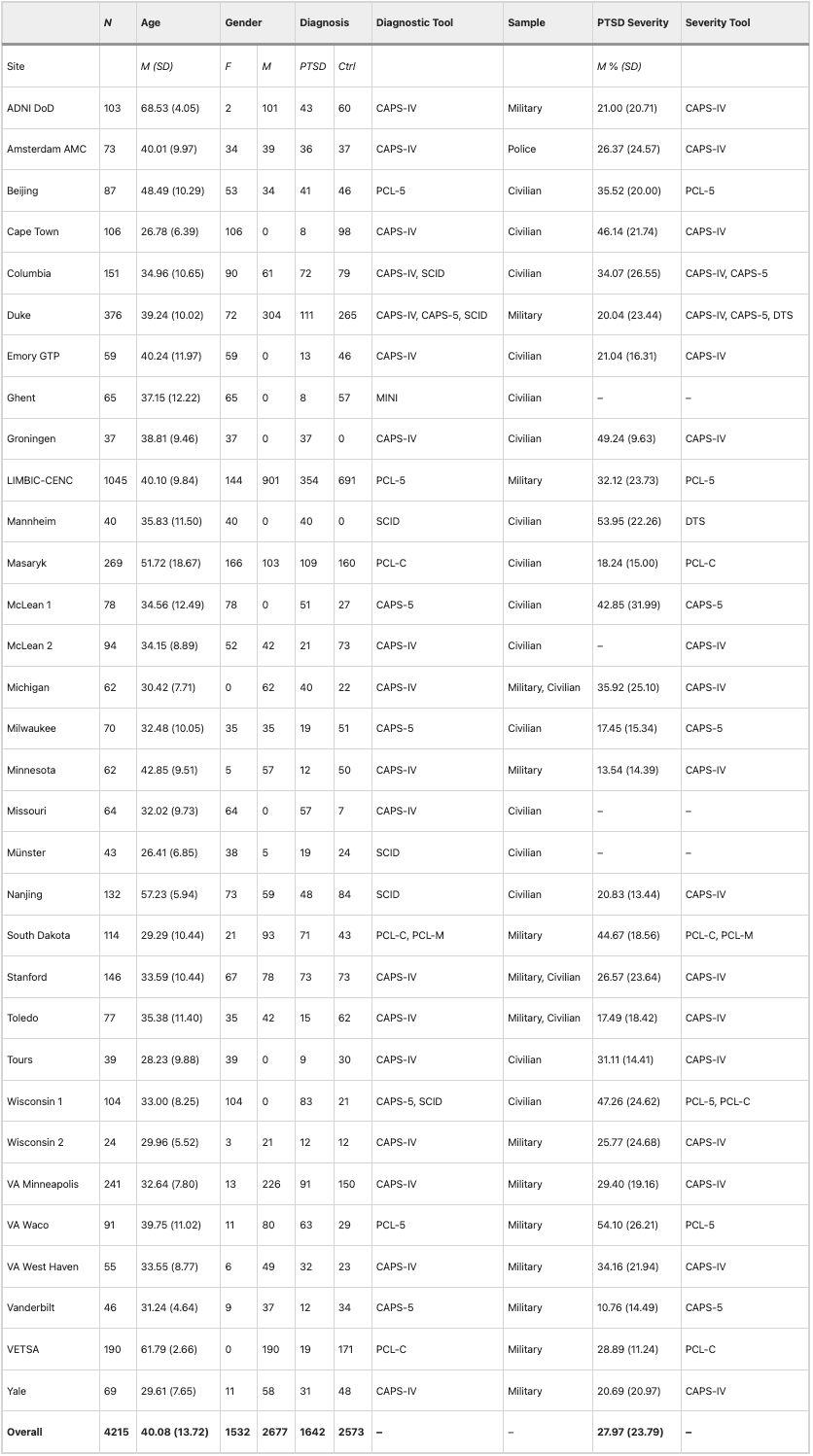
Table 1

CAPS-IV Clinician Administered PTSD Scale for DSM-IV, CAPS-5 Clinician Administered PTSD Scale for DSM-5, DTS Davidson Trauma Scale for DSM-IV, MINI Mini Neuropsychiatric Interview, PCL-C PTSD Checklist-Civilian Version, PCL-M PTSD Checklist-Military Version, PCL-5 PTSD Checklist for DSM-5, SCID Structured Clinical Interview for DSM

Image acquisition and processing

Whole-brain T1-weighted anatomical MR images were collected from each participant. Acquisition parameters for each cohort are detailed in Supplementary Table S2. Segmentation and quality control procedures were performed at Duke University. A subset of the data (n = 1045) from the Long-Term Impact of Military-Relevant Brain Injury Consortium-Chronic Effects of Neurotrauma Consortium (LIMBIC-CENC) [71] were processed at University of Utah. Cerebellar parcellation was carried out using a deep-learning algorithm, Automatic Cerebellum Anatomical Parcellation using U-Net with Locally Constrained Optimization (ACAPULCO). Images were corrected for intensity inhomogeneity using N4, blurred with a 3D Gaussian kernel (SD = 3 mm), and transformed to MNI template space. ACAPULCO then employed a cascade of two convolutional neural networks to first define a 3D-bounding box around the cerebellum and then divide it into anatomically meaningful regions. This ultimately resulted in volumetric estimates for the total cerebellum and 28 subregions, including the hemispheric anterior (lobules I-III, IV, and V), posterior (lobules VI, VIIB, VIIIA, VIIIB, IX, and crus I-II), and flocculonodular (lobule X) lobes, vermal lobules VI, VII, VIII, IX, and X, and the corpus medullare (Fig. 1). ACAPULCO achieves results comparable to other established cerebellum parcellation protocols (e.g., CERES2), but may perform better for multi-site datasets.

Fig. 1: ACAPULCO cerebellum parcellation for a representative subject.
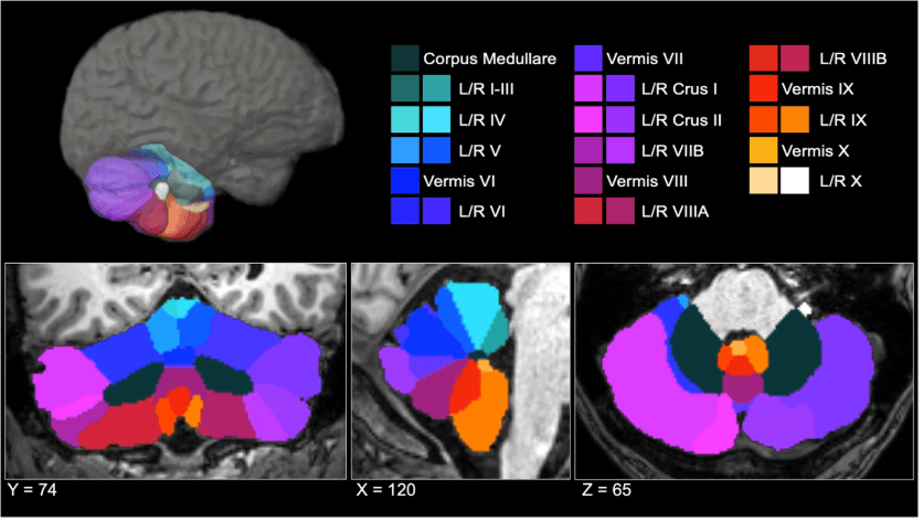
Fig 1

A three-dimensional display is presented in the upper half of the figure, along with coronal (left), sagittal (middle), and axial (right) views below. L left, R right.

Following segmentation, a two-step quality control procedure was employed, consisting of (1) removal of statistical outliers ± 2.689 SD from the site mean, and (2) visual inspection of cerebellar parcels. Each subject’s segmentation was visually inspected and given a global score by a minimum of two trained raters (AH, SL, MB, LB) on a scale from 1 (good) to 3 (poor/failed). In the event of a discrepancy between raters, the parcellation was examined by a third rater for consensus. Ratings were performed using previously published quality control procedures. Raters were trained using a graduated approach comprising didactic instruction on neuronanatomical landmarks of the cerebellum and its surrounding anatomy (e.g., cerebellar fissures, tentorium), and collaborative rating or practice examples prior to independence. Segments were considered individually; therefore, select subregional volumes (e.g., statistical outliers, circumscribed segmentation errors) for a participant could be excluded, while the remainder of their segments were retained for analysis if correct. Subjects receiving a global score of 3 were excluded from all analyses. A breakdown of ratings by site is noted in Supplementary Table S3.

Statistical analysis

To examine whether PTSD diagnosis was associated with volume differences in the grey matter volumes of the whole cerebellum, hemispheric subregions, vermis, and cerebellar white matter, we performed a series of linear mixed effects models. Statistical analyses were conducted using the lmer package in R v4.3.1. In each model, age, gender, and total intracranial volume were treated as fixed effects, and site/scanner was treated as a random effect. We considered different scanners within sites as separate sites, resulting in a total number of 49 sites coded separately in our analyses. Models were repeated implementing PTSD severity–rather than diagnosis – as a continuous predictor. Due to site measurement differences, PTSD severity was quantified as a percentage of the total score possible (see Table 1). The Benjamini-Hochberg procedure was used to adjust significance values to control the false discovery rate (p-FDR < 0.05; number of tests = 29). These adjustments were done separately for PTSD diagnosis and PTSD severity. Cohen’s d was calculated as a measure of effect size.

Given frequent co-occurrence of PTSD and likely independent effects on cerebellum volume, secondary analyses were conducted to examine the potential effects of depression, alcohol use disorder, and childhood trauma on cerebellar volumes. For sites with available covariate data (see Supplemental Material), an additional series of linear mixed effects models was conducted, including fixed effects of (1) major depressive disorder diagnosis, (2) alcohol use disorder diagnosis, and (3) total score on the Childhood Trauma Questionnaire (CTQ);

Results

Associations between PTSD diagnosis and cerebellum volumes

The effects of PTSD diagnosis on cerebellum volumes are presented in Table 2. Consistent with hypotheses, after adjusting for age, gender, and total intracranial volume, PTSD diagnosis was associated with significantly smaller total cerebellar volume, b = −981.471, t = −2.793, p-FDR = 0.005. PTSD diagnosis was also associated with smaller volume of the corpus medullare, b = −154.149, t = −2.188, p-unc = 0.026, but this did not survive multiple comparisons corrections (p-FDR = 0.096).

Table 2 Effects of PTSD diagnosis on cerebellum volume.
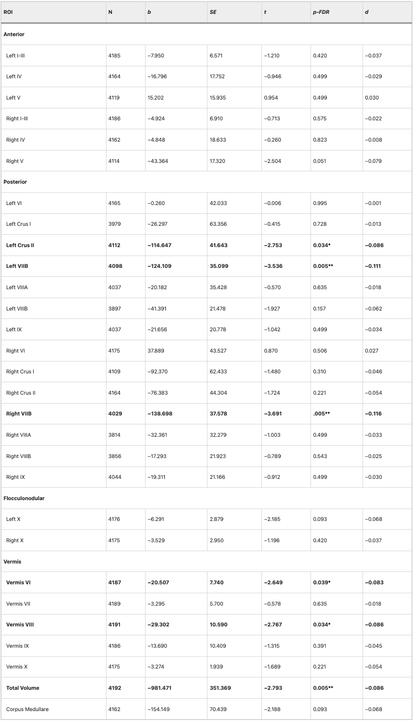
Table 2

Results of linear mixed effects models predicting cerebellar volumes including fixed effects of age, gender, PTSD diagnosis, intracranial volume, and a random effect of site. ***p-FDR < 0.001, **p-FDR < 0.01, *p-FDR < .05

Within the anterior cerebellum (lobules I-V), PTSD diagnosis was associated with a smaller volume of right lobule V, b = −43.364, t = −2.504, p-unc = 0.012, but this did not survive multiple comparisons corrections (p-FDR = 0.051).

Within the posterior cerebellum (crus, lobules VI-IX), PTSD diagnosis was associated with smaller volume of left crus II, b = −114.647, t = −2.753, p-FDR = 0.034, left lobule VIIB, b = −124.109, t = −3.536, p-FDR = 0.005, and right lobule VIIB, b = −138.698, t = −3.691, p-FDR = 0.005.

No significant effects of PTSD diagnosis were observed on volumes within the flocculonodular lobe (lobule X). There was an effect of PTSD on left lobule X volume, but this did not survive multiple comparisons corrections (p-FDR = 0.093).

There was a significant effect of PTSD diagnosis on volumes of vermal lobules VI, b = −20.507, t = −2.649, p-FDR = 0.039, and VIII, b = −29.302, t = −2.767, p-FDR = 0.034. There were no other significant effects of PTSD within the vermis.

Although these differences in cerebellar volumes between patients with PTSD and healthy controls were significant (p-FDR < 0.05), as calculated with Cohen’s d, effects were generally quite small (all d’s < −0.12). Figure 2 depicts a map of the effect sizes.

Fig. 2: Effects of PTSD diagnosis on cerebellar subregion volumes.
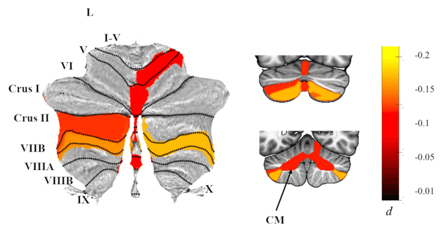
Fig 2

Atlas-based effect size (Cohen’s d) maps and MNI-based coronal slices (top: y = −72; bottom: y = −54) of the significant between-group differences for cerebellar subregion volumes in PTSD vs. Controls. Negative effect sizes reflect smaller volumes in PTSD. Regions significant at p-FDR < 0.05 are depicted in color, with the exception of right lobule V, where p-FDR = 0.051 after adjustment; right lobule V was significant p-FDR = 0.046) when examining PTSD severity instead of diagnosis. Grey-shaded subregions were non-significant. CM corpus medullary.

PTSD severity

When examining PTSD symptom severity (rather than diagnostic status), results were similar, if generally more robust (see Table 3). Specifically, PTSD symptom severity was associated with significantly smaller total cerebellum volume, b = −693.478, t = −3.719, p-FDR = 0.002, and corpus medullare volumes, b = −109.441, t = −2.915, p-FDR = 0.015. Effects were consistent across the posterior cerebellum and vermis, with significant effects of PTSD symptom severity on volumes of left crus II, b = −67.120, t = −3.044, p-FDR = 0.012, left lobule VIIB, b = −73.912, t = −3.995, p-FDR < 0.001, right lobule VIIB, b = −81.890, t = −4.085, p-FDR < 0.001, and vermal lobules VI, b = −13.931, t = −3.393, p-FDR = 0.005, and VIII, b = −17.270, t = −3.058, p-FDR = 0.012.

Table 3 Effects of PTSD severity on cerebellar volumes.
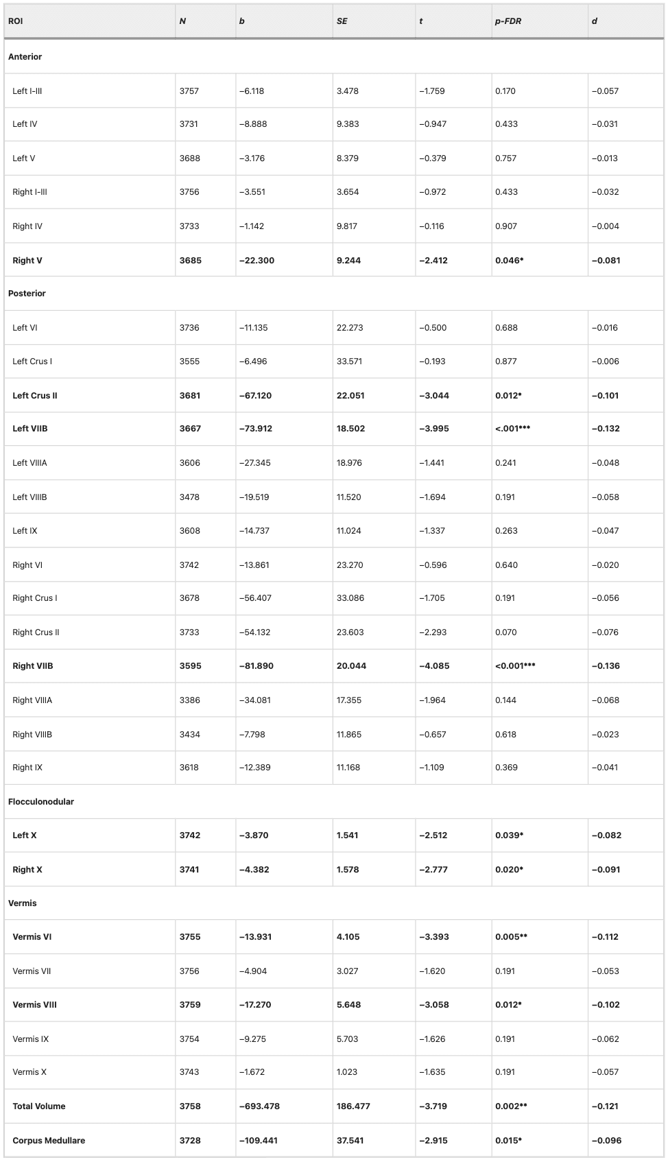
Table 3

Results of linear mixed effects models predicting cerebellar volumes including fixed effects of age, gender, PTSD severity, intracranial volume, and a random effect of site. To harmonize across sites that employed different instruments (e.g., CAPS-IV, PCL-5), PTSD severity was represented as a percentage of total points possible. ***p-FDR < 0.001, **p-FDR < 0.01, *p-FDR < .05

By contrast, the effect of PTSD on the volume of right lobule V retained significance when examining symptom severity instead of diagnosis, b = −22.300, t = −2.412, p-FDR = 0.046. Additionally, PTSD symptom severity was associated with a significantly smaller volume of the flocculonodular cerebellum, with effects observed in both hemispheres of lobule X (left: b = −3.870, t = −2.512, p-FDR = 0.039; right: b = −4.382, t = −2.777, p-FDR = 0.020).

Potential confounding variables

When including covariates assessing depression, alcohol use, and childhood trauma, effects of PTSD on cerebellar volumes were somewhat diminished (See Supplemental Material); however, when using a more liberal approach to correct for multiple comparisons, most significant effects of PTSD were retained even when accounting for depression and alcohol use disorders. Notably, detecting significant effects in these additional analyses presented a challenge to statistical power. There was high collinearity between PTSD and covariates, and—particularly in the case of childhood trauma severity—substantially reduced sample size because not all sites reported these variables. In cases where the effect of PTSD diagnosis was non-significant upon inclusion of covariates, we followed up by testing whether depression, alcohol use, or childhood trauma predicted cerebellar volumes on their own; in no instance were covariates found to independently predict cerebellar volumes when PTSD status was excluded from the model, demonstrating that our initial findings were specific to PTSD.

Depression status was available for the majority of subjects (n = 3978). When adjusting for major depressive disorder diagnosis, PTSD diagnosis remained significantly associated with smaller volume of both left and right lobule VIIB, and vermis VI. While initially significant, effects of PTSD diagnosis on right lobule V (p-FDR = 0.096) and left crus II (p-FDR = 0.133) volumes did not survive correction for multiple comparisons. PTSD symptom severity was associated with smaller total cerebellum and vermis VIII volumes. Uniquely, depression diagnosis was associated with smaller volume of right lobule X, b = −8.282, t = −2.356, p-FDR = 0.038.

When adjusting for alcohol use disorder (n = 2997), PTSD was associated with significantly smaller cerebellar volumes, including the total cerebellum (p-FDR = 0.046) and localized subregions in the posterior lobe and vermis. Specifically, PTSD diagnosis was negatively associated with volumes of the left crus II (p-FDR = 0.032), right lobule VIIB (p-FDR = 0.003), and vermal lobules VI (p-FDR = 0.034) and VIII (p-FDR = 0.050). Initially significant effects of PTSD diagnosis on right lobule V (p-FDR = 0.105) and left lobule VIIB (p-FDR = 0.056) did not survive correction for multiple comparisons.

Including CTQ severity as a covariate resulted in null effects of PTSD diagnosis; significant effects in left lobules VIIB (p-FDR = 0.280) and VIIIB (p-FDR = 0.161) were no longer significant after correction for multiple comparisons. Considering the largest sample size in these additional analyses was 1013 (approximately a quarter of the sample size in our primary analyses) and effects of PTSD diagnosis were small (Cohen’s d < 0.12), we were poorly powered to detect significant effects of PTSD when accounting for childhood trauma exposure. In addition, 77% of participants with PTSD endorsed a history of childhood trauma, contributing further challenges to identifying dissociable effects of childhood trauma and PTSD (See Supplemental Material). When we excluded PTSD from the model, however, childhood trauma was not significantly associated with cerebellar volumes in any of the regions implicated in primary analyses (e.g., total cerebellum, left and right lobules VIIB), suggesting that these effects are specific to PTSD.

Discussion

Leveraging an international, multisite dataset from ENIGMA-PGC PTSD, we conducted a mega-analysis of total and subregional cerebellar volumes in PTSD. Consistent with hypotheses based on published work, PTSD was associated with smaller total cerebellar volume. We found subregional specificity linking PTSD to smaller volumes in the posterior cerebellum, vermis, and flocculonodular cerebellum. Effects of PTSD on cerebellum volume were consistent (and generally more robust) when examining symptom severity rather than diagnostic status. Overall, these findings contribute to an emerging literature that underscores the relevance of cerebellar structure in the pathophysiology of PTSD. Although the appreciation of the cerebellum for its contributions to cognitive and affective function is relatively recent, the current results bolster a growing literature confirming the cerebellum is not exclusively devoted to motor function and may, in fact, have unique relevance to psychiatric conditions including PTSD.

Multiple neuroimaging studies have suggested that altered structure and function of the posterior cerebellum may be a neural correlate of PTSD. For instance, structural differences in lobules VIIB, VIIIA, and VIIIB were found in combat-exposed veterans with PTSD. Functionally, PTSD has been linked to increased activation during attentional and emotional tasks and decreased resting-state amplitude of low-frequency fluctuation in lobule VI. In a sample of sexual assault survivors, PTSD severity was negatively associated with activation in lobules VI, VIII, IX, and crus I during the performance of an emotional go/no-go task, and positively associated with activation in left cerebellar lobules VII-IX and crus I-II when retrieving positive memory during a mental imagery task. PTSD has also been linked to decreased global connectivity within the posterior cerebellum during symptom provocation. As the most phylogenetically recent part of the cerebellum, the posterior lobe is intricately linked with paralimbic and association cortical areas and plays an integral role in the integration of perception, emotion, and behavior. Accordingly, the posterior cerebellum contributes to the salience network (lobules VI and VII) and diverse cognitive-affective processes including working memory, attentional allocation, and associative learning. In the context of the current findings, smaller volume of lobule VIIB and crus II may be implicated in the pathophysiology of PTSD, perhaps mapping directly onto symptoms such as hypervigilance and concentration difficulties.

In the present study, PTSD was also associated with smaller volume of vermal lobules VI and VIII. The cerebellar vermis is considered part of the ‘limbic’ cerebellum and appears to play a key role in emotional processing, learning, and memory. Prior work has demonstrated that PTSD is associated with smaller volume and increased signal variability of the vermis. Importantly, structural abnormalities in the vermis may provide increased spatial specificity within existing translational models of PTSD, as converging evidence from both animals and human subjects has shown vermal activation is important for both acquisition and extinction of conditioned fear. The cerebellar vermis has strong connections to brain regions (including the brainstem, amygdala, and hypothalamus) that regulate critical survival functions. The vermis may contribute to fear learning via threat-associated autonomic changes facilitating defensive behavior, such as increases in respiration, heart rate, and blood pressure. Animal research highlights mechanistic links between vermal-midbrain connectivity and defensive behavior; in rats, for instance, lesions of the pathway between the periaqueductal gray and vermal lobule VIII provoke fear-evoked freezing behavior. Importantly, vermal connectivity is also implicated in clinical human samples, and PTSD is associated with disrupted resting-state functional connectivity from the vermis to amygdala, periaqueductal gray, and ventromedial prefrontal cortex.

Unexpectedly, PTSD was also associated (diagnosis p-FDR = 0.051, severity p-FDR = 0.046) with smaller volume of right lobule V, a subregion found within the anterior lobe of the cerebellum. Lobule V has been more consistently implicated in sensorimotor functions, including execution of hand movements and perception of tactile stimulation to the hand and foot. Prior work has found evidence of motor slowing in PTSD, and executive dysfunction is a common feature of PTSD. Importantly, many neuropsychological tests – including processing speed, set shifting, and design fluency – are dependent on speeded writing or drawing tasks. It is possible that these neuropsychological observations may be affected by both cognitive and motor contributions from the cerebellum.

PTSD symptom severity was also curiously associated with reduced volume of bilateral lobule X (which comprises the flocculonodular lobe), but its association with PTSD diagnosis was non-significant. The flocculonodular lobe is primarily implicated in ocular tracking and regulation of the vestibular system. Yet, when depression diagnosis was added to the model, there was a significant negative effect of depression on right lobule X, whereas effects of PTSD were non-significant. Structural differences in lobule X have previously been observed in major depressive disorder, and these differences have been attributed to somatic complaints, such as dizziness, that are frequently endorsed by patients with depression. PTSD and major depressive disorder are highly comorbid. Therefore, smaller lobule X volume may perhaps be unique to patients with prominent depressive features and/or a more somatic symptom profile.

In general, PTSD severity was more robustly associated with cerebellar volume differences than PTSD diagnosis. For instance, although PTSD’s effects on corpus medullare volumes did not survive correction when examining diagnosis, there was a significant association for PTSD severity. The most parsimonious explanation for this phenomenon is that continuous severity scores provide a more powerful statistical test than diagnosis. PTSD status can reflect a wide range of severity within both patient and control groups, and therefore using diagnosis is, in effect, disregarding valuable information that explains variance associated with cerebellar volume. While diagnostic status provides a clinically useful shorthand, it also fails to capture phenotypic variability within PTSD.

It is also possible the more robust results might be explained by the control group containing a mix of trauma-exposed and trauma-naïve participants. Few sites provided data for trauma-naïve participants; as such, the majority of our control group (~88%) was trauma-exposed. We chose to retain trauma-naïve individuals within the control group to benefit from increased power associated with the larger sample size, but this may have introduced additional noise (unaccounted variance) that diminished the significance of diagnosis-related statistical tests. Our severity analyses, however, excluded trauma-naïve participants, as (having no index trauma) they did not complete assessments of PTSD symptom severity. The small sample of trauma-naïve subjects precluded us from assessing whether there are cerebellar volume differences related to trauma exposure (not just PTSD), and future work to examine this question will be valuable. Although exploratory analyses suggested that most PTSD symptom domains – including re-experiencing, avoidance, and negative changes in cognition and mood – were consistently associated with cerebellar volumes (See Supplement), it is imperative that future work aims to consider PTSD beyond categorical diagnosis (including severity scores and variable symptom presentations) to create a reliable neurobiological model.

Overall, despite these significant findings suggesting associations between PTSD and smaller cerebellar volumes, effect sizes were small. As such, it is unlikely that structural cerebellar volumes alone will provide a clinically useful biomarker (e.g., for individual-level prediction). That said, the large sample size and granular parcellation in the current study provided us with increased power and precision to confidently implicate the cerebellum in PTSD. Indeed, these findings help to resolve a previously mixed literature, although the small effect sizes stand in contrast to earlier findings reporting moderate effect sizes. Yet, small sample sizes are likely to overestimate effect sizes. In the context of the small effect sizes the current study discovered, these prior studies would have required upwards of a thousand subjects for reliable, reproducible results. Prior ENIGMA-PGC studies in a subset of the current sample have identified similarly small – albeit slightly larger – effects for other brain region volumes, including the hippocampus (d = −0.17) and amygdala (d = −0.11), associated with PTSD [7]. Future work would benefit from a more systematic comparison amongst brain structures implicated in PTSD to identify the most robust neural correlates of the disorder. It is also possible that, in general, true effects are slightly larger than typically estimated in consortium datasets, which, by nature, are limited by site variability in measurement and design. Despite the advantages of larger sample sizes, statistical modeling often cannot account for other factors that may contribute to cerebellar volumes due to missing data across sites. Improved models accounting for other factors affecting cerebellar structure may provide a clearer picture of the magnitude of these effects in PTSD. Considering the cerebellum has historically been both understudied and inconsistently associated with PTSD, though, these findings provide novel insight into the pathophysiology of PTSD.

Critically, PTSD is incredibly burdensome at both the individual and societal level, causing profound distress, functional impairment, and staggering treatment costs. The insights from the current study have revealed a novel treatment target that may be leveraged to improve treatment outcomes for PTSD. In fact, prior work has shown that the cerebellum is sensitive to external modulation. For example, recent work has highlighted how non-invasive brain stimulation of the cerebellum can modulate cognitive, emotional, and social processes commonly disrupted in PTSD, including mood regulation and context-based prediction. In other work in depression, electroconvulsive therapy has been shown to increase volume of cerebellar regions including lobule VII, and these structural changes were associated with symptom reductions. Changes in cerebellum functional connectivity are also linked to reductions in PTSD symptom severity before and after cognitive processing therapy. As such, despite small effect sizes, prior work has shown that cerebellum structure and function is modifiable, and these localized cerebellum structural findings may provide useful and more precise targets for neuromodulatory, pharmacological, and even psychotherapeutic intervention. Ultimately, integrating neurobiologically-informed targets within treatment protocols may help establish treatments with stronger and more long-lasting therapeutic effects.

Limitations

This is the largest study of cerebellar volumetry in PTSD to date, however, there are several notable limitations. PTSD is a heterogeneous disorder and is highly comorbid with other psychiatric conditions (e.g., depression, substance use disorders) and environmental exposures (e.g., childhood trauma, traumatic brain injury) that are also linked to alterations in cerebellar structure. Employing a mega-analysis in a large multi-cohort consortium dataset enabled us to observe small effect sizes of PTSD on cerebellar volume in our primary analyses, but many sites did not provide diagnostic or item-level data for relevant covariates. Consequently, we were unable to investigate effects of relevant covariates at the same scale. Future studies would benefit from investigating unique and shared phenotypes of PTSD and other common comorbid psychopathologies on the cerebellum to disentangle potential dissociable effects and complex interactions more elegantly. It is also critical for future work to examine how the cerebellum may be uniquely implicated in the dissociative subtype of PTSD. Dissociative symptoms in PTSD are linked to alterations within the midbrain that facilitate passive, rather than active, defensive responses; observed differences in cerebellar functional activation and connectivity related to the dissociative subtype of PTSD may be mediated by the prominent neural pathways between the cerebellum and midbrain. The current study was also focused solely on cerebellar volumetric differences in PTSD. Multiple studies have observed disrupted cerebellar activity both at rest and during trauma-relevant tasks in patients with PTSD. Future work would benefit from improved localization of both functional and structural changes in the cerebellum that may be present in PTSD. In addition, individual differences in education may further explain cerebellar volume reductions and should be explored in future studies. Lastly, the current study is cross-sectional in nature; future longitudinal research will be imperative to better understand whether cerebellum volume confers risk for PTSD or changes as a function of the disorder.

Conclusion

In a sample of over 4000 adults from the ENIGMA-PGC PTSD Consortium, cerebellum volume was significantly smaller in patients with PTSD compared to pooled groups of trauma-exposed and trauma-naïve controls. Specific subregional volume reductions in the vermis and posterior cerebellum (crus II and lobule VIIB) align with previous work demonstrating their involvement in cognitive and affective functions relevant to PTSD, such as fear learning and regulation. Overall, these findings argue for a critical role of the cerebellum in the pathophysiology of PTSD, bolstering support for the region’s contributions to processes beyond vestibulomotor function.

Open Article as PDF

Abstract

Although the cerebellum contributes to higher-order cognitive and emotional functions relevant to posttraumatic stress disorder (PTSD), prior research on cerebellar volume in PTSD is scant, particularly when considering subregions that differentially map on to motor, cognitive, and affective functions. In a sample of 4215 adults (PTSD n = 1642; Control n = 2573) across 40 sites from the ENIGMA-PGC PTSD working group, we employed a new state-of-the-art deep-learning based approach for automatic cerebellar parcellation to obtain volumetric estimates for the total cerebellum and 28 subregions. Linear mixed effects models controlling for age, gender, intracranial volume, and site were used to compare cerebellum volumes in PTSD compared to healthy controls (88% trauma-exposed). PTSD was associated with significant grey and white matter reductions of the cerebellum. Compared to controls, people with PTSD demonstrated smaller total cerebellum volume, as well as reduced volume in subregions primarily within the posterior lobe (lobule VIIB, crus II), vermis (VI, VIII), flocculonodular lobe (lobule X), and corpus medullare (all p-FDR < 0.05). Effects of PTSD on volume were consistent, and generally more robust, when examining symptom severity rather than diagnostic status. These findings implicate regionally specific cerebellar volumetric differences in the pathophysiology of PTSD. The cerebellum appears to play an important role in higher-order cognitive and emotional processes, far beyond its historical association with vestibulomotor function. Further examination of the cerebellum in trauma-related psychopathology will help to clarify how cerebellar structure and function may disrupt cognitive and affective processes at the center of translational models for PTSD.

Summary

Experiencing trauma is common, and about 10% of people who go through trauma develop ongoing symptoms of post-traumatic stress disorder (PTSD). PTSD is a serious mental health condition that causes problems like unwanted memories, avoiding certain situations, being overly alert, and changes in mood and thinking. Research has shown that specific brain areas are different in people with PTSD compared to those who have experienced trauma but do not have PTSD.

Brain regions like the hippocampus, ventromedial prefrontal cortex (vmPFC), amygdala, insula, and anterior cingulate cortex (ACC) have consistently been found to be smaller in people with PTSD. These areas are part of a key brain network that controls thinking and emotions, which are affected in PTSD. This includes how people process threats, manage emotions, and form emotional memories.

More recently, studies using structural and functional magnetic resonance imaging (MRI) have started looking at the role of the cerebellum in PTSD. The cerebellum was once thought to mainly control movement. However, research over the last thirty years shows it also plays a large role in higher-level thinking and emotions. The human cerebellum has grown significantly over time. It makes up about 10% of the brain's total size but contains most of the brain's neurons and covers nearly 80% of the neocortex.

The cerebellum is well-connected to many other brain areas, including those involved in thinking and emotions, suggesting it does more than just control movement, which could be important for PTSD. Its connections to stress-related areas like the amygdala, hippocampus, and periaqueductal gray might make it vulnerable to the effects of trauma, potentially leading to PTSD symptoms by disrupting normal stress responses. Recent studies also indicate the cerebellum is involved in learning and remembering fear. Since PTSD involves problems with detecting and processing threats, this evidence supports including the cerebellum in current models of PTSD.

Indeed, PTSD has been linked to abnormal connections between the cerebellum and important brain regions for thinking and emotions, such as the amygdala. Studies combining data from many sources suggest that the cerebellum acts differently in people with PTSD compared to healthy individuals. In terms of brain structure, smaller cerebellum volume has been seen in both adults and children with PTSD. One large study of 84 people found that PTSD patients had smaller left cerebellar hemisphere and vermal volumes compared to trauma-exposed individuals without PTSD.

However, structural studies have not always found the cerebellum to be involved in PTSD, and limitations in past research have made it hard to understand these varied results. First, most adult studies had small sample sizes, ranging from 39 to 99 participants. In fact, three studies that found no differences combined included only 82 PTSD patients. Studies also used different ways to measure brain structure (like overall volume, detailed shape, or cortical thickness) and included different groups of people (like combat veterans, victims of violence, or first responders).

Previous research on cerebellum volume in PTSD has also often failed to consider important smaller parts of the cerebellum that are linked to different functions, such as movement, thinking, and emotions. The cerebellum is divided into three main parts by two large grooves: the anterior lobe (lobules I–V), the posterior lobe (lobules VI-IX), and the flocculonodular lobe (lobule X). The corpus medullare, which is the white matter center of the cerebellum, is a dense collection of nerve fibers that send signals to and from the cerebellum.

The anterior lobe receives signals from the spinal cord and connects with motor areas of the brain to help with movement, walking, and balance. The flocculonodular lobe is important for receiving signals related to balance and vision, and for controlling eye movements and reflexes. In contrast, the posterior cerebellum, which developed more recently, is involved in many non-motor functions. It does not receive signals from the spinal cord but connects with brain areas crucial for higher-level processes, such as the prefrontal cortex and cingulate gyrus.

Activity in the posterior lobe has been observed during tasks involving language and working memory (lobule VI, crus I), spatial processing (lobule VI), and executive functions (lobule VI and VIIB, crus I). Processing unpleasant stimuli, like painful heat or disturbing images, also involves the posterior cerebellum (lobules VI and VIIB and crus I), suggesting these areas play a role in defensive responses. The vermis, which is the central part connecting the two halves of the cerebellum, is considered part of the brain's emotion circuit and becomes active during emotional processing. Vermal lobules also interact with other regions important for learning emotional associations, such as the amygdala, hypothalamus, and periaqueductal gray. These detailed studies on brain function have identified three broad areas within the cerebellum involved in sensory-motor, cognitive, and emotional processes.

PTSD is a complex disorder linked to problems in many processes supported by the cerebellum. It is not clear whether structural differences in the cerebellum in PTSD are widespread or limited to specific smaller areas. However, most studies have used a simple approach to examine the cerebellum in PTSD, focusing only on the total volumes of the vermis and the hemispheres. While functional studies have found PTSD-related activity differences across the cerebellum, including in the vermis, crus, and lobules VI and VII, only one structural study has looked at smaller parts of the cerebellum to see if differences are specific to certain regions. Understanding the role of cerebellar structure in PTSD can help explain how chronic PTSD symptoms continue and aid in developing targeted and effective treatments.

To address this, the current study combined data from many research groups as part of the Enhancing NeuroImaging Genetics through Meta-Analysis (ENIGMA)-Psychiatric Genomics Consortium (PGC) PTSD workgroup. This approach, called a mega-analysis, combines data from multiple sites and analyzes it together, while accounting for differences between sites. A new, standardized ENIGMA cerebellum parcellation method was used to measure the volumes of cerebellar lobules using MRI data from 4215 adults, including 1642 with PTSD and 2573 without PTSD. The study looked at how PTSD affects cerebellar volumes, adjusting for age, gender, and total brain size. Based on previous research, it was predicted that PTSD would be linked to smaller total cerebellum volume. Given that functional mapping suggests the 'emotional' and 'cognitive' parts of the cerebellum are located in the vermis and posterior lobes, respectively, it was also hypothesized that PTSD would be associated with smaller volumes in these two areas.

Methods and Materials

Sample

The study included clinical, demographic, and brain imaging data from the ENIGMA-PGC PTSD working group. MRI scans from 4215 participants, including 1642 with PTSD and 2573 healthy controls, were used. Approximately 88% of the controls had experienced trauma, and 12% had not. The cerebellum was automatically divided into smaller regions. All study procedures were approved by local research ethics committees, and participants gave written consent. The current analyses were approved as exempt by the Duke University Health System Institutional Review Board.

Image acquisition and processing

Whole-brain T1-weighted MRI scans were collected from each participant. Segmentation and quality control were done at Duke University, with a subset of data processed at the University of Utah. Cerebellar parcellation (dividing the cerebellum into regions) used a deep-learning algorithm called ACAPULCO. Images were corrected for intensity differences, smoothed, and aligned to a standard brain template. ACAPULCO used two neural networks to first identify the cerebellum and then divide it into 28 meaningful anatomical regions. This provided volume estimates for the entire cerebellum and its subregions, including parts of the anterior, posterior, and flocculonodular lobes, vermal lobules, and the corpus medullare. ACAPULCO produces results similar to other established methods and may perform better with data from multiple sites.

After segmentation, a two-step quality control process was used: (1) removing statistical outliers (values far outside the average for a site) and (2) visually inspecting the cerebellar parcels. At least two trained raters visually checked each participant's segmentation and gave it a score from 1 (good) to 3 (poor/failed). If there was a disagreement, a third rater reviewed it for a final decision. Raters were trained on the anatomy of the cerebellum. Individual subregions could be excluded if they had errors, while the rest of a participant's segments were kept if correct. Participants with an overall score of 3 were removed from all analyses.

Statistical analysis

To see if a PTSD diagnosis was linked to differences in the volume of the whole cerebellum, its hemispheric subregions, the vermis, and cerebellar white matter, a series of linear mixed effects models were used. These analyses were done using the lmer package in R v4.3.1. In each model, age, gender, and total intracranial volume were considered fixed factors, and the study site/scanner was treated as a random factor. Different scanners within the same site were counted as separate sites, totaling 49 sites. The models were repeated using PTSD symptom severity, measured as a percentage of the total possible score, instead of diagnosis, as a continuous predictor. The Benjamini-Hochberg procedure was used to adjust significance values to control for false positives across multiple tests (29 tests total), with separate adjustments for PTSD diagnosis and severity. Cohen's d was used to measure the size of the effect.

Because PTSD often occurs with other conditions that might affect cerebellum volume, additional analyses were done to look at the potential effects of depression, alcohol use disorder, and childhood trauma. For sites where this data was available, more linear mixed effects models were run, including major depressive disorder diagnosis, alcohol use disorder diagnosis, and the total score on the Childhood Trauma Questionnaire (CTQ) as fixed factors.

Results

Associations between PTSD diagnosis and cerebellum volumes

The study found that a PTSD diagnosis was linked to significantly smaller total cerebellar volume, after accounting for age, gender, and total intracranial volume. This result was consistent with the initial predictions. PTSD diagnosis was also associated with smaller volume of the corpus medullare, but this finding was not strong enough after correcting for multiple comparisons.

Within the anterior cerebellum, PTSD diagnosis was linked to a smaller volume of the right lobule V, but this also did not remain significant after correcting for multiple comparisons.

In the posterior cerebellum, PTSD diagnosis was associated with smaller volumes of the left crus II, left lobule VIIB, and right lobule VIIB. These findings remained significant after correcting for multiple comparisons.

No significant effects of PTSD diagnosis were found on volumes within the flocculonodular lobe (lobule X) after correction.

There was a significant effect of PTSD diagnosis on the volumes of vermal lobules VI and VIII, which remained significant after corrections. No other significant effects were found within the vermis.

Although these volume differences between people with PTSD and healthy controls were statistically significant, the actual differences were quite small.

PTSD severity

When looking at the severity of PTSD symptoms instead of just diagnosis, the results were similar and generally stronger. Higher PTSD symptom severity was significantly linked to smaller total cerebellum volume and smaller corpus medullare volumes. The effects were consistent in the posterior cerebellum and vermis, with significant links between PTSD symptom severity and smaller volumes of left crus II, left lobule VIIB, right lobule VIIB, and vermal lobules VI and VIII.

In contrast to the diagnosis results, the link between PTSD symptom severity and the volume of right lobule V remained significant. Additionally, PTSD symptom severity was associated with significantly smaller volume in the flocculonodular cerebellum, specifically in both the left and right sides of lobule X.

Potential confounding variables

When factors like depression, alcohol use, and childhood trauma were included in the analyses, the effects of PTSD on cerebellar volumes were somewhat reduced. However, most significant effects of PTSD remained when using a less strict method to correct for multiple comparisons, even after accounting for depression and alcohol use disorders. It was difficult to detect significant effects in these additional analyses due to statistical power issues. There was a strong overlap between PTSD and these other factors, and the sample size was much smaller, especially for childhood trauma severity, as not all sites provided this data. In cases where the effect of PTSD diagnosis became non-significant after including these other factors, follow-up tests showed that depression, alcohol use, or childhood trauma did not predict cerebellar volumes on their own when PTSD status was removed from the model. This indicates that the initial findings were specific to PTSD.

Depression status was available for most participants. When adjusting for a major depressive disorder diagnosis, PTSD diagnosis was still significantly linked to smaller volumes of both left and right lobule VIIB, and vermis VI. Some effects that were initially significant for PTSD diagnosis (right lobule V and left crus II volumes) did not remain significant after correcting for multiple comparisons when depression was included. PTSD symptom severity was linked to smaller total cerebellum and vermis VIII volumes. Notably, depression diagnosis itself was linked to a smaller volume of right lobule X.

When adjusting for alcohol use disorder, PTSD was associated with significantly smaller cerebellar volumes, including the total cerebellum and localized subregions in the posterior lobe and vermis. Specifically, PTSD diagnosis was negatively associated with volumes of the left crus II, right lobule VIIB, and vermal lobules VI and VIII. Initially significant effects of PTSD diagnosis on right lobule V and left lobule VIIB did not remain significant after correction when alcohol use disorder was included.

Including childhood trauma severity as a confounding factor resulted in no significant effects for PTSD diagnosis. Significant effects in left lobules VIIB and VIIIB were no longer significant after correcting for multiple comparisons. The sample size for these additional analyses was much smaller, and the effects of PTSD diagnosis were small, meaning there was not enough power to detect significant effects of PTSD when accounting for childhood trauma exposure. Also, a high percentage of participants with PTSD reported a history of childhood trauma, making it hard to separate the effects of childhood trauma and PTSD. However, when PTSD was removed from the model, childhood trauma was not significantly linked to cerebellar volumes in the regions that were initially found to be affected, suggesting these effects are specific to PTSD.

Discussion

This study, using a large international dataset, conducted a mega-analysis of total and subregional cerebellum volumes in people with PTSD. As predicted based on earlier research, PTSD was associated with a smaller total cerebellum volume. The study found that specific smaller parts of the cerebellum, including the posterior cerebellum, vermis, and flocculonodular cerebellum, had smaller volumes in people with PTSD. The effects of PTSD on cerebellum volume were consistent, and generally stronger, when looking at the severity of symptoms rather than just whether someone had a diagnosis. Overall, these findings add to growing evidence that the structure of the cerebellum is important in the development and course of PTSD. Although the cerebellum's role in thinking and emotions has only recently been recognized, these results support that it is not just for movement and may be uniquely relevant to mental health conditions like PTSD.

Many brain imaging studies have suggested that changes in the structure and function of the posterior cerebellum may be linked to PTSD. For example, structural differences in lobules VIIB, VIIIA, and VIIIB were found in combat veterans with PTSD. Functionally, PTSD has been linked to increased activity in lobule VI during attention and emotion tasks, and decreased brain activity at rest in the same area. In a group of sexual assault survivors, PTSD severity was negatively related to activity in lobules VI, VIII, IX, and crus I during an emotional task, and positively related to activity in left cerebellar lobules VII-IX and crus I-II when recalling positive memories. PTSD has also been linked to reduced overall connections within the posterior cerebellum during symptom provocation. As the most recently evolved part of the cerebellum, the posterior lobe is closely connected to brain areas involved in emotions and higher-level thinking, and it plays a key role in combining perception, emotion, and behavior. Therefore, the posterior cerebellum contributes to the salience network (lobules VI and VII) and various cognitive-emotional processes, including working memory, attention, and associative learning. Based on the current findings, smaller volumes of lobule VIIB and crus II may be involved in the development of PTSD, possibly related to symptoms like being overly alert and having trouble concentrating.

In this study, PTSD was also associated with smaller volumes of vermal lobules VI and VIII. The cerebellar vermis is considered part of the 'emotional' cerebellum and appears to be crucial for processing emotions, learning, and memory. Previous research has shown that PTSD is linked to smaller volume and more variable signals in the vermis. Importantly, structural abnormalities in the vermis could provide more specific insights within current models of PTSD, as evidence from both animal and human studies suggests vermal activity is important for both learning and unlearning conditioned fear. The cerebellar vermis has strong connections to brain regions (like the brainstem, amygdala, and hypothalamus) that control essential survival functions. The vermis might contribute to fear learning by causing changes in the body's automatic responses during threats, such as increases in breathing, heart rate, and blood pressure. Animal research shows how connections between the vermis and midbrain are linked to defensive behavior; for instance, damage to the pathway between the periaqueductal gray and vermal lobule VIII in rats can trigger fear-induced freezing behavior. Importantly, vermal connections are also involved in human clinical samples, and PTSD is associated with disrupted resting-state functional connections from the vermis to the amygdala, periaqueductal gray, and ventromedial prefrontal cortex.

Unexpectedly, PTSD was also associated with a smaller volume of right lobule V, which is located in the anterior lobe of the cerebellum. This subregion has been more consistently linked to sensory and motor functions, such as hand movements and sensing touch. Prior research has found evidence of slower motor skills in people with PTSD, and problems with executive function are common in PTSD. Many neuropsychological tests, including those measuring processing speed, mental flexibility, and creative design, rely on quick writing or drawing. It is possible that both cognitive and motor contributions from the cerebellum could affect these test results.

PTSD symptom severity was also linked to reduced volume in both sides of lobule X (part of the flocculonodular lobe), though its association with a PTSD diagnosis was not significant. The flocculonodular lobe is mainly involved in eye movements and controlling balance. However, when a depression diagnosis was added to the model, there was a significant negative effect of depression on right lobule X, while the effects of PTSD were not significant. Structural differences in lobule X have been observed in major depressive disorder, and these differences have been linked to physical symptoms like dizziness, which are often reported by people with depression. PTSD and major depressive disorder often occur together. Therefore, a smaller lobule X volume might be specific to people with prominent depressive symptoms or more physical complaints.

Overall, PTSD severity was more strongly linked to differences in cerebellar volume than a PTSD diagnosis. For example, while the effects of PTSD on corpus medullare volumes did not remain significant after correction when looking at diagnosis, there was a significant association for PTSD severity. The simplest explanation is that continuous severity scores provide a more powerful statistical test than a simple diagnosis. A PTSD diagnosis can cover a wide range of severity within both patient and control groups, and using diagnosis effectively ignores valuable information that explains differences in cerebellar volume. While a diagnosis is a useful clinical shortcut, it does not capture the variety of symptoms within PTSD.

It is also possible that the stronger results might be because the control group included a mix of trauma-exposed and trauma-naïve participants. Few sites provided data for trauma-naïve individuals, so most of the control group (about 88%) had experienced trauma. The trauma-naïve individuals were kept in the control group to benefit from the larger sample size, but this might have introduced extra variability that reduced the significance of diagnosis-related statistical tests. However, the severity analyses excluded trauma-naïve participants because they did not complete assessments of PTSD symptom severity. The small number of trauma-naïve subjects prevented the study from assessing whether there are cerebellar volume differences related to trauma exposure alone (not just PTSD), and future research on this question would be valuable. Although exploratory analyses suggested that most PTSD symptom domains—including re-experiencing, avoidance, and negative changes in thinking and mood—were consistently linked to cerebellar volumes, future work needs to consider PTSD beyond a simple diagnosis (including severity scores and different symptom presentations) to create a reliable neurobiological model.

Despite these significant findings linking PTSD to smaller cerebellar volumes, the effect sizes were small. Therefore, it is unlikely that cerebellar structural volumes alone will serve as a clinically useful biomarker (e.g., for predicting individual outcomes). However, the large sample size and detailed parcellation in this study provided increased power and precision to confidently identify the cerebellum's involvement in PTSD. Indeed, these findings help clarify previously conflicting research, although the small effect sizes contrast with earlier reports of moderate effect sizes. However, small sample sizes tend to overestimate effect sizes. Given the small effect sizes found in the current study, previous studies would have needed thousands of participants for reliable and reproducible results. Prior ENIGMA-PGC studies using a subset of the current sample found similarly small, though slightly larger, effects for other brain regions associated with PTSD, such as the hippocampus and amygdala. Future research would benefit from a more systematic comparison among brain structures implicated in PTSD to identify the most robust brain changes associated with the disorder. It is also possible that, in general, the true effects are slightly larger than typically estimated in large consortium datasets, which are inherently limited by site-to-site variability in measurement and study design. Despite the advantages of larger sample sizes, statistical modeling often cannot account for other factors that might affect cerebellar volumes due to missing data across sites. Improved models that consider other factors affecting cerebellar structure might provide a clearer picture of the size of these effects in PTSD. Nevertheless, given that the cerebellum has historically been understudied and inconsistently linked to PTSD, these findings offer new insights into the biology of PTSD.

Crucially, PTSD places a significant burden on individuals and society, causing deep distress, functional impairment, and enormous treatment costs. The insights from this study have revealed a new treatment target that could be used to improve outcomes for PTSD. In fact, prior research has shown that the cerebellum can be influenced by external methods. For example, recent work has highlighted how non-invasive brain stimulation of the cerebellum can change cognitive, emotional, and social processes often disrupted in PTSD, including mood regulation and context-based prediction. In other research on depression, electroconvulsive therapy has been shown to increase the volume of cerebellar regions, including lobule VII, and these structural changes were linked to reduced symptoms. Changes in cerebellum functional connectivity are also linked to reductions in PTSD symptom severity before and after cognitive processing therapy. Therefore, despite small effect sizes, prior work has shown that cerebellum structure and function can be modified, and these specific structural findings in the cerebellum could provide useful and more precise targets for neuromodulatory, pharmacological, and even psychotherapeutic interventions. Ultimately, incorporating neurobiologically informed targets into treatment protocols might help establish treatments with stronger and more lasting therapeutic effects.

Limitations

This study is the largest to date on cerebellum volume in PTSD, but it has several limitations. PTSD is a complex disorder and often co-occurs with other mental health conditions (like depression, substance use disorders) and environmental exposures (like childhood trauma, traumatic brain injury) that are also linked to changes in cerebellar structure. Using a mega-analysis in a large multi-cohort dataset allowed the detection of small effect sizes of PTSD on cerebellar volume in the primary analyses. However, many sites did not provide diagnostic or detailed data for relevant co-occurring conditions. Consequently, it was not possible to investigate the effects of these relevant factors on the same scale. Future studies should investigate the unique and shared characteristics of PTSD and other common co-occurring conditions on the cerebellum to better understand separate effects and complex interactions. It is also crucial for future work to examine how the cerebellum might be uniquely involved in the dissociative subtype of PTSD. Dissociative symptoms in PTSD are linked to changes within the midbrain that promote passive, rather than active, defensive responses; observed differences in cerebellar functional activity and connectivity related to the dissociative subtype of PTSD might be explained by the strong neural pathways between the cerebellum and midbrain. This study also focused only on differences in cerebellum volume in PTSD. Many studies have observed abnormal cerebellar activity both at rest and during trauma-related tasks in people with PTSD. Future work would benefit from better pinpointing both functional and structural changes in the cerebellum that may be present in PTSD. Additionally, individual differences in education might further explain reductions in cerebellar volume and should be explored in future studies. Finally, this study looked at data at a single point in time; future longitudinal research will be essential to better understand whether cerebellum volume increases the risk for PTSD or changes as a result of the disorder.

Conclusion

In a study of over 4000 adults from the ENIGMA-PGC PTSD Consortium, the cerebellum volume was significantly smaller in people with PTSD compared to groups of controls who had either experienced trauma or not. Specific reductions in the volume of subregions in the vermis and posterior cerebellum (crus II and lobule VIIB) are consistent with previous research showing their involvement in cognitive and emotional functions relevant to PTSD, such as learning and regulating fear. Overall, these findings suggest a critical role for the cerebellum in the development and course of PTSD, strengthening the idea that this brain region contributes to processes beyond just movement.

Open Article as PDF

Abstract

Although the cerebellum contributes to higher-order cognitive and emotional functions relevant to posttraumatic stress disorder (PTSD), prior research on cerebellar volume in PTSD is scant, particularly when considering subregions that differentially map on to motor, cognitive, and affective functions. In a sample of 4215 adults (PTSD n = 1642; Control n = 2573) across 40 sites from the ENIGMA-PGC PTSD working group, we employed a new state-of-the-art deep-learning based approach for automatic cerebellar parcellation to obtain volumetric estimates for the total cerebellum and 28 subregions. Linear mixed effects models controlling for age, gender, intracranial volume, and site were used to compare cerebellum volumes in PTSD compared to healthy controls (88% trauma-exposed). PTSD was associated with significant grey and white matter reductions of the cerebellum. Compared to controls, people with PTSD demonstrated smaller total cerebellum volume, as well as reduced volume in subregions primarily within the posterior lobe (lobule VIIB, crus II), vermis (VI, VIII), flocculonodular lobe (lobule X), and corpus medullare (all p-FDR < 0.05). Effects of PTSD on volume were consistent, and generally more robust, when examining symptom severity rather than diagnostic status. These findings implicate regionally specific cerebellar volumetric differences in the pathophysiology of PTSD. The cerebellum appears to play an important role in higher-order cognitive and emotional processes, far beyond its historical association with vestibulomotor function. Further examination of the cerebellum in trauma-related psychopathology will help to clarify how cerebellar structure and function may disrupt cognitive and affective processes at the center of translational models for PTSD.

Introduction

Experiencing trauma is common, and about 10% of trauma survivors develop lasting symptoms of post-traumatic stress disorder (PTSD). PTSD is a serious mental health condition that includes symptoms like intrusive memories, avoidance, being overly alert, and negative changes in mood and thinking. Research has shown that certain brain areas are different in people with PTSD compared to those who have experienced trauma but do not have PTSD. Specifically, PTSD has been linked to smaller volumes in brain regions such as the hippocampus, ventromedial prefrontal cortex (vmPFC), amygdala, insula, and anterior cingulate cortex (ACC). These areas are part of a key brain network that supports thinking, emotions, and memory, all of which are affected in PTSD.

More studies using MRI are now looking at the role of the cerebellum in PTSD. The cerebellum has historically been known for its role in movement and balance. However, research over the past three decades shows that it also plays a large part in higher-level thinking and emotions. The human cerebellum has grown significantly over time. Although it makes up only about 10% of the brain's total size, it contains most of the brain's neurons and nearly 80% of the surface area of the neocortex. The cerebellum is richly connected to many other brain areas, including those involved in prefrontal and limbic functions. This suggests it participates in processes beyond motor control that could be important for PTSD. Its extensive connections with stress-related regions, like the amygdala and hippocampus, might make it particularly vulnerable to traumatic stress. This could lead to PTSD symptoms by disrupting normal brain responses to stress through networks between the cerebellum and other brain areas. Recent studies also indicate the cerebellum is involved in learning and remembering fear. Since PTSD involves problems with detecting and processing threats, this evidence supports including the cerebellum in current models of PTSD.

Indeed, PTSD has been associated with altered connections between the cerebellum and important cognitive and emotional brain regions, including the amygdala. Combined research from multiple studies has also suggested that cerebellar activity differs between individuals with PTSD and healthy controls. At the structural level, smaller cerebellar volume has been observed in both adult and child populations with PTSD. For example, in one large study of 84 individuals, those with PTSD had smaller volumes in the left cerebellar hemisphere and vermis compared to trauma-exposed controls. However, structural studies have not always consistently found a link between the cerebellum and PTSD, and differences in study methods have made it difficult to compare findings. Many adult studies had small sample sizes, ranging from 39 to 99 participants, and the three studies that found no differences had a total of 82 PTSD patients combined. Studies also varied significantly in the types of structural measurements used (e.g., volume, morphology, cortical thickness) and the groups studied (e.g., combat veterans, victims of violence, first responders).

Previous research on cerebellar volume in PTSD has also been limited because it often did not consider important anatomical subdivisions of the cerebellum. These subdivisions are known to be involved in different motor, cognitive, and emotional functions. The cerebellum is divided into three main parts: the anterior lobe (lobules I–V), the posterior lobe (lobules VI-IX), and the flocculonodular lobe (lobule X). The corpus medullare, the white matter core of the cerebellum, is a dense bundle of nerve fibers that sends signals to and from the cerebellum. The anterior lobe receives sensory input from the spinal cord and connects with motor areas of the brain to help with movement, gait, and balance. The flocculonodular lobe is important for processing visual and balance information and regulating eye movements and reflexes.

In contrast, the more recently evolved posterior cerebellum has many non-motor functions. It does not receive input from the spinal cord but connects with cortical areas that are crucial for higher-level processes, such as the prefrontal cortex and cingulate gyrus. Activity in the posterior lobe has been observed during tasks involving language, working memory (lobule VI, crus I), spatial processing (lobule VI), and executive function (lobule VI and VIIB, crus I). Processing unpleasant stimuli, like pain or negative images, also activates the posterior cerebellum (lobules VI, VIIB, and crus I), suggesting its role in defensive responses. The vermis, the central part connecting the two cerebellar hemispheres, is considered an extension of the Papez emotion circuit and is active during emotional processing. Vermal lobules also interact with other regions vital for emotional learning, including the amygdala, hypothalamus, and periaqueductal gray. Together, these detailed studies on brain function have identified three broad areas of the cerebellum responsible for sensorimotor, cognitive, and emotional functions.

PTSD is a complex disorder linked to problems in many processes supported by the cerebellum. It is unclear whether structural differences in the cerebellum in PTSD are widespread or limited to specific subregions. Most studies, however, have examined the cerebellum in PTSD using a broad approach, focusing only on the total volumes of the vermis and hemispheres. While functional studies have found PTSD-related differences in activity across the cerebellum, including the vermis, crus, and lobules VI and VII, only one structural study has used a more detailed approach to divide the cerebellum into smaller parts to test for specific subregional differences. Understanding the relevance of cerebellar structure in the development of PTSD could help clarify the mechanisms that maintain chronic symptoms and aid in creating targeted and effective treatments.

To address this, the current study performed a mega-analysis of total and subregional cerebellar volumes using a large dataset from the Enhancing NeuroImaging Genetics through Meta-Analysis (ENIGMA)-Psychiatric Genomics Consortium (PGC) PTSD workgroup. Unlike a meta-analysis, a mega-analysis combines and centralizes data from multiple sites and applies statistical models to the combined data while accounting for differences between sites. A new, standardized ENIGMA cerebellum parcellation method was used to measure the volumes of cerebellar lobules from structural MRI data of 4215 adults, including 1642 with PTSD and 2573 without PTSD. The study examined how PTSD affected cerebellar volumes, adjusting for age, gender, and total brain volume. Based on previous research, it was predicted that PTSD would be associated with smaller total cerebellum volume. Given that functional mapping indicates the 'limbic' and 'cognitive' areas of the cerebellum are located in the vermis and posterior lobes, respectively, the study also hypothesized that PTSD would be associated with smaller volumes within these two anatomical divisions.

Methods and Materials

Sample

The current study included clinical, demographic, and neuroimaging data from the ENIGMA-PGC PTSD working group. MRI scans from 4215 participants, consisting of 1642 PTSD patients and 2573 healthy controls, were automatically divided into cerebellar subregions. Approximately 88% of the controls had experienced trauma, while 12% had not. All study procedures were approved by local ethics committees, and participants provided written informed consent.

Image acquisition and processing

Whole-brain T1-weighted anatomical MR images were collected from each participant. Segmentation and quality control were performed at Duke University, with a subset of data processed at the University of Utah. Cerebellar parcellation was done using ACAPULCO, a deep-learning algorithm. Images were corrected for intensity variations, blurred, and transformed into a standard brain space. ACAPULCO used two convolutional neural networks to first identify the cerebellum and then divide it into 28 anatomically distinct subregions. This process provided volume estimates for the entire cerebellum and its subregions, including parts of the anterior, posterior, and flocculonodular lobes, specific vermal lobules, and the corpus medullare.

After segmentation, a two-step quality control process was implemented. This involved removing statistical outliers and visually inspecting the cerebellar segments. Each participant's segmentation was visually checked by at least two trained raters and given a score from 1 (good) to 3 (poor/failed). If there was a disagreement between raters, a third rater reviewed the segmentation. Ratings followed established quality control procedures. Raters received training on the anatomical landmarks of the cerebellum and its surrounding structures. Segments were evaluated individually, meaning specific subregional volumes could be excluded while others were retained if accurate. Participants with a global score of 3 were excluded from all analyses.

Statistical analysis

To investigate whether a PTSD diagnosis was linked to differences in the grey matter volumes of the whole cerebellum, hemispheric subregions, vermis, and cerebellar white matter, a series of linear mixed effects models were performed using R v4.3.1. In each model, age, gender, and total intracranial volume were treated as fixed effects, while site/scanner was considered a random effect. Different scanners within sites were treated as separate sites, resulting in 49 distinct sites. The models were also run using PTSD severity as a continuous predictor instead of diagnosis. PTSD severity was measured as a percentage of the total possible score due to differences in measurement across sites. The Benjamini-Hochberg procedure was applied to adjust significance values to control the false discovery rate (p-FDR < 0.05) for 29 tests, separately for PTSD diagnosis and severity. Cohen’s d was used to calculate the effect size.

Given that PTSD frequently co-occurs with other conditions that might independently affect cerebellum volume, secondary analyses were conducted. These analyses examined the potential effects of depression, alcohol use disorder, and childhood trauma on cerebellar volumes. For sites where covariate data were available, additional linear mixed effects models were run. These models included fixed effects for (1) major depressive disorder diagnosis, (2) alcohol use disorder diagnosis, and (3) total score on the Childhood Trauma Questionnaire (CTQ).

Results

Associations between PTSD diagnosis and cerebellum volumes

The study found that a PTSD diagnosis was associated with significantly smaller total cerebellar volume, after accounting for age, gender, and total brain volume. This result aligns with the initial predictions. While PTSD diagnosis was also linked to smaller volume of the corpus medullare, this finding did not remain significant after correcting for multiple comparisons.

Within the anterior cerebellum, specifically right lobule V, a smaller volume was observed in individuals with PTSD. However, this finding also did not hold up after correcting for multiple comparisons.

In the posterior cerebellum, PTSD diagnosis was associated with smaller volumes in left crus II, left lobule VIIB, and right lobule VIIB. These associations remained significant after correcting for multiple comparisons.

No significant effects of PTSD diagnosis were found in the flocculonodular lobe, though an effect on left lobule X volume did not remain significant after multiple comparisons correction.

The study also found a significant effect of PTSD diagnosis on the volumes of vermal lobules VI and VIII, which remained significant after correction. No other significant effects were found within the vermis.

Although these differences in cerebellar volumes between individuals with PTSD and healthy controls were statistically significant, the effect sizes were generally quite small (all Cohen’s d values were less than -0.12).

PTSD severity

When examining the severity of PTSD symptoms rather than diagnosis, the results were similar, and generally more pronounced. Higher PTSD symptom severity was significantly associated with smaller total cerebellum volume and corpus medullare volumes. These effects were consistent across the posterior cerebellum and vermis, with significant associations between PTSD symptom severity and smaller volumes in left crus II, left lobule VIIB, right lobule VIIB, and vermal lobules VI and VIII.

In contrast, the effect of PTSD on the volume of right lobule V remained significant when using symptom severity instead of diagnosis. Additionally, PTSD symptom severity was associated with a significantly smaller volume in both hemispheres of lobule X within the flocculonodular cerebellum.

Potential confounding variables

When factors like depression, alcohol use, and childhood trauma were included as covariates, the effects of PTSD on cerebellar volumes were somewhat reduced. However, most significant effects of PTSD remained when using a more lenient approach to correct for multiple comparisons, even after accounting for depression and alcohol use disorders. Detecting significant effects in these additional analyses was challenging due to limited statistical power. There was high overlap between PTSD and the covariates, and the sample size was significantly reduced for childhood trauma severity because not all sites reported these variables. In cases where the effect of PTSD diagnosis became non-significant after including covariates, further tests confirmed that these covariates did not independently predict cerebellar volumes when PTSD status was excluded from the model. This indicated that the initial findings were specific to PTSD.

Depression status was available for most participants. When adjusting for major depressive disorder diagnosis, PTSD diagnosis remained significantly associated with smaller volumes in both left and right lobule VIIB, and vermis VI. Some previously significant effects of PTSD diagnosis on right lobule V and left crus II volumes did not remain significant after correcting for multiple comparisons. PTSD symptom severity was associated with smaller total cerebellum and vermis VIII volumes. Uniquely, a depression diagnosis was linked to a smaller volume of right lobule X.

When adjusting for alcohol use disorder, PTSD was associated with significantly smaller cerebellar volumes, including the total cerebellum and localized subregions in the posterior lobe and vermis. Specifically, PTSD diagnosis was negatively associated with volumes of the left crus II, right lobule VIIB, and vermal lobules VI and VIII. Some initially significant effects of PTSD diagnosis on right lobule V and left lobule VIIB did not remain significant after correcting for multiple comparisons.

Including childhood trauma severity as a covariate resulted in no significant effects of PTSD diagnosis. Previously significant effects in left lobules VIIB and VIIIB were no longer significant after correcting for multiple comparisons. Given that the largest sample size in these additional analyses was much smaller than in the primary analyses, and that the effects of PTSD diagnosis were small, there was insufficient power to detect significant effects of PTSD when accounting for childhood trauma exposure. Additionally, 77% of participants with PTSD reported a history of childhood trauma, making it difficult to separate the distinct effects of childhood trauma and PTSD. However, when PTSD was excluded from the model, childhood trauma was not significantly associated with cerebellar volumes in any of the regions found in the primary analyses, suggesting that these effects are specific to PTSD.

Discussion

Using a large, international dataset from ENIGMA-PGC PTSD, a mega-analysis of total and subregional cerebellar volumes in PTSD was conducted. Consistent with predictions, PTSD was associated with a smaller total cerebellar volume. The study found specific subregional reductions in the posterior cerebellum, vermis, and flocculonodular cerebellum in individuals with PTSD. The effects of PTSD on cerebellar volume were consistent and generally stronger when examining symptom severity rather than diagnostic status. These findings contribute to the growing research that highlights the importance of cerebellar structure in the development and progression of PTSD. Although the cerebellum's role in cognitive and emotional function has only recently been recognized, these results support the idea that it is not solely dedicated to motor function and may be uniquely relevant to psychiatric conditions like PTSD.

Many brain imaging studies have indicated that altered structure and function of the posterior cerebellum may be a brain feature of PTSD. For example, structural differences in lobules VIIB, VIIIA, and VIIIB have been found in combat veterans with PTSD. Functionally, PTSD has been linked to increased activity during attention and emotional tasks and decreased resting-state activity in lobule VI. In a group of sexual assault survivors, PTSD severity was negatively associated with activity in lobules VI, VIII, IX, and crus I during an emotional task, and positively associated with activity in left cerebellar lobules VII-IX and crus I-II during a positive memory recall task. PTSD has also been linked to decreased overall connectivity within the posterior cerebellum during symptom provocation. As the most evolutionarily recent part of the cerebellum, the posterior lobe is closely connected with paralimbic and association cortical areas, playing a vital role in integrating perception, emotion, and behavior. Therefore, the posterior cerebellum contributes to the salience network (lobules VI and VII) and various cognitive and emotional processes, including working memory, attention, and associative learning. In light of the current findings, smaller volumes of lobule VIIB and crus II may be involved in the development of PTSD, possibly directly relating to symptoms such as hypervigilance and difficulty concentrating.

In this study, PTSD was also associated with smaller volumes of vermal lobules VI and VIII. The cerebellar vermis is considered part of the 'limbic' cerebellum and appears to play a key role in emotional processing, learning, and memory. Previous research has shown that PTSD is associated with smaller volume and increased signal variability in the vermis. Importantly, structural abnormalities in the vermis may offer more specific information within existing models of PTSD, as evidence from both animal and human studies indicates that vermal activity is important for both acquiring and overcoming conditioned fear. The cerebellar vermis has strong connections to brain regions, including the brainstem, amygdala, and hypothalamus, which regulate critical survival functions. The vermis may contribute to fear learning through threat-related automatic changes that facilitate defensive behaviors, such as increases in breathing, heart rate, and blood pressure. Animal research highlights specific links between vermal-midbrain connectivity and defensive behavior; for example, in rats, damage to the pathway between the periaqueductal gray and vermal lobule VIII triggers fear-induced freezing behavior. Importantly, vermal connectivity is also involved in clinical human samples, and PTSD is associated with disrupted resting-state functional connectivity from the vermis to the amygdala, periaqueductal gray, and ventromedial prefrontal cortex.

Unexpectedly, PTSD was also associated with a smaller volume of right lobule V, a subregion within the anterior lobe of the cerebellum. Lobule V has been more consistently linked to sensorimotor functions, such as hand movements and tactile perception. Previous research has found evidence of slower motor function in PTSD, and executive dysfunction is a common feature. Many neuropsychological tests, including those for processing speed and attention shifting, rely on fast writing or drawing tasks. It is possible that these observations may be influenced by both cognitive and motor contributions from the cerebellum.

PTSD symptom severity was also linked to reduced volume of bilateral lobule X (which includes the flocculonodular lobe), although its association with a PTSD diagnosis was not significant. The flocculonodular lobe is primarily involved in eye tracking and regulating the vestibular system. However, when a depression diagnosis was added to the model, there was a significant negative effect of depression on right lobule X, while the effects of PTSD were no longer significant. Structural differences in lobule X have previously been observed in major depressive disorder, attributed to physical complaints like dizziness, which are common in depressed patients. PTSD and major depressive disorder frequently occur together. Therefore, smaller lobule X volume might be unique to individuals with prominent depressive features or a more physical symptom profile.

Generally, PTSD severity showed a stronger association with cerebellar volume differences than a PTSD diagnosis. For instance, while the effects of PTSD on corpus medullare volumes did not remain significant when examining diagnosis, there was a significant association for PTSD severity. The simplest explanation for this is that continuous severity scores provide a more powerful statistical test than a binary diagnosis. PTSD status can encompass a wide range of severity within both patient and control groups. Using a diagnosis, in effect, disregards valuable information that explains variance related to cerebellar volume. While diagnostic status is a clinically useful shortcut, it does not capture the full range of individual differences within PTSD.

It is also possible that the stronger results could be explained by the control group containing a mix of participants who had experienced trauma and those who had not. Few sites provided data for trauma-naïve participants, so the majority of the control group (about 88%) had experienced trauma. Trauma-naïve individuals were included in the control group to benefit from the increased statistical power of a larger sample size, but this might have introduced additional variability that reduced the significance of diagnosis-related statistical tests. However, the severity analyses excluded trauma-naïve participants, as they did not complete assessments of PTSD symptom severity since they had not experienced an index trauma. The small sample of trauma-naïve individuals prevented an assessment of whether there are cerebellar volume differences related to trauma exposure alone, not just PTSD, making this a valuable area for future research. Although initial exploratory analyses suggested that most PTSD symptom domains, including re-experiencing, avoidance, and negative changes in thinking and mood, were consistently associated with cerebellar volumes, future work must consider PTSD beyond a categorical diagnosis (including severity scores and varied symptom presentations) to create a reliable neurobiological model.

Overall, despite these significant findings suggesting links between PTSD and smaller cerebellar volumes, the effect sizes were small. Therefore, it is unlikely that structural cerebellar volumes alone will serve as a clinically useful biomarker for individual-level prediction. Nevertheless, the large sample size and detailed division of the cerebellum in the current study provided increased power and precision to confidently link the cerebellum to PTSD. These findings help to clarify previously inconsistent research, although the small effect sizes differ from earlier reports of moderate effect sizes. However, small sample sizes tend to overestimate effect sizes. Given the small effect sizes found in the current study, previous studies would have needed thousands of participants for reliable and reproducible results. Prior ENIGMA-PGC studies using a subset of the current sample found similarly small, though slightly larger, effects for other brain region volumes, such as the hippocampus and amygdala, associated with PTSD. Future research would benefit from a more systematic comparison among brain structures implicated in PTSD to identify the most robust neural correlates of the disorder. It is also possible that, in general, true effects are slightly larger than typically estimated in consortium datasets, which are inherently limited by site variability in measurement and design. Despite the advantages of larger sample sizes, statistical modeling often cannot account for other factors that may contribute to cerebellar volumes due to missing data across sites. Improved models that consider other factors affecting cerebellar structure may provide a clearer picture of the magnitude of these effects in PTSD. Considering that the cerebellum has historically been understudied and inconsistently associated with PTSD, these findings offer new insights into the underlying biology of PTSD.

Crucially, PTSD imposes a tremendous burden on individuals and society, causing deep distress, functional impairment, and staggering treatment costs. The insights from the current study have identified a new treatment target that could be used to improve outcomes for PTSD. Indeed, previous research has shown that the cerebellum is responsive to external stimulation. For example, recent work has demonstrated how non-invasive brain stimulation of the cerebellum can modify cognitive, emotional, and social processes commonly disrupted in PTSD, including mood regulation and context-based prediction. In other studies on depression, electroconvulsive therapy has been shown to increase the volume of cerebellar regions, including lobule VII, and these structural changes were linked to reduced symptoms. Changes in cerebellar functional connectivity are also associated with reductions in PTSD symptom severity before and after cognitive processing therapy. Therefore, despite small effect sizes, prior work has shown that cerebellar structure and function can be altered, and these localized cerebellar structural findings may provide useful and more precise targets for neuromodulatory, pharmacological, and even psychotherapeutic interventions. Ultimately, incorporating neurobiologically informed targets into treatment protocols may help establish treatments with stronger and more lasting therapeutic effects.

Limitations

This study is the largest to date on cerebellar volume in PTSD; however, it has several notable limitations. PTSD is a diverse disorder that often co-occurs with other psychiatric conditions (e.g., depression, substance use disorders) and environmental exposures (e.g., childhood trauma, traumatic brain injury), which are also linked to changes in cerebellar structure. Using a mega-analysis in a large, multi-cohort dataset allowed the observation of small effect sizes of PTSD on cerebellar volume in the primary analyses. However, many sites did not provide diagnostic or item-level data for relevant covariates. Consequently, the investigation of covariate effects on the same scale was not possible. Future studies would benefit from examining the unique and shared characteristics of PTSD and other common co-occurring mental health conditions on the cerebellum to better understand distinct effects and complex interactions. It is also crucial for future work to explore how the cerebellum might be uniquely involved in the dissociative subtype of PTSD. Dissociative symptoms in PTSD are linked to alterations within the midbrain that promote passive, rather than active, defensive responses. Observed differences in cerebellar functional activation and connectivity related to the dissociative subtype of PTSD may be influenced by the prominent neural pathways between the cerebellum and midbrain. The current study focused solely on differences in cerebellar volume in PTSD. Multiple studies have observed disrupted cerebellar activity both at rest and during trauma-related tasks in patients with PTSD. Future work would benefit from improved localization of both functional and structural changes in the cerebellum that may be present in PTSD. Additionally, individual differences in education may further explain reductions in cerebellar volume and should be investigated in future studies. Finally, the current study is cross-sectional, meaning it captures a single point in time. Future longitudinal research will be essential to better understand whether cerebellum volume contributes to the risk for PTSD or changes as a result of the disorder.

Conclusion

In a sample of over 4000 adults from the ENIGMA-PGC PTSD Consortium, individuals with PTSD had significantly smaller cerebellar volumes compared to combined groups of controls who had experienced trauma and those who had not. Specific subregional volume reductions in the vermis and posterior cerebellum (crus II and lobule VIIB) are consistent with previous research showing their involvement in cognitive and emotional functions relevant to PTSD, such as fear learning and regulation. Overall, these findings suggest a critical role for the cerebellum in the development and progression of PTSD, supporting its contributions to processes beyond basic movement.

Open Article as PDF

Abstract

Although the cerebellum contributes to higher-order cognitive and emotional functions relevant to posttraumatic stress disorder (PTSD), prior research on cerebellar volume in PTSD is scant, particularly when considering subregions that differentially map on to motor, cognitive, and affective functions. In a sample of 4215 adults (PTSD n = 1642; Control n = 2573) across 40 sites from the ENIGMA-PGC PTSD working group, we employed a new state-of-the-art deep-learning based approach for automatic cerebellar parcellation to obtain volumetric estimates for the total cerebellum and 28 subregions. Linear mixed effects models controlling for age, gender, intracranial volume, and site were used to compare cerebellum volumes in PTSD compared to healthy controls (88% trauma-exposed). PTSD was associated with significant grey and white matter reductions of the cerebellum. Compared to controls, people with PTSD demonstrated smaller total cerebellum volume, as well as reduced volume in subregions primarily within the posterior lobe (lobule VIIB, crus II), vermis (VI, VIII), flocculonodular lobe (lobule X), and corpus medullare (all p-FDR < 0.05). Effects of PTSD on volume were consistent, and generally more robust, when examining symptom severity rather than diagnostic status. These findings implicate regionally specific cerebellar volumetric differences in the pathophysiology of PTSD. The cerebellum appears to play an important role in higher-order cognitive and emotional processes, far beyond its historical association with vestibulomotor function. Further examination of the cerebellum in trauma-related psychopathology will help to clarify how cerebellar structure and function may disrupt cognitive and affective processes at the center of translational models for PTSD.

Introduction

Experiencing trauma is common, and about 10% of trauma survivors develop long-lasting symptoms of posttraumatic stress disorder (PTSD). PTSD is a serious mental health condition that includes intrusive memories, avoiding certain situations, being overly alert, and changes in mood and thinking. Much research has shown differences in brain areas between people with PTSD and those who have experienced trauma but do not have PTSD. People with PTSD often have smaller volumes in brain regions like the hippocampus, prefrontal cortex, amygdala, insula, and anterior cingulate cortex. These areas are part of a brain network important for processing threats, managing emotions, and forming emotional memories, all of which are affected in PTSD.

Studies using MRI are now also looking at the cerebellum's role in PTSD. While the cerebellum was once primarily known for its role in movement, research over the past three decades shows it is also deeply involved in thinking and emotions. The human cerebellum has evolved rapidly and grown significantly. It makes up only about 10% of the brain's total size but contains most of the brain's neurons and nearly 80% of the neocortical surface. The cerebellum is richly connected to many other brain parts, including areas involved in thinking and emotions. This suggests it plays a role in processes beyond movement that could be relevant to PTSD.

The cerebellum's wide connections to stress-related brain regions, such as the amygdala and hippocampus, may make it especially vulnerable to traumatic stress. This vulnerability could lead to PTSD symptoms by disrupting normal brain responses to stress. Recent studies also show the cerebellum is involved in how people learn and remember fear. Since PTSD involves problems with detecting and processing threats, this evidence supports including the cerebellum in models that explain PTSD.

Research has linked PTSD to abnormal connections between the cerebellum and important brain areas for thinking and emotions, including the amygdala. Studies have also suggested that cerebellar activity differs in people with PTSD compared to healthy individuals. At the structural level, both adults and children with PTSD have shown smaller cerebellar volume. For example, in one large study, people with PTSD had smaller volumes in the left cerebellar hemisphere and vermis compared to trauma-exposed individuals without PTSD. However, structural studies have not always found consistent results, making it difficult to understand these varying findings. Many adult studies had small sample sizes, and the studies that found no differences had very few PTSD patients in total. Studies also differed in how they measured brain structure and the types of people included (e.g., combat veterans, first responders).

Previous research on cerebellar volume in PTSD has also often failed to consider that the cerebellum has different subdivisions, each linked to specific motor, cognitive, and emotional functions. The cerebellum is divided into three main parts: the anterior, posterior, and flocculonodular lobes. The white matter core of the cerebellum, called the corpus medullare, sends and receives signals from other brain areas. The anterior lobe is connected to the spinal cord and motor areas, supporting movement and balance. The flocculonodular lobe helps regulate balance and eye movements.

In contrast, the newer posterior cerebellum is involved in many non-motor functions. It connects with brain areas important for higher-level thinking, such as the prefrontal cortex. Activity in the posterior lobe has been observed during language tasks, spatial processing, and executive functions. Processing unpleasant stimuli, like pain or upsetting images, also involves the posterior cerebellum, suggesting its role in defensive responses. The vermis, which connects the two cerebellar hemispheres, is part of the brain's emotional circuit and becomes active during emotional processing. Vermal areas also interact with other brain regions critical for emotional learning, like the amygdala and hypothalamus. These studies show that the cerebellum has distinct areas for movement, thinking, and emotions.

PTSD is a complex disorder linked to problems in many processes that the cerebellum supports. It is unclear if structural differences in the cerebellum in PTSD are widespread or limited to specific areas. Most studies have taken a broad approach, looking only at the vermis and total hemisphere volumes. While functional studies have found PTSD-related activity differences across various parts of the cerebellum, only one structural study has examined specific subregions. Understanding the role of cerebellar structure in PTSD may help explain what causes chronic PTSD symptoms and guide the development of targeted treatments.

To address this, the current study combined data from multiple groups in a large dataset from the ENIGMA-Psychiatric Genomics Consortium (PGC) PTSD workgroup. This approach, called a mega-analysis, combines raw data from different sites and uses statistical models that account for differences between sites. The researchers used a new, standardized method to measure the volumes of cerebellar lobules using MRI data from 4215 adults, including 1642 with PTSD and 2573 without PTSD. The study examined how PTSD affects cerebellar volumes, adjusting for age, gender, and total brain size. Based on previous research, the researchers predicted that PTSD would be linked to smaller total cerebellar volume. Because the "emotional" and "cognitive" parts of the cerebellum are found in the vermis and posterior lobes, respectively, they also hypothesized that PTSD would be linked to smaller volumes in these two specific areas.

Methods and Materials

Sample

The current study included clinical, demographic, and brain imaging data from the ENIGMA-PGC PTSD working group. MRI scans from 4215 individuals, including 1642 people with PTSD and 2573 healthy controls, were used. Most control participants (about 88%) had experienced trauma, while 12% had not. The brain scans were automatically divided into cerebellar subregions. All study procedures were approved by local ethics committees, and participants provided written consent. The Duke University Health System ethics committee approved the analyses performed.

Image Acquisition and Processing

T1-weighted MRI images of the entire brain were collected from each participant. The specific settings for acquiring these images varied by cohort. Image processing and quality control were primarily done at Duke University, with a portion of the data processed at the University of Utah. The cerebellum was divided into subregions using a deep-learning method called ACAPULCO. Images were corrected for uneven brightness, smoothed, and aligned to a standard brain template. ACAPULCO used two artificial intelligence networks to first outline the cerebellum and then divide it into 28 meaningful anatomical regions. This produced volume estimates for the entire cerebellum and its subregions, including parts of the anterior, posterior, and flocculonodular lobes, specific vermal lobules, and the corpus medullare. ACAPULCO provides results similar to other established methods for dividing the cerebellum but may perform better with data collected from multiple sites.

After segmentation, a two-step quality control process was used. First, statistical outliers (values far outside the average for a site) were removed. Second, trained raters visually inspected the cerebellar segmentations. Each subject's segmentation received a global score from 1 (good) to 3 (poor/failed) by at least two raters. If there was disagreement, a third rater reviewed it to reach a consensus. Ratings followed established quality control procedures. Raters received training on the anatomy of the cerebellum and its surrounding structures before working independently. Individual segments were assessed, meaning specific subregional volumes could be excluded if they had errors, while the rest of a participant's segments were kept if correct. Subjects with a global score of 3 were excluded from all analyses.

Statistical Analysis

To determine if a PTSD diagnosis was linked to differences in the grey matter volumes of the whole cerebellum, its hemispheric subregions, the vermis, and cerebellar white matter, a series of statistical models were used. These analyses were performed using specialized software. In each model, age, gender, and total brain size were considered fixed factors, while the specific study site or scanner was treated as a random factor. Different scanners within the same site were counted as separate sites, leading to a total of 49 distinct sites in the analysis. The models were repeated, but this time using PTSD severity, measured as a percentage of the total possible score, instead of a simple diagnosis. To account for multiple statistical tests, a procedure called Benjamini-Hochberg was used to adjust the significance values, aiming for a false discovery rate of less than 5%. This adjustment was applied separately for PTSD diagnosis and severity analyses. A measure called Cohen’s d was calculated to show the size of the observed effects.

Given that PTSD often occurs with other conditions that might affect cerebellum volume, additional analyses were performed to examine the potential effects of depression, alcohol use disorder, and childhood trauma. For sites that had this extra data, more linear mixed effects models were run. These models included fixed effects for a diagnosis of major depressive disorder, a diagnosis of alcohol use disorder, and the total score from the Childhood Trauma Questionnaire (CTQ).

Results

Associations Between PTSD Diagnosis and Cerebellum Volumes

The study found that a PTSD diagnosis was linked to significantly smaller total cerebellar volume, even after accounting for age, gender, and total brain size. This finding was consistent with initial predictions. A PTSD diagnosis was also associated with smaller volume of the corpus medullare, though this finding did not remain significant after adjusting for multiple comparisons.

Within the anterior cerebellum, a PTSD diagnosis was linked to a smaller volume in the right lobule V. However, this finding also did not remain significant after adjusting for multiple comparisons.

In the posterior cerebellum, a PTSD diagnosis was associated with smaller volumes in the left crus II, left lobule VIIB, and right lobule VIIB.

No significant effects of PTSD diagnosis were found on volumes within the flocculonodular lobe. While there was an initial effect on left lobule X volume, it did not remain significant after adjusting for multiple comparisons.

A significant effect of PTSD diagnosis was observed on the volumes of vermal lobules VI and VIII. No other significant effects of PTSD were found within the vermis.

While these differences in cerebellar volumes between people with PTSD and healthy controls were statistically significant, the actual effects were quite small. A map showing the size of these effects was created.

PTSD Severity

When looking at PTSD symptom severity instead of diagnosis, the results were similar, and generally stronger. Higher PTSD symptom severity was significantly linked to smaller total cerebellum volume and corpus medullare volumes. The effects were consistent across the posterior cerebellum and vermis, with significant links between PTSD symptom severity and smaller volumes in left crus II, left lobule VIIB, right lobule VIIB, and vermal lobules VI and VIII.

In contrast, the effect of PTSD on the volume of the right lobule V remained significant when examining symptom severity. Additionally, PTSD symptom severity was associated with significantly smaller volume in both sides of lobule X, which is part of the flocculonodular cerebellum.

Potential Confounding Variables

When factors like depression, alcohol use, and childhood trauma were included in the analysis, the effects of PTSD on cerebellar volumes became somewhat weaker. However, using a less strict method to correct for multiple comparisons, most significant effects of PTSD remained even when accounting for depression and alcohol use disorders. It was challenging to detect significant effects in these additional analyses due to limited statistical power. There was a strong overlap between PTSD and these other factors, and the sample size was much smaller, especially for childhood trauma, because not all sites provided this data. In cases where the effect of PTSD diagnosis was no longer significant after including these factors, the researchers checked whether depression, alcohol use, or childhood trauma independently predicted cerebellar volumes when PTSD status was excluded from the model. None of these other factors were found to independently predict cerebellar volumes, suggesting that the initial findings were specific to PTSD.

Depression status was available for most participants. When adjusting for a major depressive disorder diagnosis, PTSD diagnosis remained significantly linked to smaller volumes in both left and right lobule VIIB, and vermis VI. However, the effects of PTSD diagnosis on right lobule V and left crus II volumes, which were initially significant, did not remain significant after adjusting for multiple comparisons. PTSD symptom severity was associated with smaller total cerebellum and vermis VIII volumes. Notably, a depression diagnosis was uniquely linked to smaller volume in the right lobule X.

When adjusting for alcohol use disorder, PTSD was significantly linked to smaller cerebellar volumes, including the total cerebellum and specific subregions in the posterior lobe and vermis. Specifically, PTSD diagnosis was negatively associated with volumes of the left crus II, right lobule VIIB, and vermal lobules VI and VIII. The initially significant effects of PTSD diagnosis on right lobule V and left lobule VIIB did not remain significant after adjusting for multiple comparisons.

Including childhood trauma severity as a factor resulted in no significant effects of PTSD diagnosis. Significant effects in left lobules VIIB and VIIIB were no longer significant after adjusting for multiple comparisons. The sample size for these additional analyses was much smaller, making it difficult to detect significant effects of PTSD when accounting for childhood trauma. Additionally, a large percentage of participants with PTSD had a history of childhood trauma, making it even harder to separate the unique effects of childhood trauma and PTSD. However, when PTSD was removed from the model, childhood trauma was not significantly linked to cerebellar volumes in any of the regions that were affected by PTSD in the main analyses, suggesting these effects are specific to PTSD.

Discussion

In a large international study, researchers examined the total and subregional volumes of the cerebellum in people with PTSD. The findings showed that PTSD was associated with a smaller total cerebellum volume, which aligns with previous research. The study also found that specific areas in the posterior cerebellum, vermis, and flocculonodular cerebellum had smaller volumes in individuals with PTSD. The effects of PTSD on cerebellum volume were consistent and generally stronger when looking at the severity of symptoms rather than just a diagnosis. Overall, these findings add to the growing evidence that the cerebellum's structure plays a crucial role in PTSD. While the cerebellum has only recently been recognized for its contributions to thinking and emotions, these results confirm that it is not solely for movement and may be particularly relevant to psychiatric conditions like PTSD.

Many brain imaging studies have indicated that changes in the structure and function of the posterior cerebellum may be linked to PTSD. For example, structural differences in certain lobules were found in combat veterans with PTSD. Functionally, PTSD has been connected to increased activity in lobule VI during tasks requiring attention and emotion, and decreased activity at rest in the same area. In a group of sexual assault survivors, PTSD severity was negatively linked to activity in specific lobules during emotional tasks and positively linked to activity in other lobules when remembering positive experiences. PTSD has also been linked to reduced overall connectivity within the posterior cerebellum during symptom provocation. The posterior lobe, the most recently evolved part of the cerebellum, is closely connected to emotional and thought-processing areas of the brain. It plays an essential role in combining perception, emotion, and behavior. Therefore, the posterior cerebellum contributes to the brain's salience network and various cognitive and emotional processes, including working memory, attention, and learning. Based on these findings, smaller volumes in lobule VIIB and crus II in people with PTSD might be involved in symptoms such as being overly alert and having trouble concentrating.

In this study, PTSD was also associated with smaller volumes in vermal lobules VI and VIII. The cerebellar vermis is considered part of the "emotional" cerebellum and appears to be critical for processing emotions, learning, and memory. Previous research has shown that PTSD is linked to smaller vermis volume and more variable signals within it. Importantly, structural abnormalities in the vermis could provide more specific insights into existing models of PTSD, as evidence from both animals and humans shows that vermal activity is important for both learning and unlearning fear. The cerebellar vermis has strong connections to brain regions, including the brainstem, amygdala, and hypothalamus, which regulate vital survival functions. The vermis may contribute to fear learning by facilitating automatic physical changes associated with threat, such as increased breathing, heart rate, and blood pressure. Animal research highlights specific links between vermal-midbrain connections and defensive behavior. For example, damage to the pathway between the periaqueductal gray and vermal lobule VIII in rats triggers fear-induced freezing behavior. Importantly, vermal connectivity is also involved in human clinical samples, and PTSD is associated with disrupted connections from the vermis to the amygdala, periaqueductal gray, and ventromedial prefrontal cortex during rest.

Surprisingly, PTSD was also associated with a smaller volume in the right lobule V, located in the anterior lobe of the cerebellum. Lobule V has primarily been linked to motor functions, such as hand movements and sensing touch. Previous research has found evidence of slower motor speed in individuals with PTSD, and problems with executive function are common in PTSD. Many neuropsychological tests, including those measuring processing speed and planning, rely on timed writing or drawing tasks. It is possible that these observations could be affected by both cognitive and motor contributions from the cerebellum.

PTSD symptom severity was also curiously linked to reduced volume in both sides of lobule X (part of the flocculonodular lobe), although its association with a PTSD diagnosis was not significant. The flocculonodular lobe is mainly involved in eye movements and regulating balance. However, when a depression diagnosis was added to the model, depression had a significant negative effect on the right lobule X, while the effects of PTSD became non-significant. Structural differences in lobule X have been observed in major depressive disorder, and these differences have been attributed to physical complaints, such as dizziness, often reported by people with depression. PTSD and major depressive disorder frequently occur together. Therefore, smaller lobule X volume might be specific to people with prominent depressive symptoms or a more physical symptom profile.

In general, PTSD severity showed stronger links to cerebellar volume differences than a PTSD diagnosis. For example, while the effects of PTSD on corpus medullare volumes did not remain significant when looking at diagnosis, there was a significant association for PTSD severity. The simplest explanation for this is that continuous severity scores provide a more powerful statistical test than a simple diagnosis. PTSD status can cover a wide range of severity within both patient and control groups. Using a diagnosis, in effect, ignores valuable information that explains differences in cerebellar volume. While a diagnostic status is clinically useful, it does not capture the variety of symptoms within PTSD.

It is also possible that the stronger results are due to the control group containing a mix of trauma-exposed and trauma-naïve participants. Few sites provided data for trauma-naïve individuals, so most of the control group (about 88%) had experienced trauma. The researchers included trauma-naïve individuals in the control group to benefit from a larger sample size and increased statistical power, but this may have introduced extra variability that reduced the significance of diagnosis-related tests. However, the severity analyses excluded trauma-naïve participants because they did not have trauma to assess PTSD symptom severity. The small number of trauma-naïve subjects made it impossible to determine if there are cerebellar volume differences related to trauma exposure alone, not just PTSD, and future research on this question would be valuable. Although exploratory analyses suggested that most PTSD symptom categories, including re-experiencing, avoidance, and negative changes in thinking and mood, were consistently linked to cerebellar volumes, future research must consider PTSD beyond just a diagnosis (including severity scores and different symptom presentations) to create a reliable biological model.

Overall, despite these significant findings suggesting links between PTSD and smaller cerebellar volumes, the effects were small. This means it is unlikely that cerebellar volumes alone will serve as a useful clinical biomarker, for example, for predicting PTSD in individuals. However, the large sample size and detailed brain segmentation in this study provided increased power and precision to confidently identify the cerebellum's involvement in PTSD. These findings help to clarify previous mixed research, even though the small effect sizes contrast with earlier studies reporting moderate effects. Small sample sizes, however, are likely to overestimate effect sizes. Given the small effect sizes found in the current study, previous studies would have needed thousands of participants for reliable and reproducible results. Earlier studies from a subset of this current sample found similarly small, though slightly larger, effects for other brain region volumes, such as the hippocampus and amygdala, associated with PTSD. Future research would benefit from a more systematic comparison among brain structures linked to PTSD to identify the most robust neural markers of the disorder. It is also possible that, in general, true effects are slightly larger than typically estimated in consortium datasets, which are limited by site-to-site variability in measurement and design. Despite the benefits of larger sample sizes, statistical modeling often cannot account for other factors that may contribute to cerebellar volumes due to missing data across sites. Improved models that account for other factors affecting cerebellar structure may provide a clearer picture of the size of these effects in PTSD. Considering that the cerebellum has historically been both understudied and inconsistently linked to PTSD, these findings offer new insights into the biology of PTSD.

Critically, PTSD causes significant distress, impairs daily life, and incurs substantial treatment costs for individuals and society. The insights from this study have identified a new treatment target that could improve PTSD treatment outcomes. Prior research has shown that the cerebellum is responsive to external stimulation. For example, recent work has highlighted how non-invasive brain stimulation of the cerebellum can influence cognitive, emotional, and social processes commonly disrupted in PTSD, such as mood regulation and context-based prediction. In studies on depression, electroconvulsive therapy has been shown to increase the volume of cerebellar regions, and these structural changes were associated with reduced symptoms. Changes in cerebellar functional connectivity are also linked to reductions in PTSD symptom severity before and after cognitive processing therapy. Therefore, despite small effect sizes, previous work has shown that cerebellum structure and function are changeable. These localized cerebellar structural findings could provide useful and more precise targets for neuromodulatory, pharmacological, and even psychotherapeutic interventions. Ultimately, incorporating biologically informed targets into treatment protocols may lead to stronger and more lasting therapeutic effects.

Limitations

While this is the largest study to date on cerebellar volume in PTSD, it has several limitations. PTSD is a complex disorder often co-occurring with other mental health conditions (like depression and substance use disorders) and environmental exposures (like childhood trauma and traumatic brain injury), which can also alter cerebellar structure. Using a mega-analysis in a large multi-site dataset allowed the researchers to observe small effects of PTSD on cerebellar volume in their main analyses. However, many sites did not provide diagnostic or detailed data for relevant co-occurring factors. As a result, the researchers could not investigate the effects of these co-occurring factors on the same scale. Future studies should examine the unique and shared characteristics of PTSD and other common co-occurring mental health conditions concerning the cerebellum to better understand distinct effects and complex interactions. It is also important for future work to explore how the cerebellum might be uniquely involved in the dissociative subtype of PTSD. Dissociative symptoms in PTSD are linked to changes in the midbrain that promote passive, rather than active, defensive responses. Observed differences in cerebellar functional activity and connectivity related to dissociative PTSD might be influenced by the strong neural pathways between the cerebellum and midbrain. This study also focused only on differences in cerebellar volume in PTSD. Many studies have observed abnormal cerebellar activity both at rest and during trauma-related tasks in people with PTSD. Future work would benefit from more precise localization of both functional and structural changes in the cerebellum that may be present in PTSD. Additionally, individual differences in education might further explain cerebellar volume reductions and should be explored in future studies. Finally, this study looked at data at a single point in time. Future long-term research will be crucial to understand whether cerebellar volume contributes to the risk of PTSD or changes as the disorder progresses.

Conclusion

In a study of over 4000 adults from the ENIGMA-PGC PTSD Consortium, the cerebellum was significantly smaller in people with PTSD compared to groups of individuals who had and had not experienced trauma. Specific reductions in the volume of the vermis and posterior cerebellum align with previous research showing their involvement in thinking and emotional functions relevant to PTSD, such as fear learning and regulation. Overall, these findings highlight a critical role for the cerebellum in the development and progression of PTSD, supporting the idea that this brain region contributes to processes beyond just movement.

Open Article as PDF

Abstract

Although the cerebellum contributes to higher-order cognitive and emotional functions relevant to posttraumatic stress disorder (PTSD), prior research on cerebellar volume in PTSD is scant, particularly when considering subregions that differentially map on to motor, cognitive, and affective functions. In a sample of 4215 adults (PTSD n = 1642; Control n = 2573) across 40 sites from the ENIGMA-PGC PTSD working group, we employed a new state-of-the-art deep-learning based approach for automatic cerebellar parcellation to obtain volumetric estimates for the total cerebellum and 28 subregions. Linear mixed effects models controlling for age, gender, intracranial volume, and site were used to compare cerebellum volumes in PTSD compared to healthy controls (88% trauma-exposed). PTSD was associated with significant grey and white matter reductions of the cerebellum. Compared to controls, people with PTSD demonstrated smaller total cerebellum volume, as well as reduced volume in subregions primarily within the posterior lobe (lobule VIIB, crus II), vermis (VI, VIII), flocculonodular lobe (lobule X), and corpus medullare (all p-FDR < 0.05). Effects of PTSD on volume were consistent, and generally more robust, when examining symptom severity rather than diagnostic status. These findings implicate regionally specific cerebellar volumetric differences in the pathophysiology of PTSD. The cerebellum appears to play an important role in higher-order cognitive and emotional processes, far beyond its historical association with vestibulomotor function. Further examination of the cerebellum in trauma-related psychopathology will help to clarify how cerebellar structure and function may disrupt cognitive and affective processes at the center of translational models for PTSD.

Summary

Many people have experienced trauma. About 1 in 10 of these people get a long-lasting problem called PTSD. PTSD can cause bad memories, avoiding things, being too alert, and changes in mood and thinking.

Scientists have found that parts of the brain in people with PTSD are different. These brain parts include the hippocampus, ventromedial prefrontal cortex (vmPFC), amygdala, insula, and anterior cingulate cortex (ACC). These areas work together for important tasks like dealing with danger, managing feelings, and remembering emotional things. These tasks can be hard for people with PTSD.

Over the past 30 years, doctors have also looked at a brain part called the cerebellum. The cerebellum helps with movement, but it also helps with thinking and feelings. It has many brain cells and connects to many other brain areas, including those for stress. This means the cerebellum might be affected by trauma and play a role in PTSD symptoms. Studies also show the cerebellum is involved in learning and remembering fear, which is important because PTSD changes how people react to danger.

Studies have shown that the cerebellum in people with PTSD may not connect well with other important brain areas, like the amygdala. Some studies found that people with PTSD have a smaller cerebellum. However, other studies did not always agree. This might be because most studies looked at small groups of people, from 39 to 99 people. Also, studies looked at different things, like the size of the brain parts or how they looked, and included different groups of people (like soldiers or people who experienced violence).

Past research on the cerebellum in PTSD did not always look closely at its smaller parts. The cerebellum has three main parts: the front, back, and a small part called the flocculonodular lobe. The front part helps with movement. The flocculonodular lobe helps with balance and eye movements. The back part, which is newer in human brains, helps with thinking and feelings. This back part works with areas that help with planning, language, and memory. It also helps with how people react to bad things. The middle part of the cerebellum, called the vermis, is important for feelings and is active when people process emotions. It also connects to areas important for learning emotional things, like fear. Scientists have found that the cerebellum has areas for movement, thinking, and feelings.

Since PTSD affects many processes that the cerebellum helps with, it's not clear if changes in the cerebellum are all over or in specific areas. Most studies on PTSD only looked at the total size of the cerebellum or its main parts. But some studies found changes in many parts of the cerebellum in people with PTSD. Understanding these changes better could help find new ways to treat PTSD.

This study looked at the size of the whole cerebellum and its smaller parts in many adults, some with PTSD and some without. The study used a special way to combine data from many different places. This allowed them to look at brain scans from 4215 adults (1642 with PTSD and 2573 without). The goal was to see if PTSD was linked to a smaller cerebellum. They thought that PTSD would be linked to a smaller total cerebellum, especially in the back part and the vermis, which are involved in feelings and thinking.

Methods and Materials

Sample

The study included information from 4215 people. This group had 1642 people with PTSD and 2573 people without it. About 88 out of 100 people without PTSD had experienced trauma, and 12 out of 100 had not. Doctors checked their brains using MRI scans, and special computer programs divided the cerebellum into smaller parts. All rules for the study were approved by local boards, and everyone agreed to be part of the study.

Image acquisition and processing

Each person had a T1-weighted MRI scan of their brain. Special computer programs were used to process the images and check their quality. A deep-learning computer program called ACAPULCO divided the cerebellum into 28 smaller parts. These parts included the front, back, and flocculonodular lobes, and different parts of the vermis.

After the computer program divided the cerebellum, trained people checked each scan to make sure the divisions were correct. If a scan was not good, it was not used.

Statistical analysis

To see if PTSD was linked to differences in cerebellum size, scientists used a type of math called linear mixed effects models. These models looked at the whole cerebellum, its main parts, and the smaller parts. They also looked at how age, gender, and total brain size affected the results. The location where the scan was taken was also considered. The scientists also looked at how severe PTSD symptoms were, not just if someone had PTSD. A special method was used to make sure the results were correct.

The scientists also did more studies to see if other problems, like depression, alcohol use, or childhood trauma, affected the cerebellum size. These problems often happen with PTSD.

Results

Associations between PTSD diagnosis and cerebellum volumes

The study found that people with PTSD had a smaller total cerebellum size. This was true even after considering age, gender, and total brain size.

For the front part of the cerebellum, people with PTSD had a smaller size in one part called right lobule V. In the back part of the cerebellum, people with PTSD had smaller sizes in left crus II, left lobule VIIB, and right lobule VIIB.

There were no big differences in the flocculonodular lobe, a small part of the cerebellum, for people with PTSD.

For the vermis, the middle part of the cerebellum, people with PTSD had smaller sizes in lobules VI and VIII.

Even though these differences were clear, the changes in cerebellum size were generally small.

PTSD severity

When scientists looked at how severe PTSD symptoms were (instead of just if someone had PTSD), the results were similar, and sometimes even stronger. More severe PTSD symptoms were linked to a smaller total cerebellum and its white matter. The same parts of the back cerebellum and vermis that were smaller in people with PTSD were also smaller in people with more severe PTSD symptoms.

The effect on right lobule V was also clear when looking at PTSD symptom severity. Also, more severe PTSD symptoms were linked to smaller sizes in both sides of lobule X, which is part of the flocculonodular lobe.

Potential confounding variables

When the study also considered depression, alcohol use, and childhood trauma, the effects of PTSD on cerebellum size were a bit less clear. But most of the original findings for PTSD were still true, especially if a less strict way of checking results was used. It was harder to get clear results in these extra analyses because there was less information about these other problems, and the sample size was smaller.

When depression was considered, PTSD was still linked to smaller sizes in left and right lobule VIIB and vermis VI. However, some other effects that were first clear for PTSD were not clear anymore. Depression itself was linked to a smaller size in right lobule X.

When alcohol use disorder was considered, PTSD was still linked to smaller cerebellum sizes, including the total cerebellum and parts of the back lobe and vermis.

When childhood trauma was considered, the effects of PTSD on cerebellum size were no longer clear. This might be because the sample size was much smaller for this part of the study. Also, most people with PTSD in the study had experienced childhood trauma, which made it hard to tell the effects apart. But when PTSD was not included in the model, childhood trauma alone was not linked to cerebellum size in the same areas. This means the findings were mainly about PTSD.

Discussion

This study, using a lot of data from many places, found that people with PTSD have a smaller overall cerebellum. This finding agrees with earlier studies. The study also found that specific parts of the cerebellum—the back part, the vermis, and the flocculonodular cerebellum—were smaller in people with PTSD. The more severe a person's PTSD symptoms were, the clearer these changes in cerebellum size were. These results show that the cerebellum is important in PTSD and helps with more than just movement, including thinking and feelings.

Many brain imaging studies have shown that changes in the back part of the cerebellum might be linked to PTSD. For example, some studies found size differences in parts of the cerebellum in soldiers with PTSD. Other studies found that the back cerebellum was more active during emotional tasks in people with PTSD. The back cerebellum is connected to areas of the brain that help with how people see things, feel, and act. It also helps with memory and attention. So, a smaller size in parts like lobule VIIB and crus II might explain symptoms like being overly alert or having trouble concentrating in people with PTSD.

This study also found that parts of the vermis (lobules VI and VIII) were smaller in people with PTSD. The vermis is a "feeling" part of the cerebellum and plays a key role in processing emotions and memories. Previous research found that the vermis can be smaller in people with PTSD. Changes in the vermis could help explain fear in PTSD, as it is involved in learning and unlearning fear. The vermis connects to brain areas that control important survival functions, like breathing and heart rate. It may also help with defense behaviors. Studies in animals and humans show that the vermis is important for fear and that its connections are different in people with PTSD.

The study also found that right lobule V, a part of the front cerebellum, was smaller in people with PTSD. This part is usually linked to movement. People with PTSD sometimes have slower movements and problems with thinking that involve quick tasks. It is possible that changes in this part of the cerebellum affect both thinking and movement.

Interestingly, more severe PTSD symptoms were linked to a smaller lobule X, a part of the flocculonodular lobe, but having a PTSD diagnosis was not. This part of the brain is mainly involved in eye movement and balance. When depression was added to the study, depression was linked to a smaller right lobule X. PTSD and depression often happen together. So, a smaller lobule X might be more common in people with depression or those who have more physical symptoms like dizziness.

In general, looking at how severe PTSD symptoms were showed clearer changes in the cerebellum than just looking at whether someone had PTSD. This is likely because symptom severity gives more detailed information. A "PTSD diagnosis" can cover a wide range of how severe the symptoms are, so using symptom severity helps to see these differences more clearly.

It is also possible that the control group, which included both people who had experienced trauma and those who had not, made the results for PTSD diagnosis less clear. Most of the control group had experienced trauma. The study kept these people to have a larger group, but this might have made it harder to see the true effects of PTSD. The severity analysis, however, did not include people who had never experienced trauma.

Overall, even though the study found clear links between PTSD and a smaller cerebellum, the actual differences in size were small. This means that cerebellum size alone might not be a good way to tell if someone has PTSD. However, this large study, which looked at the cerebellum in detail, strongly suggests that the cerebellum plays a role in PTSD. These findings help to clear up past studies that had mixed results, possibly because they looked at fewer people. Other studies have found similarly small effects for other brain areas linked to PTSD. More research is needed to compare these brain areas and see which ones are most affected in PTSD.

PTSD causes a lot of problems for individuals and society. The findings of this study point to the cerebellum as a new area that could be targeted to improve PTSD treatments. Studies have shown that the cerebellum can be changed. For example, special brain stimulation can change how the cerebellum works, affecting mood and other processes that are problems in PTSD. In people with depression, some treatments have even increased the size of cerebellum parts, and this was linked to fewer symptoms. Changes in cerebellum function are also linked to feeling better after therapy for PTSD. So, even with small changes in size, the cerebellum can be changed, and these findings could help create more targeted and effective treatments for PTSD.

Limitations

This was the biggest study on cerebellum size in PTSD so far, but it had some limitations. PTSD is a complex problem and often happens with other mental health conditions (like depression or substance use) and experiences (like childhood trauma or head injuries). These other problems can also affect the cerebellum. While this study looked at a lot of people, many sites did not have detailed information about these other problems. So, it was hard to study how these other problems affected the cerebellum on the same scale. Future studies should look at how PTSD and other common problems uniquely or together affect the cerebellum. It is also important to study how the cerebellum is involved in different types of PTSD. This study only looked at cerebellum size. Other studies have found differences in how the cerebellum works in people with PTSD. Future studies should look at both how the cerebellum changes in size and how it works. Also, a person's education might affect cerebellum size, and this should be looked at in future studies. Finally, this study only looked at people at one point in time. Future studies that follow people over time are needed to understand if cerebellum size makes someone more likely to get PTSD or if it changes as PTSD develops.

Conclusion

In a study of over 4000 adults, people with PTSD had a smaller cerebellum compared to others, including those who had experienced trauma but did not have PTSD. Specific parts of the cerebellum, like parts of the vermis and the back cerebellum (crus II and lobule VIIB), were smaller. These areas are known to help with thinking and feelings important in PTSD, such as learning and managing fear. These findings show that the cerebellum plays an important role in PTSD and does more than just control movement.

Open Article as PDF

Footnotes and Citation

Cite

Huggins, A. A., Baird, C. L., Briggs, M., Laskowitz, S., Hussain, A., Fouda, S., ... & Morey, R. (2024). Smaller total and subregional cerebellar volumes in posttraumatic stress disorder: a mega-analysis by the ENIGMA-PGC PTSD workgroup. Molecular psychiatry, 29(3), 611-623.

    Highlights