Sensory Alterations in Post-Traumatic Stress Disorder
Leland L. Fleming
Nathaniel G. Harnett
Kerry J. Ressler
SimpleOriginal

Summary

PTSD is associated with altered sensory processing and changes in sensory brain regions. This review highlights how sensory dysfunction may contribute to threat perception difficulties and symptom severity.

2024

Sensory Alterations in Post-Traumatic Stress Disorder

Keywords post-traumatic stress disorder; threat; trauma

Abstract

PTSD is characterized by difficulties in accurately evaluating the threat value of sensory stimuli. While the role of canonical fear and threat neural circuitry in this ability has been well studied, recent lines of evidence suggest a need to include more emphasis on sensory processing in the conceptualization of PTSD symptomology. Specifically, studies have demonstrated a strong association between variability in sensory processing regions and the severity of PTSD symptoms. In this review, we summarize recent findings that underscore the importance of sensory processing in PTSD, in addition to the structural and functional characteristics of associated sensory brain regions. First, we discuss the link between PTSD and various behavioral aspects of sensory processing. This is followed by a discussion of recent findings that link PTSD to variability in the structure of both gray and white matter in sensory brain regions. We then delve into how brain activity (measured with task-based and spontaneous functional imaging) in sensory regions informs our understanding of PTSD symptomology.

Introduction

Dysfunction in distinguishing between threatening vs non-threatening stimuli is a central theme in both the symptoms and our mechanistic understanding of Post-traumatic Stress Disorder (PTSD). Evaluating potentially trauma-relevant stimuli depends upon the ability to detect and perceive sensory inputs. This detection and perception involves a balance between internally and externally driven sensory information and its perceived threat to the individual [1]. Trauma exposure and subsequent development of PTSD involves an interplay between circuits for the sensorial experience and emotional appraisal of stimuli. Yet, while the contribution of threat circuitry is well appreciated, integrating sensory processing into neurobiological models of the disorder has received much less focus.

Recent work suggests that sensory circuits may be highly relevant to our understanding of risk vs. resilience following trauma exposure [2], as well as in distinguishing phenomenological presentations of PTSD such as ‘intrusion’ symptoms (e.g. – flashbacks) [3]. Intrusive symptoms and flashbacks incorporate an explicit memory component with a strong sensorial component (e.g., vividly “seeing” the trauma occur again) that elicits intense physiological reactions [1,3]. These symptoms can further become more frequent when the trauma memory is overgeneralized to non-trauma related contexts. Thus, understanding the role of sensory processing in PTSD is of crucial importance for ameliorating the sensory disturbances prevalent in the disorder.

A question that is crucial to this understanding is how variability in sensory processing, as well as the structural and functional characteristics of sensory brain regions, is linked to overall PTSD severity. Importantly, there is a fundamental need to distinguish between the variability in these features that result from PTSD itself versus those that represent risk factors for the later development of PTSD. Such an understanding has great potential to aid not only in PTSD intervention [1,4], but also has potential implications for sensory processing dysfunction in a variety of psychiatric disorders [4,5]. In the current review, we discuss recent developments in the important role of sensory processing and PTSD. We highlight recent findings showing altered sensory processing linked to PTSD and recent evidence of structural and functional brain variability associated with PTSD. We also discuss some of the latest developments in therapies for PTSD based on knowledge of sensory processing in PTSD.

Altered sensory processing and PTSD

PTSD has been linked to various deficits in sensory processing [4,6–9]. Sensory modulation, the act of increasing or decreasing activity in sensory cortex in response to stimuli, may underlie alterations to processing in specific sensory modalities including vision [6,7,10], audition [11,12], somatosensory function [13,14], and olfaction [15,16]. In PTSD, deficits in sensory modulation may cause the over/under-responsivity of sensory cortex in the presence of stimuli and may lead to symptoms such as hyperarousal states [8] or dissociative phenotypes in the form of hypo-reactivity to triggering stimuli [4]. The work done in this area has demonstrated that PTSD is associated with deficits in the processing of both emotional and non-emotional sensory stimuli [6–8].

It is well known that PTSD causes deficits in the ability to appropriately process the emotional dimensions of sensory stimuli. Other work has shown that PTSD is associated with alterations in individuals’ ability to evaluate both pleasant and aversive emotional visual stimuli [10]. Shalev and colleagues found that discrimination between neutral stimuli was impaired after those stimuli had been associated with an aversive visual stimulus [7]. Other work by Marlatte and colleagues has shown that PTSD is associated with non-emotional visual processes such as imaging spatially coherent scenes and spatial navigation through complex environments [6]. Recent work has also found evidence of sensory processing dysfunction associated with trauma early in life. For example, children exposed to continuous traumatic stress have altered sensory modulation patterns and subsequent deficits in general sensory processing independent of emotional context [8]. Still, the neurobiological underpinnings of these deficits are not fully understood.

Recent studies of brain structure and function have led to new understanding of potential mechanisms for sensory processing deficits associated with PTSD. For example, the aforementioned work by Marlatte and colleagues, identified structural features that were directly associated with PTSD-related dysfunction in non-emotional sensory processing [6]. Notably, they found that spatial processing and integration was related to white matter fiber density in tracts connecting core memory regions (e.g. – hippocampus and thalamus) with higher-order visual regions like the precuneus. Other recent work has identified potential functional mechanisms for sensory processing deficits in PTSD. Work in combat veterans shows that sensory processing dysfunction in PTSD may be related to altered synchronization of alpha activity, brain activity measured from EEG in the 8–12 Hz range, that may be involved in regulating sensory inhibition [9]. The oscillations in the alpha range are particularly important due to their observed role in gating bottom-up input between sensory cortex and frontal regions. Evidence from this work suggests that dysfunctions in the regulation of alpha connectivity in PTSD, may lead to an inability to inhibit sensory cortex, which may underlie the overactivation of sensory representation during the re-experiencing of trauma.

While these findings provide potential insight into the mechanistic underpinnings of sensory processing dysfunction in PTSD, a great deal remains yet to be understood. The fact that PTSD is associated with deficits in the sensory processing of both emotional and non-emotional stimuli raises several interesting possibilities. First, these findings may indicate the presence of a universal predisposition that may exist well before trauma exposure. In other words, it may be that individuals with pre-existing alterations in sensory processing may be more susceptible to developing PTSD-related sensory dysfunction. However, it should be noted that traits like sensory processing sensitivity (SPS) have been associated with neural patterns that are distinct from those associated with hyperarousal in PTSD [4]. If this is indeed the case, it raises the question of whether there is a causal role of trauma exposure in combination with some regions involved in fear circuitry (e.g. - PFC, amygdala, hippocampus, etc.) that explain sensory hyperarousal in PTSD [17]. One way to further uncover knowledge in this area is to examine how variability in key brain features relate to PTSD symptomology. The next sections describe these associations in more detail, focusing specifically on the relationship between PTSD and the features of brain structure, brain activity, and functional connectivity.

Neural structure of sensory cortex in PTSD

An increasing amount of evidence demonstrates an association between PTSD and the structural features of sensory cortex. Work in animals has previously linked changes in cortical structure to severe psychological stress [18]. Evidence has also been uncovered from human studies. For example, mega-analysis performed by Wang and colleagues showed that greater severity of intrusive memories was associated with lower gray matter volume (GMV) in temporal, parietal, and occipital regions [19]. These regions included the superior temporal gyrus, which helps integrate audiovisual information from emotional stimuli [20], the superior parietal gyrus (SPG) involved in dorsal visual processing stream for spatial and movement information [21] and bilateral orbitofrontal gyrus (OFG), involved in guiding sensory attention and integrating inputs from sensory and limbic structures [22]. Other studies have found decreased average cortical thickness in visual cortex associated with PTSD, with cortical thickness in the lateral occipital gyrus being lower in individuals with PTSD compared to controls [23].

Although patient-control comparisons of cortical structure between PTSD patients and non-trauma exposed individuals are important, insight can also be gained from examining how variability in cortical structure among PTSD patients leads to differential symptomology. This helps address the major question of whether the structural properties of sensory cortex associated with PTSD represent pre-trauma susceptibility vs. changes acquired in response to and following trauma exposure (Fig. 1). For example, PTSD patients with greater avoidance behaviors show greater cortical thickness in middle/lateral occipital cortex [24]. Other investigations have probed the timing of structural changes in sensory cortex in earlier phases immediately preceding the development of PTSD symptomology. For example, some insight has been gained from studying individuals with Acute Stress Disorder (ASD) [25]. ASD is characterized by PTSD-like symptomology in the immediate aftermath (up to the first month) of exposure to trauma and serves as a predictor of the later development of full-fledged PTSD. Individuals with ASD show lower GMV in temporal and occipital regions that is associated with PTSD symptom severity and symptom clusters 4 weeks later. Work from members of our own group shows significant associations between acute posttraumatic symptoms and the structure of gray and white matter in visual regions including fusiform face area, and other regions in the ventral stream [26]. Variability in these structures exhibited a curvilinear relationship with acute posttraumatic stress severity and the change in PTSD symptom severity from 1 to 12 months. Together, these findings demonstrate the importance of continued investigation of sensory cortical variability in relation to the chronological development of PTSD symptomology.
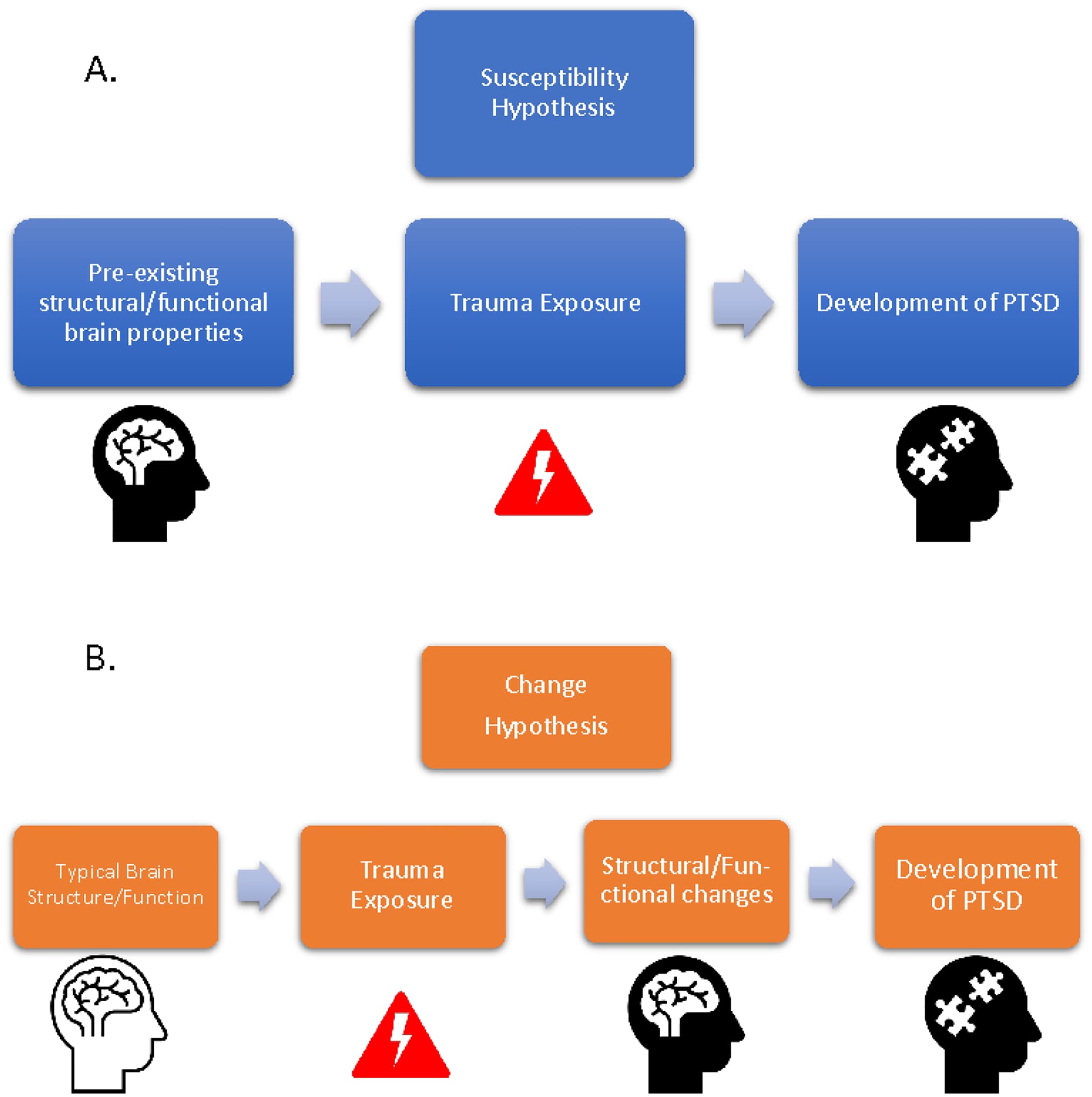
Figure 1. Conceptual Schematic

Several other studies have also identified alterations to the integrity of white matter tracts that connect sensory cortex to the rest of the brain [6,27–29]. Connections for the right inferior occipital gyrus show a more central role (i.e. - nodal centrality) in PTSD patients as well as increased overall within-network connectivity in the visual network. Additionally, recent work has shown a relationship between PTSD and white matter integrity in pathways connecting the hippocampus and thalamus with later visual regions like the precuneus [6]. Increased fractional anisotropy has also been observed in PTSD patients in the posterior cingulum bundle [27], a pathway connecting visual areas to the medial temporal lobe. However, other connections linking the temporal lobe with occipital cortex, like the inferior longitudinal fasciculus, have shown decreased structural integrity [29]. Together, these findings suggest that not all temporo-occipital connections may be altered in the same way with PTSD.

Newer approaches have also begun to emerge for studying the relationship between PTSD and brain structure in sensory cortex. For example, individual network construction based on covariance of gray matter morphology is a method that allows for the evaluation of brain networks based on T1-weighted images alone. Using this method, changes have been observed in regions of sensory networks, including the olfactory gyrus, superior and inferior occipital gyri, and middle occipital gyrus [30]. Other studies have also used gray matter structural networks to identify changes in occipital cortex, specifically in the lingual gyrus, associated with PTSD. The aforementioned work by our own group used similar approaches based on white matter diffusion data to identify variability in the structure of sensory regions that associate with PTSD [26]. The development of additional multimodal and other powerful integrative statistical methods for investigating associations between PTSD and brain structure variability will continue to be an important area for future research.

In addition to measures of cortical thickness/volume and white matter structure, another interesting measure receiving more attention that has been linked to PTSD susceptibility is cortical gyrification - a measure of the folding and patterning of the brain gyri and sulci. Functionally, these patterns of cortical folding enable the efficient wiring of local neuronal connections. In PTSD patients, relative to trauma-exposed controls, cortical gyrification was higher in regions of occipital cortex [31]. This finding suggests that cortical gyrification in occipital cortex may serve as a risk factor for the later development of PTSD following exposure to trauma.

These findings suggest that the link between brain structure and PTSD symptoms may underlie many of the subsequent changes in sensory processing that occur in individuals with PTSD (Fig. 1). However, exactly how these changes in brain morphology translate to changes in behavior remains to be understood. One potential avenue of exploration to provide further clarity is the understanding of brain activity changes following the development of PTSD.

Brain activity and PTSD

Task-based brain activity

Studying brain activity in individuals with post-traumatic stress disorder (PTSD) provides valuable insights into the underlying mechanisms associated with the condition (Fig. 2). For example, individuals with trauma exposure and/or PTSD have shown lower activity in visual cortex when examining positive visual stimuli [32], and reduced activation in secondary visual areas in response to negative visual stimuli [33]. The similarity in modulation for positive and negative stimuli matches work in individuals with anxiety and mood disorders without PTSD, where activity in visual cortex elicited by unpleasant stimuli is not significantly different from when viewing neutral stimuli [34]. This is in contrast to healthy individuals, in whom greater activity in early visual cortex is observed when viewing aversive compared to scrambled visual stimuli [7].
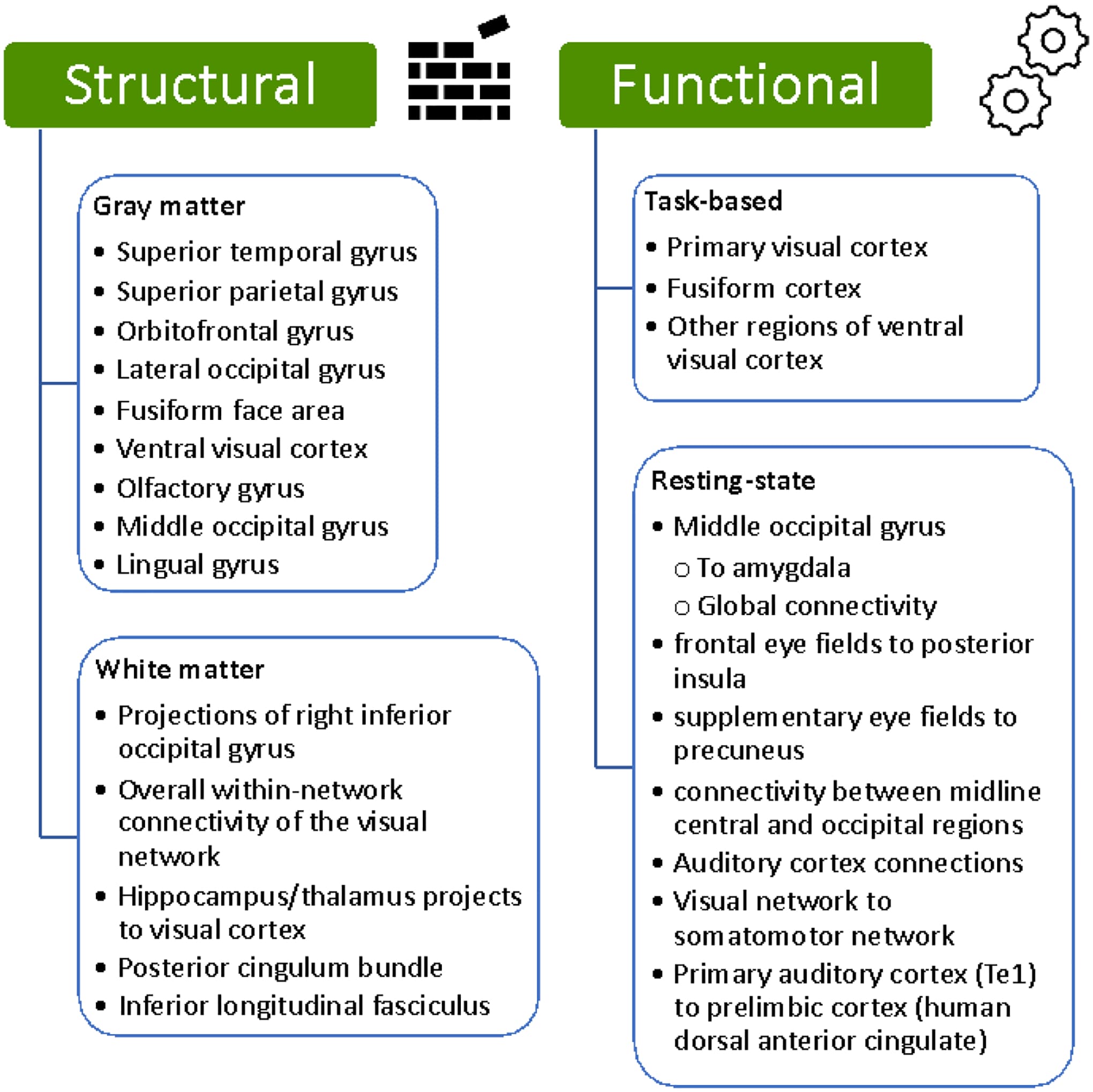
Figure 2. Summary of Recently Implicated Brain Regions

These findings match prior findings that PTSD patients exhibit difficulty with distinguishing between threatening and non-threatening stimuli [33,35]. It is also consistent with other work showing that PTSD symptoms are associated with habituation of activity in visual cortex in response to the repeated presentation of fearful face stimuli [36]). While structures in canonical fear circuitry (i.e. – the amygdala, prefrontal cortex, hippocampus, etc.) show altered patterns of activity in PTSD, the differential modulation of sensory cortex in healthy individuals that becomes less distinguishable with greater trauma exposure and PTSD, further suggests a top-down influence on sensory cortex that is mediated by subcortical and frontal regions [36].

Functional connectivity

Resting-state brain activity has also helped to inform current understanding of the neurobiology of PTSD. For example, PTSD with and without dissociation can be distinguished using the resting activity of somatosensory and motor regions [37]. Functional connectivity, or the correlation in spontaneous brain activity between regions, has also been a useful metric for understanding sensory cortex in PTSD. Notably, many recent studies have similar lines of corroborating evidence for the importance of the middle occipital gyrus (MOG) in PTSD symptomology. For example, PTSD patients show lower connectivity between the MOG and amygdala [38], in addition to altered global patterns of connectivity stemming from MOG [39,40]. This evidence bolsters the importance of not only cortico-limbic connectivity in PTSD, but also cortico-cortical connections that link sensory structures to other cortical regions including posterior cingulate cortex [41] and premotor cortex [42]. Additionally, these findings are supported by recent evidence showing reduced activity in middle occipital cortex when examining positive visual stimuli [32].

Additionally, regions involved in visual processing outside of occipital cortex, such as the frontal eye fields (FEF) and supplementary eye fields (SEF), may be involved in aspects of PTSD. Specifically, recent work suggests that functional connectivity of these areas may be related to the retrieval of trauma memories. Along these lines, Harricharan and colleagues found that PTSD patients show lower connectivity between FEF and posterior insula, SEF and precuneus [43]. These findings suggest that eye movements, or more globally – top-down control over sensory processing in PTSD, may be a potential avenue for the development of future therapies. Furthermore, the current psychotherapy approach of eye movement desensitization and reprocessing (EMDR), which is thought to primarily have efficacy through supporting emotion stabilization during exposure therapy, may also have important effects in sensory networks, which has not yet been explored in detail through a sensory neuroscience lens.

New approaches that build upon the concepts of functional connectivity have also been used to identify changes in sensory processing regions that associate with PTSD [44]. One relatively new method is meta-analytic co-activation modeling (MACM). Although slightly different from pure functional connectivity, MACM is an approach that uses meta-analysis as a means to derive functional connectivity. Using this approach, Pankey and colleagues were able to identify altered patterns of connectivity in visual processing regions that were associated with PTSD [44].

Recent work has shown that resting-state functional connectivity in sensory regions may be a helpful tool in evaluating the effectiveness of novel therapies for PTSD. For example, Zandvakili and colleagues found that after treatment with theta-burst transcranial magnetic stimulation, PTSD patients exhibited increased EEG-based functional connectivity between midline central and occipital regions [45]. In a study of youth with PTSD, functional connectivity in auditory cortex was able to predict individual responses to trauma-focused psychotherapy with up to 76% accuracy [11]. This investigation also saw increased connectivity between regions of the parietal network and the sensorimotor network. Similarly, Korgaonkar and colleagues found that functional connectivity of brain regions in the visual and somatomotor networks was related to better treatment outcomes in PTSD patients who were responsive to trauma-focused psychotherapy treatment [46].

A great deal of focus in studies of functional connectivity and PTSD has been placed on static functional connectivity, or the average functional coupling of activity between brain regions over a given time. However, additional insight has also been gained from the study of dynamic functional connectivity, which probes how functional coupling changes dynamically from moment to moment. For example, Wen and colleagues found that PTSD patients exhibited abnormal patterns of dynamic functional connectivity, specifically within regions of the visual network [47]. In addition to studies of static and dynamic functional connectivity during rest, work in task-based functional connectivity has also identified changes associated with PTSD. In a study of trauma-exposed Black women, performed in conjunction with members of our research group, negative affect symptoms in trauma-exposed individuals were associated with lower functional coupling between occipital cortex and amygdala in task-based functional connectivity [48]. This coupling, observed during a valence-rating task of emotional pictures, points towards the functional relevance of altered functional connectivity observed in PTSD patients.

Animal studies have also suggested the potential importance of functional coupling between sensory cortex and non-sensory brain regions [12,49,50]. For example, Concina and colleagues found that synchronization between primary auditory cortex (Te1) and prelimbic cortex (thought to be somewhat homologous to human dACC) plays an important role in fear discrimination [12]. Notably, the authors found that communication between prefrontal and limbic structures alone was not sufficient for fear discrimination. This finding again highlights the importance of studying cortico-cortical connectivity between sensory and prelimbic structures, in addition to connections between limbic and paralimbic structures, to fully understand their role in fear generalization and discrimination in PTSD. Other work has examined the question of whether functional coupling between sensory and non-sensory regions represents changes vs. susceptibility. For example, in a rat model of PTSD, resilience to trauma was shown to be associated with pre-trauma functional connectivity between limbic structures and regions involved in olfaction, vision, and audition [50]. Together, this work highlights potential targets for manipulating brain activity that may relate to sensory processing dysfunction in PTSD.

Developments in sensory cortex based therapies for PTSD

In addition to investigating functional and structural alterations associated with PTSD, recent work has also shown ways in which sensory cortex can be targeted to help relieve symptoms of PTSD. For example, work using repetitive transcranial magnetic stimulation (rTMS) to target activity in visual cortex has proven to be effective in relieving intrusive memories in PTSD. Herz and colleagues found that treating early visual cortex with inhibitory 1 Hz repetitive rTMS reduced the emotional intensity of intrusive memories [51]. The frequency of intrusions and the explicit visual memories themselves, however, remained intact. The finding that emotional intensity has strong clinical implications, but also contributes to fundamental knowledge about the role of early visual cortex in PTSD symptomology. This finding is consistent with evidence from the perceptual learning literature in which stimulation of early visual cortex inhibits previously learned, perceptual memory of non-emotional visual stimuli [52]. Recent work has also pinned virtual reality as a potential avenue of therapy for PTSD symptoms [53]. Such technology could potentially allow for exposure therapy with sensory stimuli in the absence of threat in a way that diminishes previously learned negative responses to stimuli associated with prior trauma. As more knowledge is uncovered about the relationship between PTSD and sensory brain structure/function, virtual reality could be a useful tool in helping to correct targeted sensory dysfunction after trauma exposure.

Conclusion

Mechanistically, there is an abundance of data in animal models that sensory processing can change, across most sensory systems, at the level of function and structure following sensory-based fear conditioning and trauma exposure [17,18,54,55]. Here, we have attempted to summarize recent studies in humans that demonstrate the presence of such changes/forms of variability associated with PTSD. With these new lines of evidence, it is becoming increasingly clear that sensory regions, their patterns of connectivity, and subsequent ties to sensory behavior may need to be incorporated more centrally into current lines of thinking around PTSD.

Moving forward, there remains a significant call to action in several areas. First, the major question that remains is: what exactly is represented by the variability in sensory systems that is associated with PTSD? While many aspects of sensory neural circuitry appear to vary with PTSD symptomology, a deeper interpretation of the causal nature of these associations must be considered. This need will perhaps be best served through more longitudinal human studies that further separate the characteristics of pre-existing susceptibility, acute responses in the immediate aftermath of trauma, and the long-term changes that occur at later post-trauma time points. Further work is also needed to build upon animal studies that have probed the susceptibility/changes in sensory cortex associated with trauma and PTSD [17,50,56]. Specifically, these efforts should focus on using pre-clinical models to better understand the mechanisms behind sensory cortex-related changes/susceptibility. For example, using more pre-post trauma designs in animal models will help reveal how variability in sensory circuits contributes to vulnerability for the development of sensory processing dysfunction and PTSD symptomatology after trauma exposure. These types of studies could then be followed by genetic/molecular manipulation of these circuits to determine whether susceptibility can be reduced by strengthening/weakening these pathways Additionally, studies that assess variability among PTSD subjects, beyond traditional case-control analyses, will provide important insight into the relationship between sensory processing and PTSD. While direct comparisons between patients and controls have been informative, much can also be learned by examining how varying degrees of symptom severity relate to variation in sensory-processing among PTSD patients. Finally, leveraging this knowledge is crucial for the development of targeted therapies and the enhancement of existing treatment options through a better understanding of their underlying mechanisms of action. As future studies continue to investigate the relationships between PTSD and sensory processing, it will aid the field in not only understanding more about the sensory mechanisms that contribute to the disorder, but also provide key insight into the dynamic relationship between emotion and sensory processing.

Highlights.

  • Sensory processing may deserve a more central role in our thinking about PTSD.

  • PTSD is linked to variability in sensory processing and properties of sensory cortex.

  • PTSD is related to local & global patterns of functional connectivity in sensory cortex.

  • PTSD is associated with structural gray/white matter differences in sensory cortex.

  • Sensory variability may represent susceptibility to, or consequences of, PTSD.

Open Article as PDF

Abstract

PTSD is characterized by difficulties in accurately evaluating the threat value of sensory stimuli. While the role of canonical fear and threat neural circuitry in this ability has been well studied, recent lines of evidence suggest a need to include more emphasis on sensory processing in the conceptualization of PTSD symptomology. Specifically, studies have demonstrated a strong association between variability in sensory processing regions and the severity of PTSD symptoms. In this review, we summarize recent findings that underscore the importance of sensory processing in PTSD, in addition to the structural and functional characteristics of associated sensory brain regions. First, we discuss the link between PTSD and various behavioral aspects of sensory processing. This is followed by a discussion of recent findings that link PTSD to variability in the structure of both gray and white matter in sensory brain regions. We then delve into how brain activity (measured with task-based and spontaneous functional imaging) in sensory regions informs our understanding of PTSD symptomology.

Introduction

Difficulty in telling the difference between things that are dangerous and things that are not is a key feature of Post-traumatic Stress Disorder (PTSD). Assessing potentially traumatic information relies on the ability to notice and interpret sensory details. This process requires a balance between internal and external sensory information and how threatening it seems to an individual. Developing PTSD after trauma involves a connection between how senses experience things and how emotions judge them. While the role of threat-related brain circuits is well understood, sensory processing has received less attention in explaining the disorder.

Recent studies suggest that sensory circuits might be very important in understanding who is at risk for PTSD after trauma and who recovers well. They also help explain specific PTSD symptoms like flashbacks. Flashbacks involve both clear memories and strong sensory experiences, such as "seeing" the trauma happen again, which can cause intense physical reactions. These symptoms can become more frequent when the memory of the trauma is wrongly applied to situations that are not actually dangerous. Therefore, understanding how sensory processing works in PTSD is essential for reducing the sensory problems common in the disorder.

A crucial question is how variations in sensory processing, and the structure and function of sensory brain areas, relate to the overall severity of PTSD. It is important to distinguish whether these variations are a result of PTSD itself or if they represent risk factors for developing the disorder later. Such knowledge could greatly help in treating PTSD and has implications for sensory processing problems in many other mental health conditions. This review discusses recent findings on the important role of sensory processing in PTSD. It highlights altered sensory processing linked to PTSD, evidence of structural and functional brain variations, and new therapies based on this understanding.

Altered Sensory Processing and PTSD

PTSD is linked to various problems in how senses process information. Sensory modulation, which is how the brain increases or decreases activity in sensory areas in response to stimuli, may explain changes in specific senses like vision, hearing, touch, and smell. In PTSD, problems with sensory modulation can lead to sensory areas being overly active or underactive when exposed to stimuli. This can cause symptoms like extreme alertness (hyperarousal) or feeling detached (dissociation) in response to triggers. Studies in this area show that PTSD is connected to problems processing both emotional and non-emotional sensory information.

It is known that PTSD causes issues with processing the emotional aspects of sensory stimuli. Other research shows that PTSD changes an individual's ability to evaluate both pleasant and unpleasant emotional visual stimuli. For instance, individuals had trouble telling neutral stimuli apart after those stimuli had been linked to an unpleasant visual experience. Other work shows that PTSD is connected to non-emotional visual processes, such as picturing coherent scenes and navigating complex environments. Recent studies have also found sensory processing problems linked to trauma experienced early in life. For example, children exposed to ongoing traumatic stress show altered sensory modulation and general sensory processing issues, regardless of emotional context. However, the brain mechanisms behind these problems are not fully understood.

Recent studies of brain structure and function have provided new insights into potential reasons for sensory processing problems in PTSD. For example, one study identified structural features directly linked to PTSD-related issues in non-emotional sensory processing. Specifically, spatial processing and integration were related to the density of white matter fibers connecting core memory regions (like the hippocampus and thalamus) with higher-level visual areas (like the precuneus). Other recent work has identified potential functional reasons for sensory processing problems in PTSD. Studies in combat veterans suggest that sensory processing dysfunction in PTSD may be linked to altered timing of alpha brain activity, which is thought to help regulate sensory inhibition. Issues in regulating alpha connectivity in PTSD may lead to an inability to quiet sensory areas, possibly explaining the overactive sensory experience during trauma re-experiencing.

These findings offer potential insights into the causes of sensory processing dysfunction in PTSD, but much remains unknown. The fact that PTSD is linked to problems in processing both emotional and non-emotional stimuli raises several interesting possibilities. It could mean that a general predisposition exists before trauma exposure. In other words, individuals with existing changes in sensory processing might be more likely to develop PTSD-related sensory problems. However, it is important to note that traits like sensory processing sensitivity have been linked to brain patterns different from those associated with hyperarousal in PTSD. If this is the case, it prompts the question of whether trauma exposure, combined with certain fear-related brain regions, plays a causal role in explaining sensory hyperarousal in PTSD. To learn more, researchers must examine how variations in key brain features relate to PTSD symptoms. The next sections describe these connections in more detail, focusing on the relationship between PTSD and brain structure, brain activity, and functional connections.

Neural Structure of Sensory Cortex in PTSD

A growing body of evidence shows a link between PTSD and the structural features of sensory brain areas. Animal studies have previously connected changes in brain structure to severe psychological stress. Evidence also comes from human studies. For example, a large combined analysis found that more severe intrusive memories were linked to less gray matter volume in temporal, parietal, and occipital regions. These regions include the superior temporal gyrus, which helps combine visual and auditory information from emotional stimuli, the superior parietal gyrus, involved in processing spatial and movement information, and the bilateral orbitofrontal gyrus, which guides sensory attention and combines input from sensory and emotional brain structures. Other studies have found decreased average cortical thickness in the visual cortex in individuals with PTSD, with less thickness in the lateral occipital gyrus compared to control groups.

While comparing brain structure between PTSD patients and non-trauma-exposed individuals is valuable, insights can also come from examining how structural variations among PTSD patients lead to different symptoms. This helps address the major question of whether the sensory cortex properties linked to PTSD are pre-trauma vulnerabilities or changes acquired after trauma exposure. For example, PTSD patients with more avoidance behaviors show greater cortical thickness in the middle and lateral occipital cortex. Other investigations have looked at the timing of structural changes in sensory cortex in the early stages before PTSD symptoms fully develop. For instance, studying individuals with Acute Stress Disorder (ASD) has provided some insight. ASD involves PTSD-like symptoms immediately after trauma (up to the first month) and can predict the later development of full PTSD. Individuals with ASD show less gray matter volume in temporal and occipital regions, which is linked to PTSD symptom severity and symptom clusters four weeks later. Research from some groups shows significant links between acute post-traumatic symptoms and the structure of gray and white matter in visual regions, including the fusiform face area and other areas in the ventral stream. Variations in these structures showed a curved relationship with acute post-traumatic stress severity and the change in PTSD symptom severity from 1 to 12 months. Together, these findings highlight the importance of continued research into sensory cortical variations in relation to the chronological development of PTSD symptoms.

Several other studies have also found changes in the integrity of white matter tracts that connect the sensory cortex to the rest of the brain. Connections for the right inferior occipital gyrus show a more central role in PTSD patients, as well as increased overall connectivity within the visual network. Additionally, recent work has shown a relationship between PTSD and white matter integrity in pathways connecting the hippocampus and thalamus with later visual regions like the precuneus. Increased fractional anisotropy, a measure of white matter integrity, has also been observed in PTSD patients in the posterior cingulum bundle, a pathway connecting visual areas to the medial temporal lobe. However, other connections linking the temporal lobe with the occipital cortex, like the inferior longitudinal fasciculus, have shown decreased structural integrity. These findings suggest that not all temporo-occipital connections may be altered in the same way with PTSD.

Newer methods have also emerged for studying the relationship between PTSD and brain structure in the sensory cortex. For example, building individual networks based on how gray matter morphology varies allows for evaluating brain networks using only T1-weighted images. Using this method, changes have been observed in regions of sensory networks, including the olfactory gyrus, superior and inferior occipital gyri, and middle occipital gyrus. Other studies have also used gray matter structural networks to identify changes in the occipital cortex, specifically in the lingual gyrus, associated with PTSD. Research by some groups used similar approaches based on white matter diffusion data to identify variations in the structure of sensory regions that are linked to PTSD. Developing additional multi-modal and other powerful integrative statistical methods for investigating connections between PTSD and brain structure variation will continue to be an important area for future research.

In addition to measures of cortical thickness/volume and white matter structure, another interesting measure receiving more attention that has been linked to PTSD susceptibility is cortical gyrification. This measures the folding and patterning of the brain's ridges and grooves. Functionally, these folding patterns allow for efficient wiring of local neuronal connections. In PTSD patients, compared to trauma-exposed control individuals, cortical gyrification was higher in regions of the occipital cortex. This finding suggests that cortical gyrification in the occipital cortex may serve as a risk factor for the later development of PTSD following trauma exposure.

These findings suggest that the link between brain structure and PTSD symptoms may explain many of the subsequent changes in sensory processing that occur in individuals with PTSD. However, exactly how these changes in brain shape translate to changes in behavior is still not fully understood. One potential area of exploration to provide further clarity is understanding how brain activity changes after PTSD develops.

Brain Activity and PTSD

Studying brain activity in individuals with PTSD provides valuable insights into the underlying mechanisms of the condition. For example, individuals exposed to trauma or with PTSD have shown lower activity in the visual cortex when looking at positive visual stimuli, and reduced activation in secondary visual areas in response to negative visual stimuli. This similar response to positive and negative stimuli matches findings in individuals with anxiety and mood disorders without PTSD, where visual cortex activity triggered by unpleasant stimuli is not significantly different from when viewing neutral stimuli. This contrasts with healthy individuals, who show greater activity in the early visual cortex when viewing unpleasant compared to scrambled visual stimuli.

These findings align with previous observations that PTSD patients have difficulty distinguishing between threatening and non-threatening stimuli. It is also consistent with other work showing that PTSD symptoms are linked to the visual cortex becoming less responsive to repeated presentations of fearful faces. While structures in typical fear circuits (such as the amygdala, prefrontal cortex, and hippocampus) show altered activity patterns in PTSD, the differing modulation of sensory cortex in healthy individuals, which becomes less distinct with greater trauma exposure and PTSD, further suggests that deeper brain regions and frontal areas influence the sensory cortex.

Resting-state brain activity has also contributed to the current understanding of PTSD neurobiology. For instance, PTSD with and without dissociation can be differentiated using the resting activity of somatosensory and motor regions. Functional connectivity, or the correlation in spontaneous brain activity between regions, has also been a useful measure for understanding the sensory cortex in PTSD. Many recent studies show similar supporting evidence for the importance of the middle occipital gyrus (MOG) in PTSD symptoms. For example, PTSD patients show lower connectivity between the MOG and amygdala, in addition to altered overall patterns of connectivity originating from the MOG. This evidence emphasizes the importance of not only connections between the cortex and limbic system in PTSD, but also connections between different cortical regions that link sensory structures to other cortical areas, including the posterior cingulate cortex and premotor cortex. Additionally, these findings are supported by recent evidence showing reduced activity in the middle occipital cortex when looking at positive visual stimuli.

Regions involved in visual processing outside the occipital cortex, such as the frontal eye fields (FEF) and supplementary eye fields (SEF), may also play a role in aspects of PTSD. Specifically, recent work suggests that the functional connectivity of these areas may be related to the retrieval of trauma memories. Along these lines, one study found that PTSD patients show lower connectivity between the FEF and posterior insula, and between the SEF and precuneus. These findings suggest that eye movements, or more broadly, top-down control over sensory processing in PTSD, could be a potential area for developing future therapies. Furthermore, the current psychotherapy approach of eye movement desensitization and reprocessing (EMDR), which is thought to primarily help stabilize emotions during exposure therapy, may also have important effects on sensory networks, which has not yet been thoroughly explored from a sensory neuroscience perspective.

New approaches that build on the concept of functional connectivity have also been used to identify changes in sensory processing regions linked to PTSD. One relatively new method is meta-analytic co-activation modeling (MACM), which uses meta-analysis to derive functional connectivity. Using this approach, researchers identified altered patterns of connectivity in visual processing regions associated with PTSD.

Recent work has shown that resting-state functional connectivity in sensory regions may be a useful tool for evaluating the effectiveness of new PTSD therapies. For example, researchers found that after treatment with theta-burst transcranial magnetic stimulation, PTSD patients showed increased EEG-based functional connectivity between midline central and occipital regions. In a study of young people with PTSD, functional connectivity in the auditory cortex was able to predict individual responses to trauma-focused psychotherapy with up to 76% accuracy. This investigation also observed increased connectivity between regions of the parietal network and the sensorimotor network. Similarly, another study found that functional connectivity of brain regions in the visual and somatomotor networks was related to better treatment outcomes in PTSD patients who responded to trauma-focused psychotherapy treatment.

Much research on functional connectivity and PTSD has focused on static functional connectivity, which is the average functional coupling of activity between brain regions over a given time. However, additional insight has also come from studying dynamic functional connectivity, which examines how functional coupling changes from moment to moment. For example, one study found that PTSD patients showed abnormal patterns of dynamic functional connectivity, specifically within regions of the visual network. In addition to studies of static and dynamic functional connectivity during rest, research on task-based functional connectivity has also identified changes associated with PTSD. In a study of trauma-exposed Black women, negative emotional symptoms in trauma-exposed individuals were linked to lower functional coupling between the occipital cortex and amygdala during task-based functional connectivity. This coupling, observed during a task where participants rated emotional pictures, highlights the functional importance of altered functional connectivity seen in PTSD patients.

Animal studies have also suggested the potential importance of functional coupling between the sensory cortex and non-sensory brain regions. For example, researchers found that synchronization between the primary auditory cortex and prelimbic cortex (thought to be similar to the human dorsal anterior cingulate cortex) plays an important role in fear discrimination. Notably, the authors found that communication between prefrontal and limbic structures alone was not enough for fear discrimination. This finding again emphasizes the importance of studying connections between sensory and prelimbic structures, in addition to connections between limbic and paralimbic structures, to fully understand their role in how fear is generalized and differentiated in PTSD. Other work has examined whether functional coupling between sensory and non-sensory regions represents changes or susceptibility. For example, in a rat model of PTSD, resilience to trauma was shown to be associated with pre-trauma functional connectivity between limbic structures and regions involved in smell, vision, and hearing. Together, this work highlights potential targets for manipulating brain activity that may relate to sensory processing dysfunction in PTSD.

Developments in Sensory Cortex Based Therapies for PTSD

In addition to investigating functional and structural changes associated with PTSD, recent work has also shown ways in which the sensory cortex can be targeted to help relieve PTSD symptoms. For example, studies using repetitive transcranial magnetic stimulation (rTMS) to target activity in the visual cortex have proven effective in reducing intrusive memories in PTSD. One study found that treating the early visual cortex with inhibitory 1 Hz repetitive rTMS reduced the emotional intensity of intrusive memories. The frequency of intrusions and the explicit visual memories themselves, however, remained intact. The finding that emotional intensity is affected has strong clinical implications and also adds fundamental knowledge about the role of the early visual cortex in PTSD symptoms. This finding is consistent with evidence from perceptual learning research, where stimulating the early visual cortex inhibits previously learned, perceptual memory of non-emotional visual stimuli. Recent work has also identified virtual reality as a potential therapy for PTSD symptoms. Such technology could allow for exposure therapy with sensory stimuli in a non-threatening way, diminishing previously learned negative responses to stimuli associated with past trauma. As more is learned about the relationship between PTSD and sensory brain structure/function, virtual reality could be a useful tool in helping to correct targeted sensory dysfunction after trauma exposure.

Conclusion

From a mechanical perspective, there is a lot of data from animal models showing that sensory processing can change, across most sensory systems, in terms of both function and structure following fear conditioning based on sensory input and trauma exposure. This document summarized recent human studies demonstrating such changes or variations associated with PTSD. With these new findings, it is becoming increasingly clear that sensory regions, their patterns of connectivity, and their links to sensory behavior may need to be included more centrally in current thinking about PTSD.

Looking ahead, several areas require significant further action. First, the main question remains: what exactly do the variations in sensory systems linked to PTSD represent? While many aspects of sensory neural circuits appear to vary with PTSD symptoms, a deeper understanding of the causal nature of these connections must be considered. This need will likely be best met through more longitudinal human studies that further separate the characteristics of pre-existing vulnerability, acute responses immediately after trauma, and the long-term changes that occur at later post-trauma time points. Further work is also needed to build upon animal studies that have explored the susceptibility and changes in the sensory cortex associated with trauma and PTSD. Specifically, these efforts should focus on using preclinical models to better understand the mechanisms behind sensory cortex-related changes and vulnerabilities. For example, using more pre- and post-trauma designs in animal models will help reveal how variations in sensory circuits contribute to vulnerability for developing sensory processing dysfunction and PTSD symptoms after trauma exposure. These types of studies could then be followed by genetic or molecular manipulation of these circuits to determine whether susceptibility can be reduced by strengthening or weakening these pathways. Additionally, studies that assess variability among PTSD subjects, beyond traditional case-control analyses, will provide important insight into the relationship between sensory processing and PTSD. While direct comparisons between patients and controls have been informative, much can also be learned by examining how varying degrees of symptom severity relate to variation in sensory processing among PTSD patients. Finally, applying this knowledge is crucial for developing targeted therapies and improving existing treatment options by better understanding their underlying mechanisms of action. As future studies continue to investigate the relationships between PTSD and sensory processing, this will help the field not only understand more about the sensory mechanisms that contribute to the disorder but also provide key insight into the dynamic relationship between emotion and sensory processing.

Highlights

  • Sensory processing should be considered a more central aspect of understanding PTSD.

  • PTSD is linked to variations in sensory processing and the properties of the sensory cortex.

  • PTSD is related to local and global patterns of functional connectivity in the sensory cortex.

  • PTSD is associated with structural differences in gray and white matter in the sensory cortex.

  • Sensory variability may indicate either a susceptibility to PTSD or a consequence of the disorder.

Open Article as PDF

Abstract

PTSD is characterized by difficulties in accurately evaluating the threat value of sensory stimuli. While the role of canonical fear and threat neural circuitry in this ability has been well studied, recent lines of evidence suggest a need to include more emphasis on sensory processing in the conceptualization of PTSD symptomology. Specifically, studies have demonstrated a strong association between variability in sensory processing regions and the severity of PTSD symptoms. In this review, we summarize recent findings that underscore the importance of sensory processing in PTSD, in addition to the structural and functional characteristics of associated sensory brain regions. First, we discuss the link between PTSD and various behavioral aspects of sensory processing. This is followed by a discussion of recent findings that link PTSD to variability in the structure of both gray and white matter in sensory brain regions. We then delve into how brain activity (measured with task-based and spontaneous functional imaging) in sensory regions informs our understanding of PTSD symptomology.

Introduction

Difficulty in distinguishing between threatening and non-threatening situations is a core feature of Post-Traumatic Stress Disorder (PTSD). Understanding how individuals evaluate potential threats involves recognizing and interpreting sensory information. This process requires a balance between internal thoughts and external sensory cues, and how an individual perceives their danger. Trauma exposure and the development of PTSD involve complex interactions between brain circuits responsible for sensory experiences and emotional evaluations. While the role of threat-related brain circuits is widely recognized, the integration of sensory processing into models of PTSD has received less attention.

Recent research suggests that sensory circuits are important for understanding who develops PTSD after trauma and who is resilient. These circuits may also help explain different ways PTSD symptoms appear, such as "intrusion" symptoms like flashbacks. Intrusive symptoms and flashbacks include a clear memory component with strong sensory details, such as vividly "seeing" a traumatic event again, which can trigger intense physical reactions. These symptoms can become more frequent when the traumatic memory is mistakenly applied to non-traumatic situations. Therefore, understanding how sensory processing contributes to PTSD is vital for addressing the sensory disturbances common in the disorder.

A critical question is how differences in sensory processing, along with the physical and functional characteristics of sensory brain regions, relate to the overall severity of PTSD. It is important to distinguish between changes in these features that result from PTSD itself and those that existed as risk factors before PTSD developed. Such understanding holds great promise for improving PTSD interventions and has implications for sensory processing difficulties seen in various other psychiatric conditions. This discussion explores recent findings on the important role of sensory processing in PTSD, highlighting how altered sensory processing is linked to the disorder, and discussing structural and functional brain differences associated with PTSD. It also covers new developments in PTSD therapies based on knowledge of sensory processing.

Altered Sensory Processing and PTSD

PTSD has been linked to various problems in how sensory information is processed. Sensory modulation, which involves increasing or decreasing activity in the brain's sensory areas in response to stimuli, may explain changes in processing for specific senses like vision, hearing, touch, and smell. In PTSD, problems with sensory modulation can lead to sensory brain areas being either over-responsive or under-responsive to stimuli. This can result in symptoms such as hyperarousal (being overly alert) or dissociative states (feeling detached or numb) in response to triggering events. Research in this area shows that PTSD is associated with difficulties in processing both emotional and non-emotional sensory stimuli.

It is well-known that PTSD affects the ability to properly process the emotional aspects of sensory stimuli. Other studies show that PTSD is associated with changes in how individuals evaluate both pleasant and unpleasant emotional visual stimuli. For example, some research found that distinguishing between neutral stimuli became harder after those stimuli had been paired with an unpleasant visual stimulus. Other work has shown that PTSD affects non-emotional visual processes, such as mentally picturing coherent scenes and navigating complex environments. Recent findings also indicate sensory processing problems associated with early life trauma. For instance, children exposed to ongoing traumatic stress show altered sensory modulation patterns and general sensory processing deficits, regardless of emotional context. However, the exact brain mechanisms behind these problems are still not fully understood.

Recent brain structure and function studies have provided new insights into the potential causes of sensory processing deficits in PTSD. For example, some research identified structural features directly linked to PTSD-related problems in non-emotional sensory processing. Specifically, they found that spatial processing and integration were related to the density of white matter fibers connecting core memory regions (like the hippocampus and thalamus) with higher-order visual areas such as the precuneus. Other recent work has identified potential functional brain mechanisms for sensory processing deficits in PTSD. Studies in combat veterans suggest that sensory processing problems in PTSD might be related to altered synchronization of alpha activity (brain activity in the 8–12 Hz range measured by EEG), which may be involved in regulating sensory inhibition. Alpha oscillations are particularly important because of their role in controlling "bottom-up" sensory input between sensory brain areas and frontal regions. Evidence suggests that problems in regulating alpha connectivity in PTSD may lead to an inability to inhibit sensory areas, which could cause the overactivation of sensory representations during the re-experiencing of trauma.

While these findings offer potential insights into the underlying mechanisms of sensory processing dysfunction in PTSD, much remains to be understood. The fact that PTSD is linked to sensory processing deficits for both emotional and non-emotional stimuli raises several interesting possibilities. These findings might indicate a universal predisposition that exists before trauma exposure. In other words, individuals with pre-existing alterations in sensory processing might be more susceptible to developing PTSD-related sensory dysfunction. However, it is worth noting that traits like sensory processing sensitivity (SPS) have been associated with brain patterns that are different from those linked to hyperarousal in PTSD. If this is the case, it prompts the question of whether trauma exposure, combined with certain fear-related brain regions (such as the prefrontal cortex, amygdala, and hippocampus), plays a causal role in explaining sensory hyperarousal in PTSD. Further understanding in this area can be achieved by examining how variations in key brain features relate to PTSD symptoms. The following sections describe these associations in more detail, focusing specifically on the relationship between PTSD and brain structure, brain activity, and functional connectivity.

Neural Structure of Sensory Cortex in PTSD

A growing body of evidence shows a link between PTSD and the structural features of sensory brain regions. Animal studies have previously connected changes in brain structure to severe psychological stress. Similar evidence has also emerged from human studies. For example, a large-scale analysis found that more severe intrusive memories were associated with lower gray matter volume in temporal, parietal, and occipital brain regions. These regions include the superior temporal gyrus, which helps combine auditory and visual information from emotional stimuli; the superior parietal gyrus, involved in processing spatial and movement information; and the bilateral orbitofrontal gyrus, which helps guide sensory attention and integrate inputs from sensory and emotional brain structures. Other studies have found decreased average cortical thickness (the thickness of the outer layer of the brain) in the visual cortex associated with PTSD, with thickness in the lateral occipital gyrus being lower in individuals with PTSD compared to control groups.

While comparing brain structure between PTSD patients and individuals not exposed to trauma is important, additional insights can be gained by examining how variations in cortical structure among PTSD patients lead to different symptoms. This helps address the major question of whether the structural properties of sensory brain regions associated with PTSD represent a pre-trauma vulnerability or changes acquired in response to trauma exposure. For example, PTSD patients with greater avoidance behaviors show greater cortical thickness in the middle and lateral occipital cortex. Other investigations have explored the timing of structural changes in sensory brain regions immediately before PTSD symptoms fully develop. Insights have come from studying individuals with Acute Stress Disorder (ASD), which involves PTSD-like symptoms in the first month after trauma and predicts later development of full PTSD. Individuals with ASD show lower gray matter volume in temporal and occipital regions, which is associated with PTSD symptom severity and symptom clusters four weeks later. Work by some research groups shows significant associations between acute post-traumatic symptoms and the structure of gray and white matter in visual regions, including the fusiform face area and other areas in the ventral stream. Variability in these structures showed a curved relationship with the severity of acute post-traumatic stress and the change in PTSD symptom severity from 1 to 12 months. Collectively, these findings highlight the importance of continued research into sensory cortical variability in relation to the chronological development of PTSD symptoms.

Several other studies have also identified alterations to the integrity of white matter tracts that connect sensory brain regions to the rest of the brain. Connections for the right inferior occipital gyrus show a more central role (i.e., nodal centrality) in PTSD patients, as well as increased overall connectivity within the visual network. Additionally, recent work has shown a relationship between PTSD and white matter integrity in pathways connecting the hippocampus and thalamus with later visual regions like the precuneus. Increased fractional anisotropy (a measure of white matter integrity) has also been observed in PTSD patients in the posterior cingulum bundle, a pathway connecting visual areas to the medial temporal lobe. However, other connections linking the temporal lobe with the occipital cortex, such as the inferior longitudinal fasciculus, have shown decreased structural integrity. Together, these findings suggest that not all temporo-occipital connections may be altered in the same way with PTSD.

Newer approaches have also emerged for studying the relationship between PTSD and brain structure in sensory areas. For example, individual network construction based on the covariance of gray matter morphology allows for the evaluation of brain networks using only T1-weighted images. Using this method, changes have been observed in regions of sensory networks, including the olfactory gyrus, superior and inferior occipital gyri, and middle occipital gyrus. Other studies have also used gray matter structural networks to identify changes in the occipital cortex, specifically in the lingual gyrus, associated with PTSD. Research by some groups used similar approaches based on white matter diffusion data to identify variability in the structure of sensory regions that are associated with PTSD. The development of additional multimodal and powerful integrative statistical methods for investigating associations between PTSD and brain structure variability will continue to be an important area for future research.

In addition to measures of cortical thickness/volume and white matter structure, another interesting measure receiving more attention that has been linked to PTSD susceptibility is cortical gyrification—a measure of the folding and patterning of the brain's ridges and grooves. Functionally, these patterns of cortical folding allow for the efficient wiring of local neuronal connections. In PTSD patients, compared to trauma-exposed control individuals, cortical gyrification was higher in regions of the occipital cortex. This finding suggests that cortical gyrification in the occipital cortex may serve as a risk factor for the later development of PTSD following trauma exposure. These findings indicate that the link between brain structure and PTSD symptoms may underlie many of the subsequent changes in sensory processing that occur in individuals with PTSD. However, exactly how these changes in brain shape translate into changes in behavior remains to be understood. One potential area of exploration to provide further clarity is understanding changes in brain activity following the development of PTSD.

Brain Activity and PTSD

Task-based Brain Activity

Studying brain activity in individuals with PTSD provides valuable insights into the mechanisms associated with the condition. For example, individuals with trauma exposure or PTSD have shown lower activity in the visual cortex when viewing positive visual stimuli and reduced activation in secondary visual areas in response to negative visual stimuli. The similar modulation for positive and negative stimuli aligns with research in individuals with anxiety and mood disorders without PTSD, where activity in the visual cortex triggered by unpleasant stimuli is not significantly different from when viewing neutral stimuli. This contrasts with healthy individuals, who show greater activity in early visual cortex when viewing unpleasant compared to scrambled visual stimuli.

These findings align with previous observations that PTSD patients have difficulty distinguishing between threatening and non-threatening stimuli. It is also consistent with other work showing that PTSD symptoms are associated with reduced brain activity in the visual cortex (habituation) in response to repeated presentations of fearful face stimuli. While structures in the typical fear circuitry (such as the amygdala, prefrontal cortex, and hippocampus) show altered patterns of activity in PTSD, the differing modulation of sensory cortex in healthy individuals, which becomes less distinct with greater trauma exposure and PTSD, further suggests a top-down influence on sensory cortex mediated by subcortical and frontal brain regions.

Functional Connectivity

Resting-state brain activity has also contributed to the current understanding of the neurobiology of PTSD. For example, PTSD with and without dissociation can be distinguished using the resting activity of somatosensory and motor regions. Functional connectivity, or the correlation in spontaneous brain activity between regions, has also been a useful measure for understanding sensory cortex in PTSD. Many recent studies have similar supporting evidence for the importance of the middle occipital gyrus (MOG) in PTSD symptoms. For instance, PTSD patients show lower connectivity between the MOG and amygdala, in addition to altered global patterns of connectivity originating from the MOG. This evidence strengthens the importance of not only connections between the cortex and limbic system in PTSD, but also connections between different cortical regions that link sensory structures to other cortical areas, including the posterior cingulate cortex and premotor cortex. Additionally, these findings are supported by recent evidence showing reduced activity in the middle occipital cortex when examining positive visual stimuli.

Brain regions involved in visual processing outside of the occipital cortex, such as the frontal eye fields (FEF) and supplementary eye fields (SEF), may also play a role in aspects of PTSD. Specifically, recent work suggests that the functional connectivity of these areas may be related to retrieving trauma memories. Along these lines, some researchers found that PTSD patients show lower connectivity between the FEF and posterior insula, and between the SEF and precuneus. These findings suggest that eye movements, or more broadly, top-down control over sensory processing in PTSD, could be a potential avenue for developing future therapies. Furthermore, the current psychotherapy approach of eye movement desensitization and reprocessing (EMDR), which is thought to primarily work by supporting emotional stabilization during exposure therapy, may also have important effects in sensory networks, which has not yet been explored in detail from a sensory neuroscience perspective.

New approaches that build upon the concepts of functional connectivity have also been used to identify changes in sensory processing regions associated with PTSD. One relatively new method is meta-analytic co-activation modeling (MACM). Although slightly different from pure functional connectivity, MACM uses meta-analysis to derive functional connectivity. Using this approach, researchers were able to identify altered patterns of connectivity in visual processing regions that were associated with PTSD.

Recent work has shown that resting-state functional connectivity in sensory regions may be a helpful tool in evaluating the effectiveness of new therapies for PTSD. For example, some researchers found that after treatment with theta-burst transcranial magnetic stimulation, PTSD patients exhibited increased EEG-based functional connectivity between midline central and occipital regions. In a study of young people with PTSD, functional connectivity in the auditory cortex was able to predict individual responses to trauma-focused psychotherapy with up to 76% accuracy. This investigation also observed increased connectivity between regions of the parietal network and the sensorimotor network. Similarly, other researchers found that functional connectivity of brain regions in the visual and somatomotor networks was related to better treatment outcomes in PTSD patients who responded to trauma-focused psychotherapy treatment.

Developments in Sensory Cortex Based Therapies for PTSD

In addition to investigating functional and structural alterations associated with PTSD, recent work has also shown ways in which the sensory cortex can be targeted to help relieve PTSD symptoms. For example, research using repetitive transcranial magnetic stimulation (rTMS) to target activity in the visual cortex has proven effective in relieving intrusive memories in PTSD. Some researchers found that treating the early visual cortex with inhibitory 1 Hz repetitive rTMS reduced the emotional intensity of intrusive memories. The frequency of intrusions and the explicit visual memories themselves, however, remained intact. The finding that emotional intensity is affected has strong clinical implications and contributes to fundamental knowledge about the role of the early visual cortex in PTSD symptoms. This finding is consistent with evidence from perceptual learning research, where stimulation of the early visual cortex inhibits previously learned, perceptual memories of non-emotional visual stimuli. Recent work has also identified virtual reality as a potential therapy avenue for PTSD symptoms. Such technology could potentially allow for exposure therapy with sensory stimuli in a safe environment, helping to diminish previously learned negative responses to stimuli associated with prior trauma. As more knowledge is uncovered about the relationship between PTSD and sensory brain structure and function, virtual reality could be a useful tool in helping to correct targeted sensory dysfunction after trauma exposure.

Conclusion

From a mechanistic perspective, a significant amount of data from animal models indicates that sensory processing can change, across most sensory systems, at both functional and structural levels following sensory-based fear conditioning and trauma exposure. This discussion has summarized recent human studies that demonstrate the presence of such changes or variations associated with PTSD. With these new lines of evidence, it is becoming increasingly clear that sensory regions, their patterns of connectivity, and their subsequent links to sensory behavior may need to be incorporated more centrally into current theories about PTSD.

Moving forward, there remains a significant need for action in several areas. First, the major question that persists is: what exactly do the variations in sensory systems associated with PTSD represent? While many aspects of sensory neural circuitry appear to vary with PTSD symptoms, a deeper interpretation of the causal nature of these associations must be considered. This need will perhaps be best served through more longitudinal human studies that further differentiate characteristics of pre-existing vulnerability, acute responses immediately after trauma, and long-term changes that occur at later post-trauma time points. Further work is also needed to build upon animal studies that have explored the vulnerability or changes in sensory cortex associated with trauma and PTSD. Specifically, these efforts should focus on using pre-clinical models to better understand the mechanisms behind sensory cortex-related changes and susceptibility. For example, using more pre- and post-trauma designs in animal models will help reveal how variability in sensory circuits contributes to vulnerability for the development of sensory processing dysfunction and PTSD symptoms after trauma exposure. These types of studies could then be followed by genetic or molecular manipulation of these circuits to determine whether susceptibility can be reduced by strengthening or weakening these pathways. Additionally, studies that assess variability among PTSD subjects, beyond traditional case-control analyses, will provide important insight into the relationship between sensory processing and PTSD. While direct comparisons between patients and controls have been informative, much can also be learned by examining how varying degrees of symptom severity relate to variation in sensory processing among PTSD patients. Finally, leveraging this knowledge is crucial for the development of targeted therapies and the enhancement of existing treatment options through a better understanding of their underlying mechanisms of action. As future studies continue to investigate the relationships between PTSD and sensory processing, it will aid the field in not only understanding more about the sensory mechanisms that contribute to the disorder but also provide key insight into the dynamic relationship between emotion and sensory processing.

Highlights

  • Sensory processing may deserve a more central role in understanding PTSD.

  • PTSD is linked to variability in sensory processing and the properties of sensory brain regions.

  • PTSD is related to both local and global patterns of functional connectivity in the sensory cortex.

  • PTSD is associated with structural differences in gray and white matter in the sensory cortex.

  • Sensory variability may represent either a susceptibility to, or a consequence of, PTSD.

Open Article as PDF

Abstract

PTSD is characterized by difficulties in accurately evaluating the threat value of sensory stimuli. While the role of canonical fear and threat neural circuitry in this ability has been well studied, recent lines of evidence suggest a need to include more emphasis on sensory processing in the conceptualization of PTSD symptomology. Specifically, studies have demonstrated a strong association between variability in sensory processing regions and the severity of PTSD symptoms. In this review, we summarize recent findings that underscore the importance of sensory processing in PTSD, in addition to the structural and functional characteristics of associated sensory brain regions. First, we discuss the link between PTSD and various behavioral aspects of sensory processing. This is followed by a discussion of recent findings that link PTSD to variability in the structure of both gray and white matter in sensory brain regions. We then delve into how brain activity (measured with task-based and spontaneous functional imaging) in sensory regions informs our understanding of PTSD symptomology.

Introduction

A main problem in Post-traumatic Stress Disorder (PTSD) is the difficulty people have telling the difference between things that are truly dangerous and things that are not. How a person judges potential threats depends on their ability to notice and understand what they are seeing, hearing, or feeling. This process requires a balance between information coming from inside the body and from the outside world, and how threatening it seems. Developing PTSD after a traumatic event involves a connection between how the senses experience things and how emotions evaluate them. While the brain circuits related to fear are well-known, less attention has been given to how sensory processing fits into our understanding of this disorder.

Recent research suggests that sensory circuits are very important for understanding why some people develop PTSD after trauma and others do not. They also help explain different ways PTSD appears, such as "intrusion" symptoms like flashbacks. Flashbacks and intrusive symptoms include clear memories with strong sensory details, such as vividly "seeing" the trauma again. These experiences can cause strong physical reactions. Such symptoms can happen more often when the memory of the trauma spreads to situations that are not actually related to the trauma. Therefore, understanding the role of sensory processing in PTSD is vital for improving the sensory problems common in the disorder.

A key question is how differences in sensory processing, and the structure and function of brain areas related to the senses, connect to the overall severity of PTSD. It is important to separate these differences into those caused by PTSD itself and those that might be risk factors for developing PTSD later. This understanding could greatly help in treating PTSD. It could also have meaning for sensory processing problems seen in many other mental health conditions. This review discusses recent findings about the important role of sensory processing in PTSD. It highlights new evidence showing changes in sensory processing and brain structure/function linked to PTSD. The discussion also covers the latest developments in PTSD therapies that use this knowledge of sensory processing.

Altered sensory processing and PTSD

PTSD has been linked to various problems in how people process sensory information. Sensory modulation is the brain's way of increasing or decreasing activity in sensory areas when faced with stimuli. Problems with sensory modulation may cause changes in how specific senses work, including sight, hearing, touch, and smell. In PTSD, issues with sensory modulation can lead to sensory areas of the brain being overly active or underactive when stimuli are present. This can result in symptoms like feeling constantly on edge (hyperarousal) or feeling detached (dissociative phenotypes) when exposed to things that trigger memories. Research in this area shows that PTSD involves problems in processing both emotional and non-emotional sensory information.

It is well known that PTSD affects a person's ability to properly process the emotional aspects of sensory input. Studies have shown that PTSD is connected to changes in how individuals evaluate both pleasant and unpleasant visual stimuli. For example, some research found that it was harder to tell the difference between neutral things after they had been linked to an unpleasant visual experience. Other work has shown that PTSD is connected to non-emotional visual processes, such as picturing clear scenes and finding one's way through complex places. Recent studies have also found evidence of sensory processing problems linked to trauma experienced early in life. For instance, children exposed to ongoing traumatic stress show altered sensory modulation and later problems in general sensory processing, regardless of emotional context. However, the brain mechanisms behind these problems are still not fully understood.

Recent studies of brain structure and function have provided new understanding of possible ways sensory processing problems linked to PTSD might occur. For example, the previously mentioned research identified brain structural features directly related to PTSD-caused problems in non-emotional sensory processing. They noted that spatial processing (understanding space) and integration were linked to the density of white matter fibers in pathways connecting important memory areas (like the hippocampus and thalamus) with higher-level visual areas (like the precuneus). Other recent work has found potential functional reasons for sensory processing problems in PTSD. Studies in combat veterans suggest that sensory processing issues in PTSD might be related to altered synchronization of alpha brain activity. Alpha activity is measured from the brain using EEG in the 8–12 Hz range and may help control sensory inhibition. Alpha waves are especially important because they are thought to regulate how sensory information flows from the senses to the front of the brain. Evidence suggests that problems in how alpha waves connect in PTSD may lead to an inability to calm sensory areas. This might explain why sensory experiences are so strong during trauma re-experiencing.

While these findings offer insight into the possible mechanisms behind sensory processing problems in PTSD, much more needs to be understood. The fact that PTSD is linked to problems in processing both emotional and non-emotional stimuli raises several interesting possibilities. First, these findings might point to a general predisposition that exists long before trauma exposure. In other words, individuals with existing changes in sensory processing might be more likely to develop PTSD-related sensory problems. However, it is important to note that traits like sensory processing sensitivity have been linked to brain patterns that are different from those seen in PTSD hyperarousal. If this is true, it raises the question of whether trauma exposure, combined with certain brain regions involved in fear (such as the prefrontal cortex, amygdala, and hippocampus), plays a causal role in explaining sensory hyperarousal in PTSD. One way to learn more is to study how differences in key brain features relate to PTSD symptoms. The next sections describe these connections in more detail, focusing on PTSD and features of brain structure, brain activity, and how brain parts connect.

Neural structure of sensory cortex in PTSD

A growing amount of evidence shows a link between PTSD and the structural features of brain regions responsible for sensory processing. Animal studies have previously connected changes in the brain's outer layer (cortex) structure to severe psychological stress. Evidence has also come from human studies. For example, a large combined analysis of many studies showed that more severe intrusive memories were linked to lower gray matter volume in areas of the temporal, parietal, and occipital lobes. These areas include the superior temporal gyrus, which helps combine visual and auditory information from emotional stimuli. Other areas are the superior parietal gyrus, involved in processing spatial and movement information, and the orbitofrontal gyrus on both sides of the brain, which guides sensory attention and combines input from sensory and emotional brain structures. Other studies have found thinner average cortex in the visual brain areas of people with PTSD, with the lateral occipital gyrus being thinner in individuals with PTSD compared to those without the disorder.

Comparing the brain structure of PTSD patients to non-trauma-exposed individuals is important. However, understanding how differences in cortex structure among PTSD patients lead to different symptoms also provides insight. This helps answer a major question: do the structural features of sensory brain areas linked to PTSD represent a susceptibility that existed before trauma, or changes that happened during and after trauma exposure? For example, PTSD patients who show more avoidance behaviors have thicker cortex in the middle/lateral occipital area. Other investigations have looked at when structural changes happen in sensory brain areas, specifically in the early stages before PTSD symptoms fully develop. For instance, studying individuals with Acute Stress Disorder (ASD) has provided some insight. ASD involves PTSD-like symptoms right after trauma (up to the first month) and predicts whether full PTSD will develop later. Individuals with ASD show lower gray matter volume in temporal and occipital regions, which is linked to the severity of PTSD symptoms and symptom clusters four weeks later. Research from our own group shows strong links between immediate post-traumatic symptoms and the structure of gray and white matter in visual areas, including the fusiform face area and other regions involved in visual processing. Differences in these structures showed a curved relationship with how severe acute post-traumatic stress was and how much PTSD symptoms changed from 1 to 12 months. Together, these findings highlight the importance of continuing to study sensory cortex differences in relation to how PTSD symptoms develop over time.

Several other studies have also found changes in the health of white matter tracts, which are the connections that link sensory brain areas to the rest of the brain. Connections for the right inferior occipital gyrus show a more central role (meaning it's a key hub) in PTSD patients, along with more overall connections within the visual network. Additionally, recent work has shown a link between PTSD and the health of white matter pathways connecting the hippocampus and thalamus with later visual areas like the precuneus. Increased fractional anisotropy (a measure of white matter integrity) has also been seen in PTSD patients in a part of the brain called the posterior cingulum bundle, a pathway connecting visual areas to the medial temporal lobe. However, other connections linking the temporal lobe with the occipital cortex, such as the inferior longitudinal fasciculus, have shown decreased structural health. These findings together suggest that not all connections between the temporal and occipital lobes change in the same way with PTSD.

Newer ways of studying the relationship between PTSD and brain structure in sensory areas have also begun to emerge. For example, building individual brain networks based on how gray matter shape varies allows researchers to evaluate brain networks using only specific MRI images. Using this method, changes have been observed in parts of sensory networks, including the olfactory gyrus (smell), superior and inferior occipital gyri (vision), and middle occipital gyrus (vision). Other studies have also used gray matter structural networks to find changes in the occipital cortex, specifically in the lingual gyrus, linked to PTSD. The previously mentioned work by our group used similar methods based on white matter data to find differences in the structure of sensory regions that are connected to PTSD. Developing more multimodal (combining different types of data) and powerful statistical methods for studying the connections between PTSD and brain structure differences will remain an important area for future research.

Besides measures of cortical thickness/volume and white matter structure, another interesting measure getting more attention that has been linked to PTSD risk is cortical gyrification. This measures the folding and patterns of the brain's ridges and grooves. Functionally, these folding patterns allow for efficient wiring of local brain cell connections. In PTSD patients, compared to trauma-exposed individuals without PTSD, cortical gyrification was higher in parts of the occipital cortex. This finding suggests that cortical gyrification in the occipital cortex might be a risk factor for later developing PTSD after experiencing trauma. These findings suggest that the link between brain structure and PTSD symptoms might explain many of the later changes in sensory processing that occur in individuals with PTSD. However, exactly how these changes in brain shape translate into changes in behavior is still not understood. One way to gain more clarity is to understand brain activity changes after PTSD develops.

Brain activity and PTSD

Studying brain activity in individuals with Post-traumatic Stress Disorder (PTSD) offers valuable insights into the condition's underlying mechanisms. For instance, people exposed to trauma or living with PTSD have shown less activity in their visual cortex when looking at positive images and reduced activity in other visual areas when viewing negative images. This similar response to both positive and negative stimuli is also seen in individuals with anxiety and mood disorders without PTSD. In contrast, healthy individuals show greater activity in early visual cortex when looking at unpleasant images compared to jumbled ones. These findings support earlier observations that PTSD patients struggle to tell the difference between dangerous and non-dangerous stimuli. They also align with other work showing that PTSD symptoms are linked to the visual cortex becoming less responsive to repeated images of fearful faces. While core fear-related brain circuits (like the amygdala, prefrontal cortex, and hippocampus) show altered activity in PTSD, the differing responses of the sensory cortex in healthy individuals—which become less clear with more trauma exposure and PTSD—suggest that other brain regions influence the sensory cortex.

Another way to understand brain activity in PTSD is through functional connectivity, which looks at how different brain regions communicate by measuring the correlation in their spontaneous activity. Many recent studies point to the middle occipital gyrus (MOG) as an important area for PTSD symptoms. For example, PTSD patients show less connectivity between the MOG and the amygdala, as well as changes in the MOG's overall connection patterns. This highlights the importance of not only connections between the cortex and emotional centers but also connections among different cortical regions, linking sensory areas to others like the posterior cingulate cortex and premotor cortex. Additionally, brain regions involved in visual processing outside the occipital cortex, such as the frontal eye fields (FEF) and supplementary eye fields (SEF), may play a role in PTSD, particularly in recalling trauma memories. Research suggests that PTSD patients have less connectivity between FEF and posterior insula, and between SEF and the precuneus. This indicates that eye movements, or more broadly, the brain's top-down control over sensory processing in PTSD, could be a target for future treatments. Current therapies like eye movement desensitization and reprocessing (EMDR) might also have significant effects on sensory networks, an area needing more detailed study.

New methods, such as meta-analytic co-activation modeling, have also been used to identify changes in sensory processing regions linked to PTSD. Studies have shown that resting-state functional connectivity in sensory regions can help predict how well new PTSD therapies will work. For example, after a specific type of brain stimulation, PTSD patients showed increased functional connectivity between central and occipital regions. In young people with PTSD, functional connectivity in the auditory cortex could predict their response to trauma-focused therapy with high accuracy. Similar findings suggest that functional connectivity in visual and sensorimotor networks is linked to better treatment outcomes for PTSD patients responsive to trauma-focused psychotherapy. Beyond static connections, research into dynamic functional connectivity (how connections change moment by moment) also shows that PTSD patients have unusual patterns within the visual network. Task-based functional connectivity studies also show that negative emotional symptoms in trauma-exposed individuals are linked to weaker connections between the occipital cortex and the amygdala during tasks involving emotional pictures. Animal studies further emphasize the importance of connections between sensory cortex and non-sensory brain regions in fear discrimination and resilience to trauma, suggesting these connections could be targets for intervention.

Developments in sensory cortex based therapies for PTSD

Beyond investigating changes in brain function and structure related to PTSD, recent research has also found ways to target the sensory cortex to help relieve PTSD symptoms. For example, repetitive transcranial magnetic stimulation (rTMS), which targets activity in the visual cortex, has been effective in reducing intrusive memories in PTSD. Studies found that treating the early visual cortex with a specific inhibitory rTMS reduced the emotional intensity of intrusive memories. While the frequency of intrusions and the visual memories themselves remained unchanged, the reduction in emotional intensity has strong clinical importance. This finding also adds to our basic understanding of the early visual cortex's role in PTSD symptoms, aligning with research showing that stimulating this area can inhibit previously learned visual memories.

Recent work also points to virtual reality as a possible therapy for PTSD symptoms. This technology could allow for exposure therapy where sensory stimuli are presented in a safe environment, without threat. This might reduce negative responses previously learned in connection with past trauma. As more is learned about the link between PTSD and sensory brain structure/function, virtual reality could become a valuable tool for correcting specific sensory problems after trauma exposure.

Conclusion

From a mechanical standpoint, a lot of animal research shows that sensory processing can change in most sensory systems, both in how they work and how they are structured, after fear conditioning and trauma exposure related to the senses. This document has aimed to summarize recent human studies that show these types of changes or differences linked to PTSD. With this new evidence, it is becoming increasingly clear that sensory brain regions, their connection patterns, and their links to sensory behavior may need to be included more centrally in current understanding of PTSD.

Moving forward, several important areas need more research. First, a major question remains: what exactly do the differences in sensory systems linked to PTSD represent? While many aspects of sensory brain circuits seem to vary with PTSD symptoms, a deeper understanding of whether these connections are causal needs to be explored. This will likely be best achieved through more long-term human studies that better separate existing vulnerabilities, immediate responses right after trauma, and long-term changes that occur much later. More work is also needed to build upon animal studies that have explored susceptibility to or changes in the sensory cortex linked to trauma and PTSD. Specifically, these efforts should focus on using animal models to better understand the mechanisms behind sensory cortex-related changes or vulnerabilities. For example, using "before-and-after trauma" study designs in animal models will help show how differences in sensory circuits contribute to a person's risk of developing sensory processing problems and PTSD symptoms after trauma. These types of studies could then be followed by genetic or molecular changes to these circuits to see if vulnerability can be reduced by strengthening or weakening these pathways. Additionally, studies that assess differences among PTSD patients, beyond simple comparisons between patients and healthy individuals, will provide important insight into the relationship between sensory processing and PTSD. While direct comparisons have been helpful, much can also be learned by examining how different levels of symptom severity relate to variations in sensory processing among PTSD patients. Finally, using this knowledge is crucial for developing targeted therapies and improving existing treatments by better understanding how they work. As future studies continue to investigate the relationships between PTSD and sensory processing, it will help the field not only understand more about the sensory mechanisms that contribute to the disorder but also provide key insight into the dynamic relationship between emotion and sensory processing.

Highlights

  • Sensory processing should play a more central role in understanding PTSD.

  • PTSD is linked to differences in sensory processing and the features of sensory brain regions.

  • PTSD is related to both local and overall patterns of brain connections in sensory areas.

  • PTSD is connected to structural differences in the gray and white matter of sensory brain regions.

  • Sensory differences might indicate a person's risk for PTSD or be a result of PTSD.

Open Article as PDF

Abstract

PTSD is characterized by difficulties in accurately evaluating the threat value of sensory stimuli. While the role of canonical fear and threat neural circuitry in this ability has been well studied, recent lines of evidence suggest a need to include more emphasis on sensory processing in the conceptualization of PTSD symptomology. Specifically, studies have demonstrated a strong association between variability in sensory processing regions and the severity of PTSD symptoms. In this review, we summarize recent findings that underscore the importance of sensory processing in PTSD, in addition to the structural and functional characteristics of associated sensory brain regions. First, we discuss the link between PTSD and various behavioral aspects of sensory processing. This is followed by a discussion of recent findings that link PTSD to variability in the structure of both gray and white matter in sensory brain regions. We then delve into how brain activity (measured with task-based and spontaneous functional imaging) in sensory regions informs our understanding of PTSD symptomology.

Introduction

People with Post-traumatic Stress Disorder (PTSD) often have trouble telling the difference between things that are truly dangerous and things that are not. This problem is a big part of what PTSD feels like and how we understand it. To know if something is a threat, a person needs to sense and understand information from the world around them. This involves balancing what the mind feels inside with what the eyes, ears, and other senses pick up.

When someone goes through a scary event and then gets PTSD, it changes how their brain handles sensory information and emotions. While we know a lot about how the brain reacts to threats, we need to learn more about how the senses play a part in PTSD.

New studies show that sensory parts of the brain might help us understand why some people get PTSD after a bad event and others don't. These sensory parts are also important for symptoms like flashbacks. Flashbacks are very strong, real-feeling memories of the scary event, often with clear sights, sounds, or feelings, that cause strong body reactions. They can happen more often when the memory of the trauma spreads to everyday, non-scary situations. So, understanding how sensory processing works in PTSD is key to helping people with these kinds of problems.

Altered sensory processing and PTSD

PTSD is linked to many problems with how people's brains process information from their senses. The brain's ability to turn up or down its response to sensory signals might be changed in different senses, such as sight, hearing, touch, and smell. For people with PTSD, this can mean their senses react too much (like being easily startled) or not enough (like feeling numb to things that should be upsetting). These changes can lead to symptoms like feeling overly alert. Studies show that PTSD causes problems in processing both emotional and non-emotional sensory information.

People with PTSD often have trouble correctly understanding the emotional meaning of sensory signals. For example, some studies found that people with PTSD had difficulty telling the difference between neutral things after those things were linked to something scary they saw. Other work showed that PTSD affects how people process non-emotional visual things, like seeing full scenes or finding their way in a complex place. Even children who have lived through constant trauma show changes in how they process senses, even when emotions are not involved. But we still don't fully know how these problems happen in the brain.

Recent studies of brain structure and activity have started to explain why people with PTSD have sensory problems. For example, specific brain structures were linked to problems with non-emotional sensory processing in PTSD. These included parts of the brain that connect memory areas with visual areas. Other studies found that changes in brain waves might stop the brain from properly calming down sensory input. This could explain why sensory memories are so strong when people relive a traumatic event.

While these findings give us clues, there is still much to learn about sensory problems in PTSD. The fact that PTSD affects how people process both emotional and non-emotional senses raises important questions. It might mean that some people already have sensory processing differences before trauma, which makes them more likely to develop PTSD. Or, it could be that trauma itself, along with brain areas involved in fear, causes these sensory issues. To understand this better, we need to look at how differences in key brain features are linked to the severity of PTSD symptoms.

Neural structure of sensory cortex in PTSD

More and more proof shows that PTSD is linked to how sensory parts of the brain are built. Studies in animals have already shown that severe stress can change brain structure. In humans, research found that people with more intrusive memories (like flashbacks) had less gray matter in parts of the brain that help with sight, sound, space, and attention. Other studies found that the outer layer of the brain (cortex) in visual areas was thinner in people with PTSD.

Comparing the brain structure of people with PTSD to those without it is important. But it's also helpful to look at how differences in brain structure among people with PTSD relate to their specific symptoms. This helps answer a big question: are these brain structure differences a risk factor before trauma, or do they happen because of trauma? For example, people with PTSD who avoid things more tend to have a thicker cortex in certain visual areas. Studies looking at people right after a trauma (Acute Stress Disorder) found lower gray matter in some brain areas, and these changes predicted who would later develop full PTSD. Our own research found links between early trauma symptoms and the structure of visual brain areas. These findings show how important it is to keep studying how sensory brain areas change as PTSD develops over time.

Other studies have found changes in the important white matter pathways that connect sensory parts of the brain to the rest of the brain. Some visual connections might play a bigger role in people with PTSD, and some pathways connecting visual areas to memory areas show changes. However, not all connections linking visual and memory areas are changed in the same way.

New ways of looking at brain structure have also come up. For example, by looking at brain shape, scientists can see changes in sensory networks, including areas for smell and sight. Other studies using similar methods also found changes in visual brain areas linked to PTSD. Using more advanced ways to study the brain's structure will continue to be important for future research.

Besides thickness and volume, another interesting measure is cortical gyrification, which is how much the brain's surface folds. These folds help brain connections work well. In people with PTSD, certain visual areas of the brain showed more folding than in trauma-exposed people without PTSD. This suggests that more folding in these areas might be a risk factor for developing PTSD after a scary event.

These findings suggest that changes in brain structure might be a reason for many of the sensory processing problems seen in people with PTSD. But we still need to understand exactly how these brain shape changes lead to changes in behavior. Looking at changes in brain activity after PTSD develops might help us understand this better.

Brain activity and PTSD

The way the brain works, or its activity, also gives us important clues about PTSD. For example, when people with PTSD look at positive pictures, visual parts of their brain show less activity. They also show less activity in some visual areas when looking at negative pictures. This is different from healthy people, who often show more activity in visual areas when seeing scary things. These findings support the idea that people with PTSD have trouble telling threatening things from non-threatening ones.

While areas of the brain known for fear and emotion show different activity patterns in PTSD, the way sensory areas react also suggests that other brain parts influence them.

How different brain parts talk to each other (called functional connectivity) is another important way to understand sensory areas in PTSD. Many recent studies point to the middle occipital gyrus, a part of the brain involved in sight, as very important for PTSD symptoms. For instance, people with PTSD show less connection between this visual area and the amygdala, a brain area involved in fear. Changes are also seen in how this visual area connects to other parts of the brain. This highlights the importance of connections between sensory areas and emotional/memory areas, as well as connections between different sensory areas.

Brain areas involved in eye movements may also play a role in PTSD, especially in remembering trauma. Some studies found that people with PTSD had weaker connections in these eye-movement areas. This suggests that controlling eye movements, or overall top-down control over senses, could be a path for new treatments. For example, a therapy called EMDR, which uses eye movements, might work by affecting these sensory networks in ways we don't fully understand yet.

New methods that look at how brain areas work together have also found changes in sensory processing regions linked to PTSD. Some studies even used brain connectivity in sensory areas to predict how well people with PTSD would respond to therapy. For example, after a special brain treatment, people with PTSD showed more connections between certain brain regions. In young people with PTSD, brain connections in hearing areas could predict therapy success with high accuracy.

Most studies look at static functional connectivity, meaning the average connections over time. But looking at how connections change moment to moment (dynamic functional connectivity) also shows abnormal patterns in visual areas for people with PTSD. Studies also show that during tasks, like rating emotional pictures, people with trauma had weaker connections between visual areas and the amygdala.

Animal studies also show how important the connections between sensory areas and other brain regions are. For instance, in rats, how well hearing areas connect with prefrontal areas helps them learn to tell safe sounds from scary ones. Some animal studies also explored whether these connections are a risk factor for trauma or a result. This work points to possible targets in the brain to help fix sensory processing problems in PTSD.

Developments in sensory cortex based therapies for PTSD

Beyond studying what changes in the brain with PTSD, recent work also shows ways to target sensory areas to help ease symptoms. For example, a treatment using magnets (rTMS) on the visual part of the brain has helped lessen the emotional intensity of intrusive memories in PTSD. People still had the memories, but they didn't feel as strong emotionally. This finding is important because it shows how the visual part of the brain plays a role in PTSD symptoms.

Virtual reality is also being explored as a possible therapy for PTSD. This technology could allow people to experience trauma-related sensory information in a safe setting, which might help reduce the fear linked to past trauma. As we learn more about how PTSD affects sensory brain areas, virtual reality could become a useful tool to fix specific sensory problems after trauma.

Conclusion

Studies in animals show that many sensory systems in the brain can change in how they work and how they are built after a scary event. This paper summarized recent human studies that also show these kinds of changes or differences in people with PTSD. With this new evidence, it is clear that sensory areas of the brain, how they connect, and how they affect sensory behavior should be a more central part of how we think about PTSD.

Looking forward, there are still important questions to answer. The main one is: what do these differences in sensory systems in PTSD really mean? While many parts of the sensory brain seem to change with PTSD symptoms, we need to better understand if these changes are causes or effects. We need more long-term human studies to see what brain features existed before trauma, what changes happen right after trauma, and what changes appear much later. Also, we need more animal studies to understand the exact ways sensory areas change with trauma. This includes looking at how differences in sensory circuits make someone more or less likely to develop sensory problems and PTSD symptoms after trauma. Then, we could try to change these circuits to see if we can reduce the risk.

Finally, using this knowledge is key to creating new treatments and making existing ones better by understanding how they work. As studies continue to look into the links between PTSD and sensory processing, it will help us understand more about how our senses contribute to the disorder and how emotions and senses work together.

Highlights

  • Sensory processing should be a more important part of understanding PTSD.

  • PTSD is linked to differences in how people process senses and in sensory brain areas.

  • PTSD is related to how sensory brain areas connect to each other and to the rest of the brain.

  • PTSD is linked to differences in the gray and white matter structure of sensory brain areas.

  • Sensory differences might be a risk factor for PTSD, or they might be a result of PTSD.

Open Article as PDF

Footnotes and Citation

Cite

Fleming, L. L., Harnett, N. G., & Ressler, K. J. (2024). Sensory alterations in post-traumatic stress disorder. Current opinion in neurobiology, 84, 102821. https://doi.org/10.1016/j.conb.2023.102821

    Highlights