Psychedelics and Neuroplasticity: A Systematic Review Unraveling the Biological Underpinnings of Psychedelics
Cato de Vos
Natasha Mason
Kim Kuypers
SimpleOriginal

Summary

Preclinical and clinical studies show psychedelics rapidly induce neuroplasticity at molecular and structural levels, linking changes in BDNF and dendritic complexity to long-term therapeutic potential.

2021

Psychedelics and Neuroplasticity: A Systematic Review Unraveling the Biological Underpinnings of Psychedelics

Keywords Psychedelics; Neuroplasticity; Psilocybin; LSD; BDNF; 5-HT2A receptor; Systematic Review; Therapeutic effects; Molecular neuroplasticity; Cellular neuroplasticity

Abstract

Clinical studies suggest the therapeutic potential of psychedelics, including ayahuasca, DMT, psilocybin, and LSD, in stress-related disorders. These substances induce cognitive, antidepressant, anxiolytic, and antiaddictive effects suggested to arise from biological changes similar to conventional antidepressants or the rapid-acting substance ketamine. The proposed route is by inducing brain neuroplasticity. This review attempts to summarize the evidence that psychedelics induce neuroplasticity by focusing on psychedelics' cellular and molecular neuroplasticity effects after single and repeated administration. When behavioral parameters are encountered in the selected studies, the biological pathways will be linked to the behavioral effects. Additionally, knowledge gaps in the underlying biology of clinical outcomes of psychedelics are highlighted. The literature searched yielded 344 results. Title and abstract screening reduced the sample to 35; eight were included from other sources, and full-text screening resulted in the final selection of 16 preclinical and four clinical studies. Studies (n = 20) show that a single administration of a psychedelic produces rapid changes in plasticity mechanisms on a molecular, neuronal, synaptic, and dendritic level. The expression of plasticity-related genes and proteins, including Brain-Derived Neurotrophic Factor (BDNF), is changed after a single administration of psychedelics, resulting in changed neuroplasticity. The latter included more dendritic complexity, which outlasted the acute effects of the psychedelic. Repeated administration of a psychedelic directly stimulated neurogenesis and increased BDNF mRNA levels up to a month after treatment. Findings from the current review demonstrate that psychedelics induce molecular and cellular adaptations related to neuroplasticity and suggest those run parallel to the clinical effects of psychedelics, potentially underlying them. Future (pre)clinical research might focus on deciphering the specific cellular mechanism activated by different psychedelics and related to long-term clinical and biological effects to increase our understanding of the therapeutic potential of these compounds.

Introduction

Classic serotonergic hallucinogens (psychedelics) are a class of psychoactive compounds that produce mind-altering effects through agonism of the serotonergic receptors (5-HT), especially the 5-HT2A receptor. Psilocybin, lysergic acid diethylamide (LSD), N, N-dimethyltryptamine (DMT), and the DMT-containing brew ayahuasca are prototypical examples of recreationally used psychedelics that have been shown to influence humans' physiological, cognitive, and emotional state, including mood changes and increased conscious processing of emotions. Psychedelics are considered physiologically safe as they do not provoke physical toxicity. Effects depend on the dose, type of substance, route of administration, body weight, tolerance, age, species, and metabolism, where high doses frequently intensify subjective effects compared to lower doses. Other significant predictors of psychedelic effects are the mental state (set) and environment (setting), mood, and personality.

When looking closer, these psychedelics differ slightly in their pharmacologic characteristics. Psilocybin, found in specific fungi like the Psilocybe Cubensis, is degraded quickly into its active metabolite psilocin after ingestion. Both psilocin and psilocybin exhibit affinity for a range of serotonin receptors (5-HT1A/B/D/E, 2B, 5, 6, 7) with high affinity for the 5-HT2A receptor. Psychological effects start around 10–40 min after ingestion and last for 2–6 h. Linear pharmacokinetics over the 0.3–0.6 mg/kg oral dose range were demonstrated. LSD exhibits affinity for 5-HT1A/D, 2A/B/C, and 5-HT6, the dopamine D1 and D2, and α-adrenergic receptors. It displays a shared agonism for 5-HT2A and dopamine D2 receptors. The acute physiological effect of a moderate dose of LSD, 75–150 μg p.o. for humans, shows dose-proportional pharmacokinetic effects that last 6–12 h, with the maximum plasma concentration after 1.5 h. DMT and its analog 5-MeO-DMT are agonists of 5-HT1A/D, 2A, and 5-HT6 receptors, and 5-HT1A, and 2A/B/C receptors, respectively. Ayahuasca contains next to DMT non-psychedelic β-alkaloids that act as inhibitors of monoamine oxidase A. These compounds allow DMT to pass through the digestive tract and reach the brain unmetabolized. When DMT is administered without the other ayahuasca components, effects arise within minutes after ingestion when inhaled or injected, and last for 15 min. After intake of ayahuasca, effects are noticeable 30 min after ingestion, lasting for 3 h, with a peak at 1.5–2 h, corresponding with the peak in DMT plasma concentration, indicating a significant role for DMT in the pharmacology of ayahuasca.

Next to their acute effects, studies have demonstrated that psychedelics also induce changes in processes, as mentioned above, beyond their expected blood plasma lifetime. Naturalistic research, for example, has shown enhancement of emotional and cognitive processes after oral self-administration of psilocybin and ayahuasca, in a social setting, lasting up to 4 weeks after the experience, compared to baseline. In placebo-controlled experimental studies, LSD, ayahuasca, and psilocybin improved depressive, anxiolytic, and addictive symptoms in patients after one to two doses, measurable 3 weeks to 6 months after administration [for a review, see]. Given the persisting nature of the psychological effects beyond the presence of the substance in the blood, a biological adaptation is suggested.

Biological adaptations that can underlie psychedelics' persisting behavioral and cognitive changes include changes in neuroplasticity. Neuroplasticity is the brain's ability to change throughout life and consists of changes in cell structure, structural plasticity, and changes in the efficacy of synaptic transmission, also called functional plasticity. Structural and functional plasticity are interconnected processes at a molecular and (sub)cellular level (Figure 1).

Figure 1. Mechanisms of neuroplasticity.

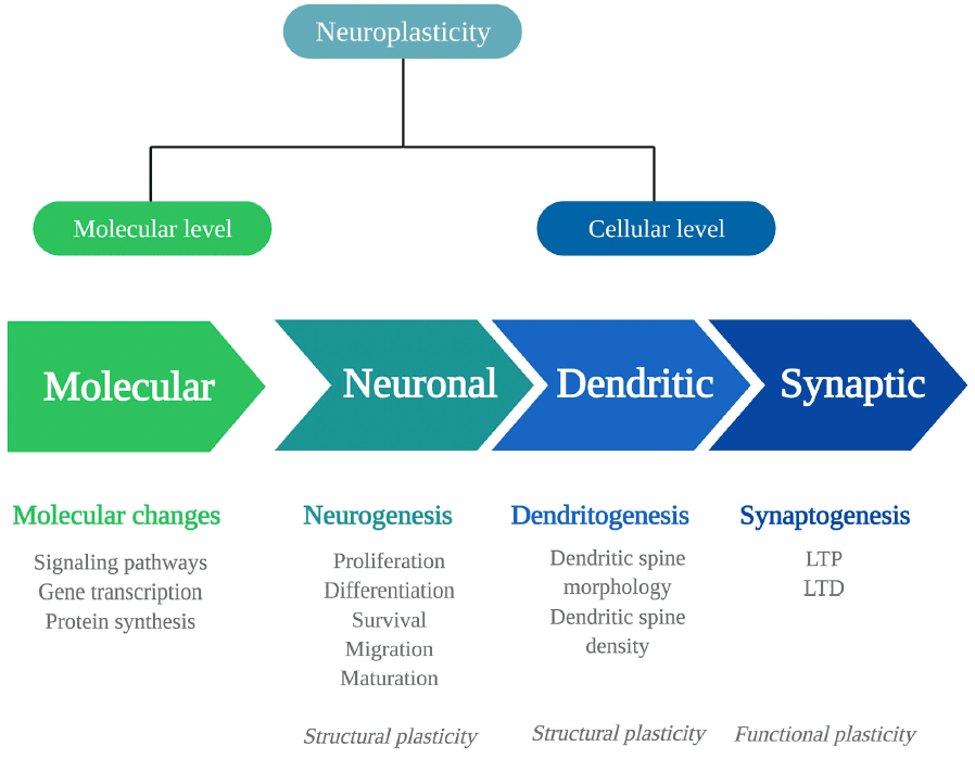
To fully understand the extent of psychedelics' effects on these levels, more detail is given first about the levels at which neuroplasticity can occur and the signaling substances involved.

At a molecular level, neuroplastic changes occur via signaling pathways, that is, cascades of intracellular proteins transmitting signals from receptors to the DNA. Signaling pathways are activated by Ca2+ influx through depolarization or N-methyl-D-aspartate receptor (NMDAR) activation. They include the Ca2+/calmodulin-dependent protein kinase (CaMK2), extracellular regulated kinase 1/2 (ERK1/2) mitogen-activated protein kinase (MAP)/ERK, and the brain-derived neurotrophic factor/tropomyosin receptor kinase B (BDNF/TrkB) pathway. In the nucleus, the cyclic AMP-responsive element-binding protein (CREB) or the nuclear factor kappa B protein complex (NF-kB) is activated, allowing modulation of gene transcription and protein synthesis of plasticity processes. For example, immediate early genes (IEGs), such as c-Fos, Arc, Egr1/2, C/EBP-β, Fosb, Junb, Sgk1, Nr4a1, and Dusp1, are rapidly expressed upon neuronal activity and are essential for synaptic plasticity. These changes in the expression of plasticity-related genes can influence neuroplasticity at the cellular level.

At a cellular level, changes can be structural or functional, and both types have different levels that will be listed here. Structural plasticity includes neuronal plasticity, dendritic plasticity, and synaptic plasticity. Neuronal plasticity consists of neurogenesis, the generation of neurons, and occurs in distinctive phases. First, proliferating progenitor cells are generated in the hippocampal subgranular zone and differentiate into dentate granule neurons. The proliferating cells that survive the elimination via apoptotic cell death migrate and mature into newborn granule cells and fully integrate into the hippocampal network. Dendritic plasticity includes changes in the number or the complexity of dendritic spines, where a high number of spines and complex dendritic branches reflect more synaptic strength. Of note, the extensive release of γ-aminobutyric acid (GABA) or glutamate causes dendritic spine formation.

At the synapse, the strength of synapses is related to learning and memory formation. It can change in two directions, either increasing, known as long-term potentiation (LTP), and decreasing, called long-term depression (LTD). This type of synaptic plasticity alters the neuron's structure and its functional properties. Synaptic plasticity is regulated by various factors, with the protein BDNF as the primary regulator; BDNF is expressed highly throughout the central nervous system, particularly in the hippocampus.

BDNF is involved in multiple levels of neuroplasticity like synaptic modulation, adult neurogenesis, and dendritic growth. Interestingly, studies have shown that BDNF levels are diminished in pathological populations suffering from anxiety, depression, and addiction. Preclinical and clinical research has shown that these markers are increased and enhanced by selective serotonin reuptake inhibitors (SSRIs) used to manage the symptoms of these disorders. The rapid-acting dissociative agent ketamine, which has shown its efficacy in treating depression, is known to increase BDNF levels. It has been suggested that the persisting therapeutic effects of psychedelics are attributable to a similar biological mechanism.

Psychedelics' influence on neuroplasticity is investigated in preclinical (in vitro/in vivo) and clinical studies (Figure 2). In vitro studies using rodent cell lines include neuronal stem cells (NSCs) derived from the subgranular zone of the hippocampus of embryonic mice. Human cell lines include induced pluripotent stem cells (iPSCs), cerebral organoids that consist of artificially grown cells of synthesized tissues resembling the cortex, and the neuroblastoma cell line SH-SY5Y. The latter can be used to model neuronal function and differentiation, and neurodegeneration by inducing chemical damage (oxygen deprivation) via in vitro administration of the dopamine analog and neurotoxin 6-hydroxydopamine (6-OHDA). In vivo studies in rodents use electrophysiology, the measurement of gene transcription and protein levels, and receptor knockout models to test the contribution of—for example—a specific receptor in the drug effects. Moreover, a well-established in vivo technique to identify neurogenesis is immunostaining [“immunohistochemistry” (IHC)] with the mitotic marker 5-bromo-2′-deoxyuridine (BrdU) or Ki-67 to determine progenitor cell growth and division (proliferation). These techniques can be used in healthy, intact animals or after surgical damage to the brain using suturing of an internal carotid artery to test neuroplasticity changes after drug administration and brain damage.

Figure 2. Evaluation of psychedelics' influence on cellular and molecular neuroplasticity in preclinical and clinical studies.

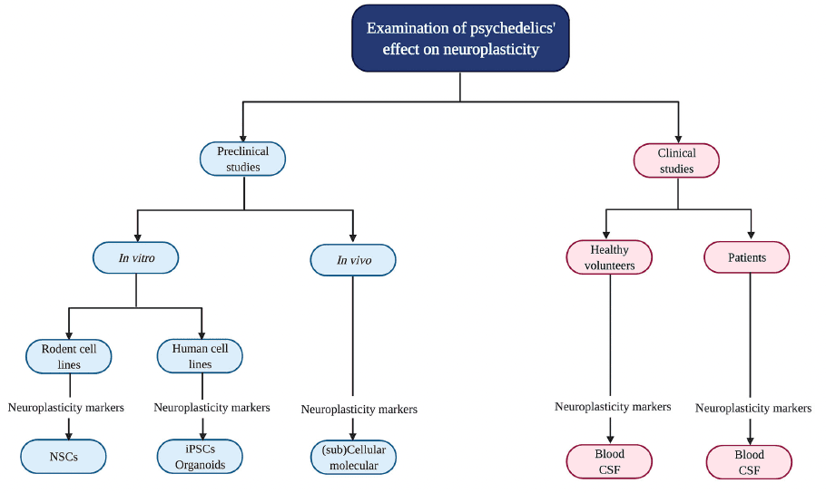
In clinical studies, biological samples are collected from healthy volunteers and patients suffering from a psychopathology like treatment-resistant depression (TRD) to determine BDNF levels. Clinical symptoms are assessed with, for example, the Montgomery-Åsberg Depression Rating Scale (MADRS) to test for depression severity.

To summarize, it is hypothesized that neurobiological changes, specifically enhanced neuroplasticity, underlie psychedelics' therapeutic effects. The techniques mentioned above can be used to assess changes in plasticity after the administration of psychedelics compared to baseline, a placebo, or a control group. Understanding the biological pathways of psychedelics' acute and persisting effects is essential to grasp these compounds' full therapeutic potential. Although psychedelics do not have an established therapeutic use in psychiatry yet, promising preliminary findings of their therapeutic potential support further investigation and give insight into psychiatric disorders' biological underpinnings. To address this knowledge gap and answer the question of what effects (serotonergic) psychedelics have on molecular and cellular neuroplasticity, a systematic review was performed focusing exclusively on classical serotonergic psychedelics (including psilocybin, LSD, ayahuasca, DMT, and its closely related analogue 5-methoxy-N,N-dimethyltryptamine, 5-MeO-DMT). The listed substances were chosen because of their shared agonism at 5-HT2A receptors. In line with SSRIs and ketamine, it was hypothesized that psychedelics enhance molecular and cellular neuroplasticity.

Methods

According to PRISMA guidelines, a literature search was performed using the database PubMed in October 2020. Two search strings were combined with the Boolean command “AND.” The first string included MeSH terms referring to neuroplasticity: neuronal plasticity, functional neuroplasticity, structural neuroplasticity, spine density, receptor density, axonal arbor, neuritogenesis, synaptogenesis, synapse formation, neurogenesis, BDNF, proliferation, maturation, survival, migration, neuronal migration; the second string included terms to describe the psychedelics that were focal in this review: classical psychedelics, psychedelics, hallucinogens, psilocybin, 4-phosphoryloxy-N,N-dimethyltryptamine, psilocin, 4-hydroxy-N,N-dimethyltryptamine, LSD, lysergic acid diethylamide, DMT, N,N dimethyltryptamine, 5-MeO-DMT, 5-methoxy-N,N dimethyltryptamine.

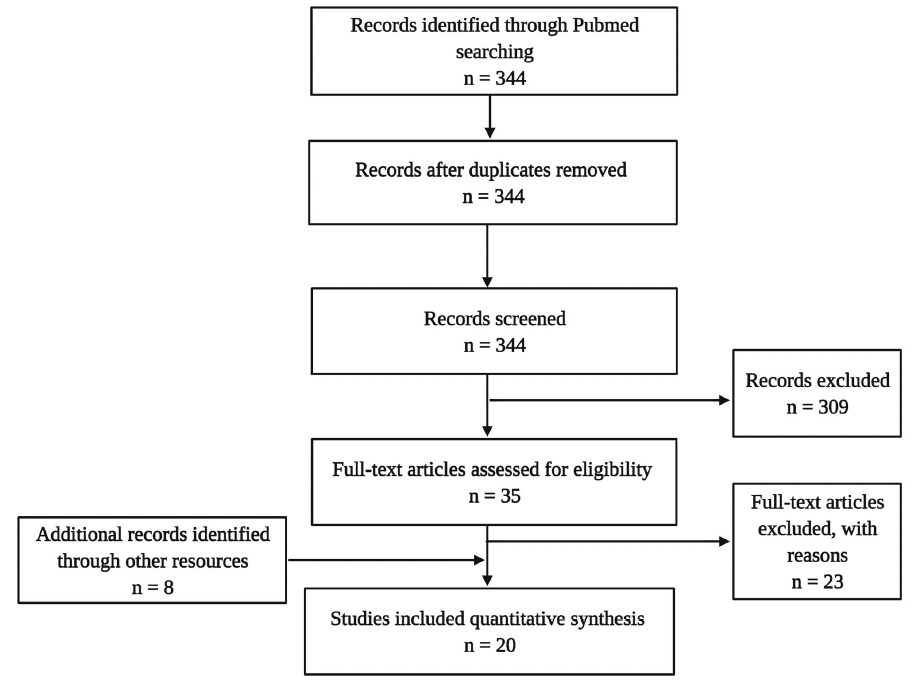
The literature search targeting the title and abstract gave 344 hits in total. This sample underwent de-duplication (n = 0) and a selection process using the following inclusion criteria: published in a peer-reviewed journal in the English language, including one of the target psychedelics, and assessing neurobiological parameters (e.g., cellular or molecular). This led to a sample of 35 articles, from which 23 were excluded because no molecular or cellular parameters of neuroplasticity were assessed. Additionally, eight articles were identified through other sources (cross-references), eventually resulting in a final dataset of 16 experimental studies in animals and four in humans (Figure 3).

Figure 3. Flow diagram illustrating the selection and review processes of the systematic review.

Results

The preclinical and clinical research findings are discussed in two separate sections; the methodological details of reviewed studies are presented in Tables 1, 2. A distinction is made between single and repeated dose administration and between acute, subacute, and long-term effects. Acute effects are measured within 24 h after administration of the psychedelic, subacute effects are measured between 24 h and 1 week after administration, and effects are considered long-term when they are observed after more than 1-week post-treatment.

Table 1. Preclinical studies investigating psychedelics' effects on molecular and (sub)cellular neuroplasticity.Table 1 (continued)Table 1 (continued)Table 2. Clinical studies investigating cellular and molecular effects of serotonergic psychedelics.

Preclinical Studies

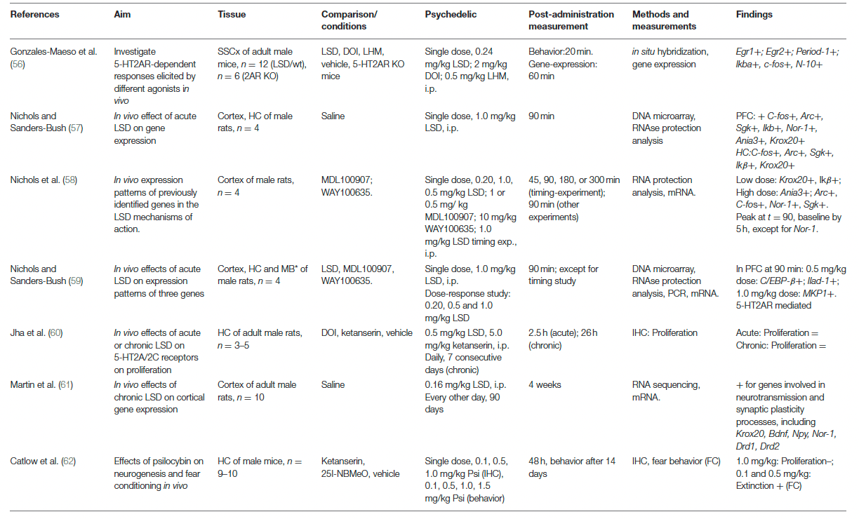
The effects of psychedelics on molecular and cellular neuroplasticity in preclinical studies are presented from a molecular to a subcellular level. They are separated by in vitro and in vivo studies. Evidence from preclinical studies (n = 15 out of 16) suggests that psychedelics induce structural and functional synaptic modulations at a molecular and cellular level (Table 1).

In vitro

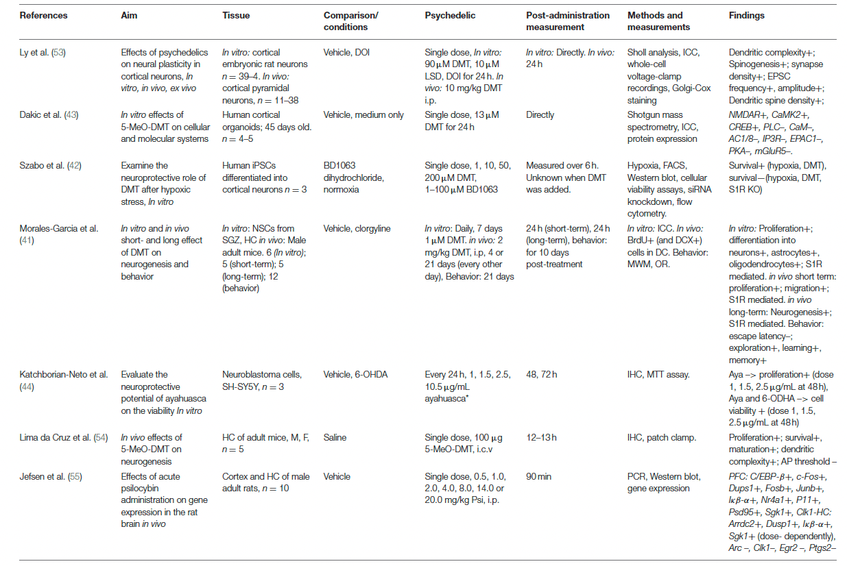
Evidence from in vitro studies (n = 5) suggests that psychedelics stimulate molecular and cellular neuroplasticity. The acute effect of a single dose of (5-MeO-)DMT on neuroplasticity was investigated by three in vitro studies. Administration of DMT (10 μM) and LSD (90 μM) to cortical rat neurons (n = 39–41) for 24 h resulted in increased dendritic complexity, expressed by an increased number and total length of dendrites compared to vehicle-treated controls. The dendritic spine-promoting properties were found to be 5-HT2AR-mediated, as they were blocked by ketanserin. LSD was the most potent psychedelic regarding neuritogenesis compared to the tested psychedelics. In human cerebral organoids (n = 4–5), 5-MeO-DMT (13 μM) administered for 24 h directly resulted in the stimulated synthesis of proteins involved in plasticity-related intracellular signaling pathways such as NMDAR, alpha-amino-3-hydroxy-methyl-5-4-isoxazolpropionic receptor (AMPAR), and Eprhin B2. These findings show acute changes in molecular processes related to structural and functional neuroplasticity induced by 5-MeO-DMT.

Besides the stimulation of neuroplasticity in “optimal” (healthy) conditions, evidence shows that a single dose of DMT (1, 10, 50, 200 μM) exhibits acute neuroprotective properties in cultured human iPSCs cells that were differentiated into cortical neurons (n = 3) and exposed to severe neuronal stress. DMT stimulated neurogenesis by increasing the neuronal survival rate from 19% (untreated cells) to 31% (10 μM) and 64% (50 μM), 6 h after exposure to severe hypoxic stress. Moreover, selective silencing of the plasticity-promoting intracellular sigma-1 receptor (S1R) decreased the survival of iPSCs by 93%, indicating that the S1R mediated the DMT-induced survival. Together, these findings show that a single dose of 5-MeO-DMT, DMT, and LSD in vitro directly stimulate dendritic and neuronal plasticity that resulted from intracellular changes. Subacute and long-term effects of a single dose of a psychedelic on molecular and cellular plasticity were not investigated in vitro.

Acute effects of repeated administration of DMT and ayahuasca were investigated in vitro, and suggest stimulation of neuroplasticity at a (sub)cellular level. Repeated daily administration of DMT (1 μM, 7 days) to neural stem cells (n = 6) from adult mice's subgranular zone of the dentate gyrus showed stimulation of proliferation, and differentiation to neurons, astrocytes, and oligodendrocytes. In cultured human neuroblastoma (SH-SY5Y) cells (n = 3) exposed to neurotoxic stress using 6-OHDA, cell viability was differentially changed after treatment with ayahuasca (1, 1.5, 2.5, or 10 μg/mL) every 24 h, and incubation for 48 and 72 h. Low doses of ayahuasca (1.5 and 2.5 μg/mL) increased cell viability (±70%) compared to non-stressed controls after 48 h, whereas cell viability was decreased after a high dose (10.5μg/mL) at 72 h post-administration. These findings suggest acute stimulative effects of neural plasticity by DMT, dose-dependent neuroprotective properties, and possibly proliferative effects of ayahuasca in stressed cell cultures at low doses. Together, the findings from in vitro studies support plasticity-promoting characteristics of DMT, ayahuasca, and LSD when administered once or multiple times. Subacute and long-term effects of repeated administration of psychedelics on molecular and cellular plasticity were not investigated in vitro.

In vivo

Evidence from in vivo studies (n = 13 out of 16) also indicates neuroplastic effects of psychedelics. Findings from in vivo studies investigating acute effects of a single dose of 5-MeO-DMT, DMT, psilocybin, and LSD (37%) suggest altered cellular plasticity of structural and functional nature. A single dose of 5-MeO-DMT (100 μg, i.c.v.) stimulated neurogenesis and spinogenesis in mice (n = 5), 12 h post-treatment. The proliferation of neuronal progenitor cells, and the survival of newborn granule cells were increased in the ventral hippocampus, a brain area involved in emotion and stress regulation, compared to vehicle-treated controls. 5-MeO-DMT in this study also stimulated spinogenesis of granule cells in the hippocampus. The dendritic spines grew more quickly toward the complex morphology of a mature neuron. Electrophysiological analysis showed a lower action potential threshold of synapses in the hippocampus, indicating that the synapses are more prone to receive synaptic input and suggesting stimulated functional plasticity. A single dose of psilocybin (0.5, 1, 2, 4, 8, 14, 20 mg/kg, i.p.) administered to rats (n = 10) altered the expression of plasticity-promoting genes in the prefrontal cortex (PFC) and hippocampus, 90 min after treatment. Psilocybin stimulated the expression of IEGs in the PFC, and induced stimulating and inhibiting effects in the hippocampus. Because more target genes were regulated in the PFC, the authors suggested a stronger stimulation in the PFC over the hippocampus by psilocybin. Moreover, in both areas, most IEGs were affected in a dose-dependent manner, with higher doses inducing more stimulation of gene expression. A single dose of LSD (0.20, 0.24, 0.5, 1.0 mg/kg i.p.) stimulated the mRNA expression of plasticity-promoting genes in the cortex of rats and mice, within 1–2 h after administration and in a time- and dose-dependent manner. Conversely, in a different study with rats (n = 5) that were administered LSD (0.5 mg/kg, i.p.), no changes were found in hippocampal proliferation 2.5 h after administration compared to vehicle-treated controls indicated by BrdU+ cells. Taken together, it is shown that a single treatment with psychedelics acutely regulates molecular processes of plasticity-promoting gene expression, neuroplasticity at the cellular level of neurogenesis, and dendritic plasticity. Psychedelics' subacute effects of a single dose of DMT (10 mg/kg, i.p.) were investigated in rat cortical pyramidal neurons (n = 11–37 neurons from three animals). Spontaneous excitatory postsynaptic currents (EPSCs) and dendritic spine density were stimulated 24 h after administration, indicating stimulated structural and functional plasticity. Psychedelics' long-term effects of a single dose were not assessed on molecular and cellular plasticity in vivo.

Evidence from in vivo studies investigating subacute and long-term, but not acute, effects of repeated administration of psychedelics (n = 3) show that DMT and LSD stimulate neurogenesis. The subacute effects of repeated (4 consecutive days) or prolonged (21 days, every other day) DMT (2 mg/kg, i.p.) treatment were assessed on neurogenesis in mice, 24 h after the administration stopped. Short-term treatment (n = 5) resulted in BrdU+ cells in the hippocampus, indicating enhanced proliferation and migration of neuronal precursors, and long-term (n = 12) increased neuronal survival in the subgranular zone. Repeated LSD (0.5 mg/kg, i.p.) administration, daily for seven consecutive days to rats (n = 3–5) did not result in changes in BrdU+ cells compared to vehicle-treated controls, 26 h after administration, indicating that repeated LSD administration did not affect neurogenesis. Together, these findings show that repeated administration of DMT, but not LSD, subacutely resulted in stimulated hippocampal neurogenesis. The long-term effects of repeated doses of psychedelics were examined for LSD, at a molecular level. Chronic treatment with LSD (0.16 mg/kg i.p., every other day for 90 days) increased the expression of plasticity-related genes in the mPFC of rats (n = 10), 4 weeks after treatment cessation. Among the upregulated genes were Bdnf, Egr2, Nor-1, Nr2a, and Npy. Nr2a encodes for NMDA receptor subunits, and the NPY protein stimulates neurogenesis and has anxiolytic effects. These findings show that repeated LSD administration stimulates the expression of plasticity-related genes 4 weeks after treatment. Taken together, the findings from in vivo studies support plasticity-promoting properties of psychedelics at a molecular and cellular level after a single or multiple dose administration.

Behavior

Four studies (out of 16) investigated the relationship between biological effects of psychedelics (5-MeO-DMT, DMT, ayahuasca, LSD, and psilocybin), and behavioral changes. While the acute effects of a single dose of a psychedelic on biological markers and behavior were not assessed, the long-term effects of a single dose of psilocybin were investigated. Mice (n = 6) that were administered a single, low dose of psilocybin (0.1 mg/kg, i.p) showed a non-significant increase in proliferation of hippocampal progenitor cells 14 days later. In contrast, higher doses (1.0 mg/kg, i.p.) led to a significant decrease in proliferation, 14 days post-treatment. To investigate if the hippocampus mediates behavioral changes 48 h after treatment with psilocybin mice (n = 9–10) underwent fear conditioning. Psilocybin-treated mice exhibited increased extinction compared to saline-treated controls for all doses, indicating a quicker learning response to fear. At a biological level, psilocybin induced a dose-dependent effect on neurogenesis, with a low dose increasing, and a high dose decreasing neurogenesis. Behavioral effects of psilocybin demonstrated a dose-independent stimulation of fear extinction, suggesting that alterations to hippocampal neurogenesis are not related to fear extinction after psilocybin administration.

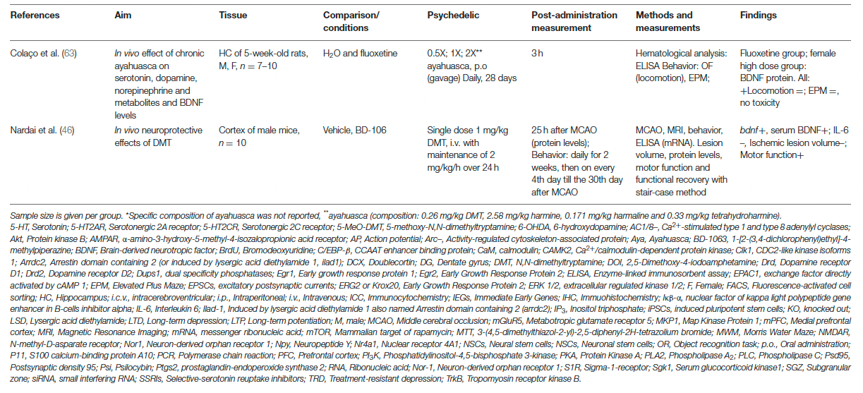
The relationship between psychedelics' effect on biological markers and behavior was also investigated after repeated psychedelic administration. Three studies examined the immediate and long-term effects of repeated administration of ayahuasca, LSD, and DMT. Ayahuasca was administered daily to rats (n = 7–10) for 28 days, in a dose that was 0.5, 1, or 2 times the human, ritualistic oral dose, which is 0.26 mg/kg DMT p.o., 2.58 mg/kg harmine, 0.171 mg/kg harmaline, and 0.33 mg/kg tetrahydroharmine. These administration patterns did not change hippocampal BDNF protein levels and resulted in increased anxiety behavior in male rats treated with the middle dose, 1 h after the last treatment. After 3 h, female rats treated with the high dose exhibited increased hippocampal BDNF protein levels but did not show changed anxiety behavior. These findings indicate direct sex- and dose-specific effects of repeated ayahuasca on molecular neuroplasticity and anxiety behavior.

While subacute effects of repeated doses of psychedelics were not investigated, long-term effects of chronic DMT (2 mg/kg, i.p.) administration (21 days, every other day) to mice (n = 10–12) were tested. Findings showed enhanced neurogenesis that corresponded with improved spatial learning and memory tasks for 10 days post-treatment. In a study in rats (n = 10) that had received surgical brain damage and were treated with DMT (1 mg/kg, i.p., followed by a maintenance dose of 2 mg/kg/h for 24 h) afterwards, DMT led to stimulated cortical (mRNA) and plasma BDNF (protein) levels 1 h after DMT treatment cessation. On a behavioral level, rats (n = 8) showed an increased motor function that lasted up to 30 days after treatment. Moreover, animals (n = 10) treated with DMT combined with an S1R antagonist (BD-1,063) exhibited a higher lesion volume than DMT-treated animals 24 h after brain damage was inflicted, suggesting that the effects of DMT are S1R-mediated. These findings show that DMT has an immediate stimulative impact on molecular plasticity processes and promotes recovery behavior up to a month after induced brain damage. Taken together, the findings from behavior studies support the plasticity-promoting characteristics of ayahuasca and DMT at a molecular and cellular level, accompanied by plasticity-related changes in behavior.

Clinical Studies

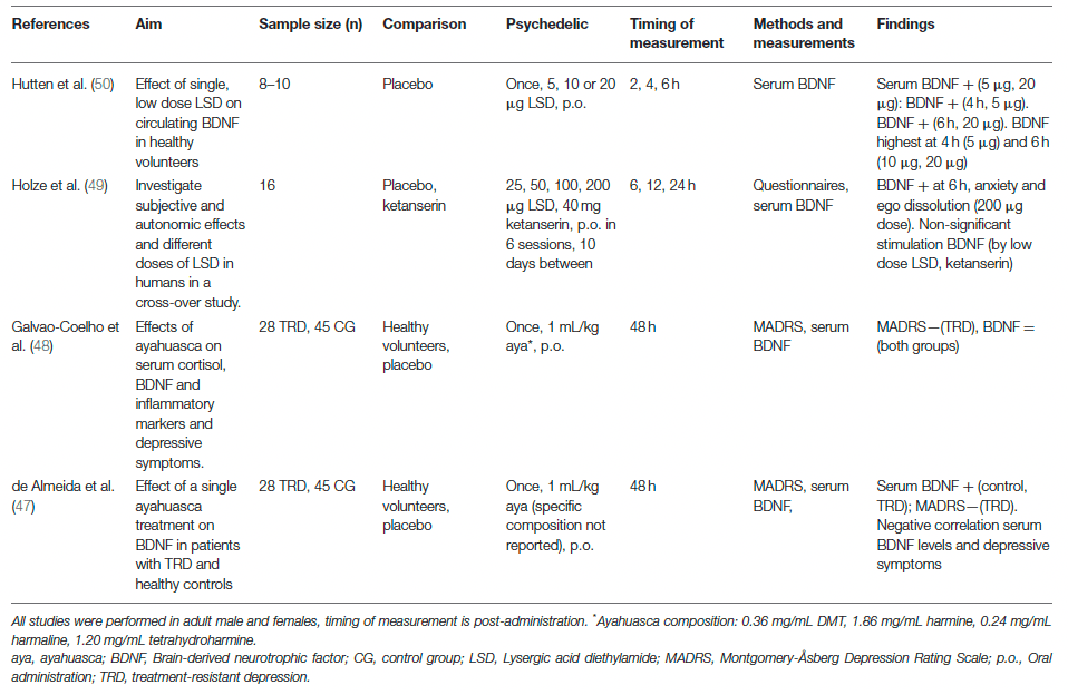
Evidence from four randomized, placebo-controlled studies investigating the acute and subacute, but not long-term effects, of a single dose of psychedelics on a molecular level show that a single treatment with ayahuasca or LSD can, but does not always, increase circulating BDNF in healthy volunteers and TRD patients (Table 2). Clinical research investigating psychedelics' effect on cellular neuroplasticity is lacking.

A single, low dose of LSD (5, 10, and 20 μg) administered to healthy volunteers (n = 24) resulted in increased serum BDNF levels compared to placebo, 6 h after treatment. Blood samples taken every 2 h, until 6 h after administration, showed elevated plasma BDNF levels at 4 h after administration for the 5 μg, and at 6 h for the 20 μg dose. BDNF levels were highest at 4 h after treatment for the 5 μg dose and at 6 h after treatment for the 10 μg and 20 μg doses, suggesting dose-specific stimulation of BDNF. In a cross-over study in healthy participants (n = 18) that were treated with single doses of LSD (25, 50, 100, and 200 μg) over six sessions, with 10 days in-between administrations, findings showed that blood plasma BDNF levels were dose-dependently elevated compared with placebo. Six hours after administering 200 μg, participants reported ego dissolution and anxiety, in parallel with increased plasma BDNF. The subjective response was partially prevented by administering a 5-HT2A/C receptor antagonist (ketanserin) 1 h before LSD treatment, as demonstrated by a “25 μg-dose response” after administration of 200 μg of LSD plus ketanserin.

The subacute neuroplastic properties of a single, oral dose of ayahuasca (1 mL/kg, p.o., ayahuasca composition not reported) were assessed in patients suffering from TRD (n = 28), and in healthy controls (n = 45) naïve to ayahuasca, in a controlled environment. Blood serum BDNF levels were increased at 48 h after administration compared to baseline in both groups, and they correlated negatively with the MADRS scores in TRD patients treated with ayahuasca. These findings suggest that lower depressive symptomatology was associated with higher BDNF levels. It was suggested that patients with the most persistent depression benefited the most from ayahuasca treatment. Since the specific composition of the ayahuasca brew and thus the DMT dose was not reported in the study, it is difficult to compare the outcomes of this experiment with similar studies. Conversely, in a follow-up study, a single oral dose of ayahuasca (1 mL/kg, p.o., 0.36 mg/mL DMT, 1.86 mg/mL harmine, 0.24 mg/mL harmaline) administered to TRD patients (n = 28) and healthy controls (n = 45) in a controlled setting did not affect serum BDNF levels at 48 h after administration.

The limited number of studies investigating molecular biological and behavioral correlates of psychedelics' effects shows that psychedelics acutely and subacutely stimulate molecular plasticity and decrease depressive symptoms in healthy and TRD patients, with effects lasting up to 48 h after administration. The acute biological and behavioral effects of repeated administration on molecular and cellular plasticity were not investigated in a clinical setting.

Discussion

To understand the acute, subacute (24 h−1 week post-treatment), and longer-term effects of (serotonergic) psychedelics on molecular and cellular neuroplasticity, preclinical and clinical studies were evaluated. Evidence from preclinical studies shows that psychedelics acutely stimulate structural neuroplasticity processes at a molecular and (sub)cellular level after a single dose. Subacute effects of a single dose of a psychedelic on molecular and cellular neuroplasticity have not yet been investigated, and one study investigating the long-term effects of psilocybin showed decreased neurogenesis weeks after a single dose. Repeated administration of psychedelics is shown to stimulate neurogenesis acutely and molecular plasticity, subacutely. Moreover, a limited number of (pre)clinical studies that investigated the relationship between biological and behavioral adaptations showed that the stimulation of molecular and neuronal was accompanied by increased learning behavior. Under stressful conditions, neuronal plasticity and molecular plasticity processes were found to be stimulated in rodents, and ayahuasca-induced increases in plasma BDNF levels correlated with diminished depressive symptoms in clinical populations, subacutely. Similarly, findings from clinical studies showed that blood BDNF levels were directly elevated in healthy participants that were treated with a single dose of LSD. Long-term and repeated administration effects on molecular and cellular plasticity were not investigated. Overall, the limited evidence that is presented is consistent with our hypothesis that psychedelics stimulate molecular and cellular structural neuroplasticity.

Of note, the antidepressant effects of ayahuasca may also be produced by its non-psychedelic β-alkaloids harmine, tetrahydroharmine, and harmaline present in the ayahuasca brew. Findings from in vitro and in vivo studies show that these compounds stimulate neurogenesis, BDNF, and have antidepressant effects. Neuroplastic changes induced by ayahuasca may result from DMT, β-alkaloids, or an interaction between these compounds, something that should be taken into consideration when interpreting findings from biological studies using ayahuasca.

Four main findings stand out from our review. The first concerns dose differences between preclinical and clinical studies and their translation from animal to human. Clinically, LSD doses varied between 5 and 200 μg. To compare preclinical and clinical doses, the conversion formula for the animal dose = human dose × (37/3) was used. For example, a high dose of 200 μg p.o. LSD for a human with an average weight of 70 kg equals 0.00285 mg/kg, and converts to a dose of 0.021 mg/kg LSD for rats, and 0.041 mg/kg LSD for mice. Using the molarity formula (M = m/MW * 1/V where m = mass in grams, MW = molecular weight of LSD and V = volume of the diluent in liters), assuming a mouse weight of 25 g and making 0.025 L solution, an approximation of the in vitro LSD dosage would be 0.126 μM based on a human dose of 200 μg per 70 kg. This is remarkably lower than the 10 μM LSD used by Ly et al., and presumably even lower for the other, lower, clinical doses. Conversion from in vitro to in vivo doses and vice versa is more complicated than these calculations. Animal doses of LSD varied between 0.16 and 1.0 mg/kg i.p. in rats and mice, indicating that LSD doses were higher in animal studies than in clinical studies. This difference is even larger due to the first-pass metabolism reducing the systemic exposure of LSD after oral administration used in clinical studies. With intraperitoneal administration in rodents, this degradation is avoided. These findings suggest that the highest doses given in clinical studies resemble the lowest doses of LSD in preclinical studies, highlighting an important factor that should be considered in the translation of preclinical findings to humans. Further research into the neuropharmacokinetics of psychedelics could bridge this gap between optimal preclinical and clinical doses.

The second significant finding concerns sex-differences in response to psychedelics, which were shown in a preclinical study where male, but not female rats showed increased anxiety behavior directly after prolonged ayahuasca administration. This could be related to sex-specific changes in neuroplasticity. The female sex hormone estrogen exhibits antidepressant effects through stimulation of BDNF and synaptic plasticity, in a manner that is distinct for males and females. In that line, female rats showed greater sensitivity to the antidepressant effects of ketamine than male rats, and effects were abolished in rats whose ovaries had been removed and restored when estrogen and progesterone were supplemented. The antidepressive effects of ketamine and psychedelics are both suggested to result from changes in neuroplasticity, and these findings indicate a potential role for gonadal hormones in the sex-specific response to these substances. Neurobiological research in animal models is biased toward males. These facts highlight the importance of investigating both sexes in preclinical research to further elucidate sex differences in psychopathologies and improve translation to clinical populations.

The third finding concerns the measurement of BDNF in clinical studies. All clinical studies reported peripheral BDNF levels, an indirect measure of BDNF levels in the brain. It would be more precise to examine cerebrospinal fluid (CSF) BDNF levels as this directly reflects brain activity. While the collection of CSF is invasive, only a limited number of studies have investigated BDNF CSF levels; two studies found a positive correlation between CSF and plasma BDNF levels in first-episode psychotic and depressed patients. Furthermore, while previously it was not clear whether clinical response was related with plasma BDNF levels, evidence suggests that there is a positive relation with clinical improvement being linked with improved neuroplasticity. Nonetheless, further research is recommended to investigate the effect of psychedelics on CSF BDNF levels in clinical populations and its relation with BDNF plasma levels.

The fourth finding of this review concerns the sample size of some in vivo studies, which was low. A sample size of six animals per group is considered an adequate sample size in animal research by many researchers but reduces the statistical power. This is a well-known problem in (neuro)biological research. Researchers are to justify the number of animals used in their experiments, which should be designed to minimalize the number of animals used. This could explain the low sample size in in vivo studies reviewed here, and is essential to consider because it reduces the statistical power and limiting the reliability of conclusions.

The observed psychedelics-induced changes in neuroplasticity are suggested to result from the neurobiological pathways activated by 5-HT2AR upon activation by psychedelics, affecting the serotonergic and glutamatergic system (Figure 4). Psychedelics primarily act on 5-HT2ARs expressed on glutamatergic pyramidal cells in cortical and deep cortical layers (V and VI). Via activation of 5-HT2AR, psychedelics activate intracellular signaling pathways such as PLC, PLA, and Src. Activation of Src is suggested to be essential for psychedelics' hallucinogenic effects, as its inhibition prevented hallucinogenic effects of LSD. Activation of these pathways' intracellular signaling leads to Ca2+ and glutamate release that stimulates synaptic plasticity. Increased glutamate in the cortex release can further stimulate synaptic plasticity via AMPAR on pyramidal neurons in cortical layer V and subsequent transportation (trafficking) of AMPAR to the postsynaptic cell membrane. This increases AMPAR density, resulting in more extracellular glutamate and BDNF release in the cortex. The potential of classic psychedelics to alter glutamate in the human cortex, albeit in a region-dependent manner, has been demonstrated. Indirectly, psychedelics influence plasticity via the expression of BDNF and other plasticity-related genes and proteins, including IEGs. Cortical bdnf mRNA was upregulated by LSD and ayahuasca. IEGs are implicated in synaptic plasticity and synaptogenesis and many IEGs encode for proteins involved in specific signaling cascades. For instance, Arc is localized at dendrites and involved in cytoskeletal rearrangements, Egr2 has coupled activity with the NMDAR, Sgk promotes cell survival and the Neuron-derived orphan receptor 1 (Nor1; NR4A3) is important for LTP.

Figure 4. Proposed mechanism of action of cellular and molecular effects of serotonergic psychedelics.

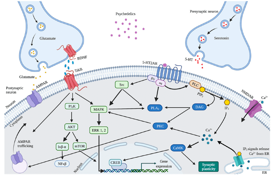
Alternatively, enhanced neuroplasticity can be attributed to differences in receptor affinity, given that psychedelics are not pure 5-HT2AR agonists, which could explain the different effects on neuroplasticity between psychedelics. DMT exhibits, besides the 5-HT2AR, also high affinity for the S1R, which is highly expressed in the hippocampus and is a stimulator of synaptic plasticity. Activation of S1R by DMT has been suggested to stimulate synaptic plasticity in addition to 5-HT2AR-induced modulation and is likely responsible for ayahuasca's antidepressant effects. Moreover, LSD was shown to stimulate S1R indirectly via activation of the neurosteroid dehydroepiandrosterone (DHEA), which stimulated synaptic plasticity and neurogenesis. In like manner, activation of S1R by SSRIs enhances BDNF expression. These findings support the hypothesis that the psychedelics'-induced stimulation of neuroplasticity underlies a mechanism similar to SSRIs.

The dissociative ketamine is another substance whose antidepressant effects are suggested to result from enhanced BDNF and synaptic plasticity. As an NMDAR antagonist, ketamine blocks postsynaptic NMDAR located on glutamatergic neurons in the cortex. It deactivates the eukaryotic Elongation Factor-2 (eEF2) kinase, which subsequently alleviates its block on BDNF translation, resulting in heightened BDNF levels. In addition, ketamine is hypothesized to block NMDAR on GABA interneurons, releasing the inhibition of glutamate release. This activates AMPAR on glutamatergic cells and subsequently increases BDNF and glutamate in the cortex. Ketamine and psychedelics activate cortical AMPAR and subsequently stimulate BDNF and synaptic efficacy. This could explain the (rapid) antidepressant and anxiolytic effect of psychedelics and ketamine, and provides insight into the biological underpinnings of these substances and their therapeutic potential.

This systematic review is the first to explain psychedelics' rapid antidepressant and cognitive effects, by investigating molecular and cellular changes related to neuroplasticity. The data reviewed here contributes to a clearer understanding of the underlying biological mechanisms of serotonergic psychedelics and emphasizes the need for scientific research in this field, because psychedelics are not only beneficial in populations suffering from psychopathologies, but also for those without, enhancing social and cognitive skills such as empathy and creativity, but also general well-being. Further research is essential to establish the specific (intra)cellular mechanism activated by different psychedelics, their long-term effects, and their relation with altered behavior. The current findings support research exploring psychedelics' potential in the treatment of psychopathologies.

Open Article as PDF

Abstract

Clinical studies suggest the therapeutic potential of psychedelics, including ayahuasca, DMT, psilocybin, and LSD, in stress-related disorders. These substances induce cognitive, antidepressant, anxiolytic, and antiaddictive effects suggested to arise from biological changes similar to conventional antidepressants or the rapid-acting substance ketamine. The proposed route is by inducing brain neuroplasticity. This review attempts to summarize the evidence that psychedelics induce neuroplasticity by focusing on psychedelics' cellular and molecular neuroplasticity effects after single and repeated administration. When behavioral parameters are encountered in the selected studies, the biological pathways will be linked to the behavioral effects. Additionally, knowledge gaps in the underlying biology of clinical outcomes of psychedelics are highlighted. The literature searched yielded 344 results. Title and abstract screening reduced the sample to 35; eight were included from other sources, and full-text screening resulted in the final selection of 16 preclinical and four clinical studies. Studies (n = 20) show that a single administration of a psychedelic produces rapid changes in plasticity mechanisms on a molecular, neuronal, synaptic, and dendritic level. The expression of plasticity-related genes and proteins, including Brain-Derived Neurotrophic Factor (BDNF), is changed after a single administration of psychedelics, resulting in changed neuroplasticity. The latter included more dendritic complexity, which outlasted the acute effects of the psychedelic. Repeated administration of a psychedelic directly stimulated neurogenesis and increased BDNF mRNA levels up to a month after treatment. Findings from the current review demonstrate that psychedelics induce molecular and cellular adaptations related to neuroplasticity and suggest those run parallel to the clinical effects of psychedelics, potentially underlying them. Future (pre)clinical research might focus on deciphering the specific cellular mechanism activated by different psychedelics and related to long-term clinical and biological effects to increase our understanding of the therapeutic potential of these compounds.

Psychedelics and Neuroplasticity: Exploring the Brain's Biological Changes

Introduction

Psychedelics are a group of substances that alter the mind, primarily by acting on serotonin 2A (5-HT2A) receptors in the brain. Examples include psilocybin, lysergic acid diethylamide (LSD), N,N-dimethyltryptamine (DMT), and the DMT-containing brew ayahuasca. These compounds are considered physically safe and generally do not cause toxicity. Their effects depend on factors like dose, the specific substance, and an individual's mental state and environment. While their acute effects vary in duration, ranging from minutes to hours depending on the substance and its form (e.g., psilocybin is quickly broken down, LSD has a longer duration, ayahuasca allows DMT to reach the brain), a common characteristic is their ability to induce lasting changes in mood, emotion processing, and cognition that extend beyond the substance's presence in the body.

These long-lasting psychological effects suggest underlying biological adaptations in the brain, particularly changes in neuroplasticity. Neuroplasticity refers to the brain's capacity to change and reorganize throughout life. This includes structural plasticity, which involves changes in brain cell structures, and functional plasticity, which relates to changes in the efficiency of communication between brain cells (synapses). These processes are interconnected at the molecular and cellular levels.

At the molecular level, brain changes occur through signaling pathways—cascades of proteins that transmit signals from receptors to DNA, influencing gene transcription and protein production. Key pathways involve substances like brain-derived neurotrophic factor (BDNF), which is crucial for brain changes. BDNF is involved in many aspects of neuroplasticity, including synaptic changes, the creation of new neurons (neurogenesis), and dendritic growth. Low BDNF levels are often seen in individuals with conditions like anxiety, depression, and addiction. Medications like selective serotonin reuptake inhibitors (SSRIs) and ketamine, known for their antidepressant effects, have been shown to increase BDNF levels.

At the cellular level, structural changes include neurogenesis, the formation of new neurons, particularly in the hippocampus. Dendritic plasticity involves changes in the number and complexity of dendritic spines, which are important for synaptic strength. Synaptic plasticity, which is the ability of connections between neurons (synapses) to strengthen or weaken, is crucial for learning and memory. Researchers investigate psychedelics' influence on neuroplasticity using various methods. Preclinical studies employ cell lines and animal models, using techniques like measuring gene and protein levels, and observing new neuron growth. Clinical studies involve collecting biological samples from human volunteers and patients to measure markers like BDNF and assess changes in mood or symptoms. The hypothesis is that these neurobiological changes, specifically enhanced neuroplasticity, contribute to the therapeutic effects of psychedelics.

Methods

To identify relevant research, a literature search was conducted in October 2020 using the PubMed database. The search combined terms related to neuroplasticity, such as "neuronal plasticity" and "BDNF," with terms for classical psychedelics, including "psilocybin," "LSD," and "DMT."

The initial search yielded 344 results. After removing duplicates, articles were selected based on inclusion criteria: peer-reviewed, in English, focusing on one of the target psychedelics, and assessing neurobiological parameters. This process led to 35 articles. Twenty-three of these were excluded because they did not assess molecular or cellular neuroplasticity. Additionally, eight articles were found through other sources. Ultimately, 16 experimental studies in animals and four in humans were included in the final analysis.

Results

The findings from preclinical and clinical research are presented in two separate sections, detailing methodological specifics in tables.

Preclinical Studies

Evidence from preclinical studies, specifically in vitro (cell culture) experiments, indicates that psychedelics stimulate molecular and cellular neuroplasticity. For instance, single doses of DMT and LSD increased the complexity of neuron branches, and 5-MeO-DMT stimulated the synthesis of proteins involved in plasticity pathways. DMT also showed neuroprotective qualities in stressed cells, improving cell survival. Repeated administration of DMT stimulated the growth and differentiation of neural stem cells, while ayahuasca improved cell viability in stressed cultures at lower doses. These findings suggest that both single and repeated doses of DMT, ayahuasca, and LSD promote plasticity in cell models.

In vivo (animal models) studies also support the neuroplastic effects of psychedelics. A single dose of 5-MeO-DMT stimulated the formation of new neurons and changes in neuron structure in mice. Psilocybin and LSD acutely altered the expression of genes related to plasticity in brain regions like the prefrontal cortex and hippocampus, often in a dose-dependent manner. While a single dose of LSD did not immediately change neurogenesis in one study, subacute effects of single-dose DMT showed stimulated structural and functional plasticity in rat brain cells. Long-term effects of a single dose were not evaluated in animals.

Regarding behavior, four preclinical studies explored the link between psychedelics' biological effects and behavioral changes. A single low dose of psilocybin in mice non-significantly increased progenitor cell proliferation long-term, while higher doses decreased it. Psilocybin also improved fear extinction, but this behavioral effect did not directly correlate with hippocampal neurogenesis changes. Repeated administration of ayahuasca in rats showed sex- and dose-specific effects on anxiety and BDNF levels. Chronic DMT administration in mice enhanced neurogenesis, correlating with improved spatial learning and memory. In rats with brain damage, DMT stimulated BDNF levels and improved motor function, with effects lasting up to a month, suggesting its neuroprotective actions are linked to a specific receptor (S1R). These studies suggest that plasticity-promoting effects of ayahuasca and DMT in animals are accompanied by beneficial behavioral changes.

Clinical Studies

Clinical studies, though limited in number, have investigated the effects of single-dose psychedelics on molecular neuroplasticity in humans. A single low dose of LSD in healthy volunteers increased circulating BDNF levels within hours, with higher doses showing a dose-dependent effect. Ayahuasca's subacute neuroplastic properties were assessed in patients with treatment-resistant depression (TRD) and healthy controls. One study found increased blood serum BDNF levels 48 hours after ayahuasca administration, correlating with reduced depressive symptoms in TRD patients. However, a follow-up study with a similar design did not find changes in BDNF levels. Clinical research on cellular neuroplasticity is currently lacking. Overall, these findings indicate that psychedelics can acutely stimulate molecular plasticity and may reduce depressive symptoms in clinical populations, with effects lasting up to 48 hours.

Discussion

This review evaluated preclinical and clinical studies to understand the acute, subacute (24 hours to 1 week post-treatment), and long-term effects of serotonergic psychedelics on molecular and cellular neuroplasticity. Preclinical evidence largely shows that psychedelics acutely stimulate structural neuroplasticity at molecular and cellular levels after a single dose. Repeated administration also stimulated neurogenesis acutely and molecular plasticity subacutely. Limited studies also suggest that these biological changes are linked to improved learning behaviors. Under stress, neuronal and molecular plasticity processes were stimulated in rodents. In clinical studies, single doses of LSD directly increased circulating BDNF levels, and ayahuasca-induced BDNF increases in plasma correlated with reduced depressive symptoms subacutely. These findings generally support the hypothesis that psychedelics stimulate molecular and cellular structural neuroplasticity.

Several important considerations emerge from this review. First, the antidepressant effects of ayahuasca might also be influenced by its non-psychedelic beta-alkaloids (harmine, tetrahydroharmine, harmaline), which are known to stimulate neurogenesis, BDNF, and have antidepressant properties. Therefore, neuroplastic changes from ayahuasca could stem from DMT, these alkaloids, or their interaction. Second, significant dose differences exist between preclinical animal studies and human clinical studies, with animal doses generally being much higher when scaled. This highlights a need for better translation of optimal doses between research settings. Third, sex differences in response to psychedelics were observed in preclinical studies; for example, male and female rats showed different anxiety responses to ayahuasca, possibly due to hormonal influences. This emphasizes the importance of including both sexes in preclinical research. Fourth, clinical studies primarily measured peripheral BDNF levels, which are an indirect measure of brain activity; exploring BDNF in cerebrospinal fluid might provide a more direct assessment. Finally, some in vivo studies had small sample sizes, which could affect statistical power and the reliability of conclusions.

The observed changes in neuroplasticity are thought to result from neurobiological pathways activated by psychedelics, primarily through their action on 5-HT2A receptors on brain cells. This activation leads to a cascade of intracellular signaling, causing the release of calcium and glutamate, which stimulates synaptic plasticity. Increased glutamate in the cortex can further enhance synaptic plasticity and BDNF release. Psychedelics also indirectly influence plasticity by affecting the expression of BDNF and other genes crucial for brain changes. Additionally, psychedelics are not exclusive to 5-HT2A receptors; for example, DMT has a high affinity for the sigma-1 receptor (S1R), also known to stimulate synaptic plasticity. This S1R activation, alongside 5-HT2AR activity, may contribute to the antidepressant effects, similar to how SSRIs activate S1R to enhance BDNF expression. Like the dissociative agent ketamine, which also has antidepressant effects by enhancing BDNF and synaptic plasticity, psychedelics appear to activate similar pathways, leading to rapid antidepressant and anxiolytic effects.

This systematic review provides a clearer understanding of the biological mechanisms underlying the rapid antidepressant and cognitive effects of serotonergic psychedelics. The findings emphasize the need for continued scientific research in this area, as psychedelics show potential not only for treating mental health conditions but also for enhancing general well-being, empathy, and creativity in healthy individuals. Further research is essential to pinpoint the specific cellular mechanisms activated by different psychedelics, their long-term effects, and how they relate to behavioral changes. The current evidence strongly supports further exploration of psychedelics' potential in psychiatric treatment.

Open Article as PDF

Abstract

Clinical studies suggest the therapeutic potential of psychedelics, including ayahuasca, DMT, psilocybin, and LSD, in stress-related disorders. These substances induce cognitive, antidepressant, anxiolytic, and antiaddictive effects suggested to arise from biological changes similar to conventional antidepressants or the rapid-acting substance ketamine. The proposed route is by inducing brain neuroplasticity. This review attempts to summarize the evidence that psychedelics induce neuroplasticity by focusing on psychedelics' cellular and molecular neuroplasticity effects after single and repeated administration. When behavioral parameters are encountered in the selected studies, the biological pathways will be linked to the behavioral effects. Additionally, knowledge gaps in the underlying biology of clinical outcomes of psychedelics are highlighted. The literature searched yielded 344 results. Title and abstract screening reduced the sample to 35; eight were included from other sources, and full-text screening resulted in the final selection of 16 preclinical and four clinical studies. Studies (n = 20) show that a single administration of a psychedelic produces rapid changes in plasticity mechanisms on a molecular, neuronal, synaptic, and dendritic level. The expression of plasticity-related genes and proteins, including Brain-Derived Neurotrophic Factor (BDNF), is changed after a single administration of psychedelics, resulting in changed neuroplasticity. The latter included more dendritic complexity, which outlasted the acute effects of the psychedelic. Repeated administration of a psychedelic directly stimulated neurogenesis and increased BDNF mRNA levels up to a month after treatment. Findings from the current review demonstrate that psychedelics induce molecular and cellular adaptations related to neuroplasticity and suggest those run parallel to the clinical effects of psychedelics, potentially underlying them. Future (pre)clinical research might focus on deciphering the specific cellular mechanism activated by different psychedelics and related to long-term clinical and biological effects to increase our understanding of the therapeutic potential of these compounds.

Psychedelics and Neuroplasticity: A Systematic Review

Introduction

Psychedelics are psychoactive compounds known for their mind-altering effects, primarily by interacting with serotonin receptors, especially the 5-HT2A receptor. Common examples include psilocybin, LSD, DMT, and ayahuasca. These substances can influence an individual's physical, mental, and emotional state, leading to shifts in mood and a heightened awareness of emotions. Psychedelics are generally considered safe for the body, as they do not typically cause physical toxicity. The effects observed can vary widely, influenced by factors such as the dose, specific substance, how it is administered, and individual characteristics like body weight, tolerance, age, and metabolism. Higher doses often lead to more intense subjective experiences. An individual's mental state and the surrounding environment are also crucial in determining the outcome of a psychedelic experience.

The various psychedelics exhibit distinct pharmacological profiles. For example, psilocybin, found in certain mushrooms, rapidly converts to its active form, psilocin, after consumption. Both compounds strongly bind to the 5-HT2A receptor, among others, with effects typically emerging within 10-40 minutes and lasting 2-6 hours. LSD interacts with a broader range of serotonin and dopamine receptors, and its effects can persist for 6-12 hours after a moderate dose. DMT and its analogue 5-MeO-DMT also act on serotonin receptors. Ayahuasca, a brew containing DMT, includes other compounds called beta-alkaloids that prevent DMT from being broken down in the gut, allowing it to reach the brain. Without these additional compounds, inhaled or injected DMT acts very quickly, within minutes, but lasts only about 15 minutes. When consumed as ayahuasca, effects appear around 30 minutes and can last for 3 hours, with DMT reaching peak levels in the blood during this time.

Beyond their immediate effects, psychedelics have been observed to induce lasting changes in psychological and cognitive processes, well after the substance has left the bloodstream. Research indicates improvements in emotional and cognitive functions following psilocybin and ayahuasca use, with effects sometimes lasting up to four weeks. Clinical trials have also shown that single or double doses of LSD, ayahuasca, and psilocybin can alleviate symptoms of depression, anxiety, and addiction, with benefits lasting from weeks to several months. These prolonged psychological effects suggest that the substances trigger a more enduring biological adaptation within the brain. A key biological adaptation believed to contribute to these persistent behavioral and cognitive shifts is neuroplasticity, which refers to the brain's lifelong ability to modify its structure and the strength of connections between neurons. These structural and functional changes are deeply linked at the molecular and cellular levels.

Neuroplasticity operates at several interconnected levels. At the molecular level, changes involve signaling pathways, which are chains of proteins that transmit signals from receptors to the cell's DNA. These pathways are activated by various cellular events, including calcium influx, and involve key molecules such as CaMK2, ERK1/2, and the BDNF/TrkB pathway. Within the cell nucleus, proteins like CREB and NF-kB are activated, regulating gene expression and protein production vital for plasticity. For instance, immediate early genes (IEGs) are quickly expressed when neurons are active and are crucial for strengthening synaptic connections. These molecular changes then influence neuroplasticity at the cellular level. Cellular changes can be structural or functional. Structural plasticity includes neurogenesis, the creation of new neurons, particularly in the hippocampus. It also involves dendritic plasticity, which refers to changes in the number or complexity of dendritic spines, small protrusions on neurons that receive signals; more spines and complex branches typically indicate stronger synaptic connections. Functional plasticity, such as long-term potentiation (LTP) and long-term depression (LTD), describes the strengthening or weakening of synaptic connections, respectively, and is fundamental for learning and memory. Brain-Derived Neurotrophic Factor (BDNF) is a crucial protein that regulates many aspects of neuroplasticity, including synaptic modulation, neurogenesis, and dendritic growth.

BDNF plays a central role in multiple facets of neuroplasticity. Notably, reduced BDNF levels are often observed in individuals with anxiety, depression, and addiction. Medications like Selective Serotonin Reuptake Inhibitors (SSRIs), commonly used for these conditions, have been shown to increase BDNF levels. Similarly, the fast-acting antidepressant ketamine is known to elevate BDNF. This has led to the hypothesis that psychedelics might exert their long-lasting therapeutic effects through a comparable biological mechanism, by influencing BDNF and enhancing neuroplasticity.

Research into how psychedelics affect neuroplasticity involves both preclinical (laboratory-based) and clinical (human) studies. Preclinical studies utilize in vitro methods with cell lines, such as neuronal stem cells or human induced pluripotent stem cells, and in vivo methods in rodents, employing techniques like electrophysiology to measure brain activity, gene and protein level analysis, and models where specific receptors are removed to understand their role. Neurogenesis, the creation of new neurons, is often identified using specific markers like BrdU or Ki-67. Clinical studies involve collecting biological samples, such as blood, from healthy individuals or patients with conditions like treatment-resistant depression, to measure substances like BDNF. Patient symptoms are also evaluated using standard scales. The central hypothesis is that psychedelics' therapeutic benefits stem from their ability to enhance neuroplasticity. This systematic review aims to explore the effects of classical serotonergic psychedelics on molecular and cellular neuroplasticity, specifically those with shared activity at the 5-HT2A receptor, anticipating that they will promote neuroplastic changes similar to SSRIs and ketamine.

Methods

A systematic literature search was conducted in October 2020 using the PubMed database, adhering to PRISMA guidelines. The search combined terms related to neuroplasticity (e.g., neuronal plasticity, neurogenesis, BDNF) with terms describing classical psychedelics (e.g., psilocybin, LSD, DMT). The initial search yielded 344 results. After removing duplicates, articles were selected based on specific criteria: publication in a peer-reviewed English journal, focus on one of the target psychedelics, and assessment of neurobiological parameters related to neuroplasticity. This process narrowed the selection to 35 articles. Further exclusion of 23 articles, which did not assess molecular or cellular neuroplasticity, and the addition of 8 articles identified from other sources, resulted in a final dataset of 16 experimental animal studies and 4 human studies.

Results

Findings from preclinical and clinical research are presented separately. The studies evaluated are categorized by whether a single or repeated dose was administered, and by the duration of effects: acute (within 24 hours), subacute (24 hours to 1 week), or long-term (beyond 1 week post-treatment).

Preclinical Studies

Preclinical studies, comprising 15 out of 16 total animal investigations, indicate that psychedelics influence both structural and functional synaptic changes at the molecular and cellular levels.

In in vitro studies, psychedelics showed a capacity to stimulate molecular and cellular neuroplasticity. A single dose of DMT and LSD acutely increased the complexity and length of dendrites in rat cortical neurons, a process mediated by the 5-HT2A receptor. LSD proved particularly potent in promoting new neurite growth. In human cerebral organoids, 5-MeO-DMT boosted the synthesis of proteins involved in plasticity pathways. Furthermore, single doses of DMT demonstrated acute neuroprotective effects in stressed human induced pluripotent stem cells, enhancing neuronal survival through a mechanism potentially involving the sigma-1 receptor (S1R). Repeated administration of DMT to neural stem cells stimulated proliferation and differentiation into various neural cell types. Ayahuasca also demonstrated dose-dependent neuroprotective and possibly proliferative effects in stressed human neuroblastoma cells. Overall, in vitro research supports the plasticity-promoting attributes of DMT, ayahuasca, and LSD with both single and repeated exposures. Subacute and long-term effects were not widely explored in these studies.

In vivo studies also demonstrated neuroplastic effects. A single dose of 5-MeO-DMT acutely enhanced neurogenesis and spinogenesis in mice, leading to more complex dendritic spines and increased synaptic sensitivity in the hippocampus. Psilocybin and LSD acutely influenced the expression of plasticity-related genes in brain regions like the prefrontal cortex and hippocampus in a dose-dependent manner, though one study with LSD did not find changes in hippocampal neurogenesis. A single dose of DMT also showed subacute effects, stimulating both structural and functional plasticity in rat cortical neurons 24 hours post-administration. Regarding repeated administration, DMT treatment, both short-term and long-term, stimulated hippocampal neurogenesis in mice, enhancing the proliferation, migration, and survival of new neurons. However, repeated LSD administration did not show similar effects on neurogenesis in rats. Interestingly, chronic LSD treatment did lead to an increase in the expression of plasticity-related genes, including Bdnf and genes for NMDA receptor subunits, in the prefrontal cortex of rats even four weeks after treatment cessation. These in vivo findings generally support the notion that psychedelics promote molecular and cellular plasticity following single or multiple doses.

Several preclinical studies also explored the link between psychedelics' biological effects and behavioral changes. While acute behavioral effects were not assessed, a single low dose of psilocybin in mice non-significantly increased hippocampal progenitor cell proliferation after 14 days, while higher doses significantly decreased it. Behaviorally, psilocybin dose-independently enhanced fear extinction learning. This suggests that the observed neurogenesis changes might not directly correlate with this specific behavioral outcome. Studies on repeated administration revealed complex relationships. Daily ayahuasca administration in rats did not change hippocampal BDNF levels and led to increased anxiety in male rats at a moderate dose, while females at a higher dose showed increased BDNF without behavioral change. This highlights sex- and dose-specific effects. Long-term chronic DMT administration in mice enhanced neurogenesis, which corresponded with improvements in spatial learning and memory tasks lasting 10 days post-treatment. In rats with brain damage, DMT boosted BDNF levels and improved motor function for up to 30 days, with these effects seemingly mediated by the S1R. These findings indicate that ayahuasca and DMT can promote plasticity at molecular and cellular levels, often accompanied by related changes in behavior.

Clinical Studies

Four randomized, placebo-controlled clinical studies examined the acute and subacute molecular effects of single doses of psychedelics. These studies primarily focused on circulating BDNF levels in healthy volunteers and patients with treatment-resistant depression (TRD), with little research on cellular neuroplasticity in humans. Single, low doses of LSD consistently increased serum BDNF levels in healthy volunteers, with BDNF levels showing dose- and time-dependent elevation. One study also noted that higher LSD doses led to subjective effects like ego dissolution and anxiety, which correlated with increased plasma BDNF and could be partially blocked by a 5-HT2A/C receptor antagonist. For ayahuasca, one study in TRD patients and healthy controls found increased serum BDNF 48 hours after a single dose, with higher BDNF correlating with reduced depressive symptoms in TRD patients. However, a subsequent study by the same group, using a reported ayahuasca composition, did not observe similar changes in BDNF levels. Overall, the available clinical evidence suggests that psychedelics can acutely and subacutely stimulate molecular plasticity markers, such as BDNF, which may correspond with reduced depressive symptoms, with effects observed up to 48 hours post-administration. The acute biological and behavioral effects of repeated psychedelic administration have not been thoroughly investigated in clinical settings.

Discussion

This review examined preclinical and clinical studies to understand how serotonergic psychedelics impact molecular and cellular neuroplasticity over acute, subacute, and long-term periods. Preclinical evidence largely supports that psychedelics acutely stimulate structural neuroplasticity at a molecular and cellular level after a single dose. While subacute effects of single doses are less explored, one long-term study on psilocybin showed decreased neurogenesis weeks later. Repeated administration of psychedelics appears to acutely stimulate neurogenesis and subacutely enhance molecular plasticity. A limited number of studies also indicated that these biological changes correlate with improved learning behaviors, and that under stress, rodents show stimulated neuronal and molecular plasticity. In clinical settings, ayahuasca-induced BDNF increases correlated with reduced depressive symptoms, and LSD acutely elevated BDNF in healthy individuals. Overall, the available evidence aligns with the hypothesis that psychedelics promote molecular and cellular structural neuroplasticity.

It is important to consider that ayahuasca's antidepressant effects might not solely be due to DMT. The brew also contains non-psychedelic beta-alkaloids like harmine, tetrahydroharmine, and harmaline, which have independently been shown in studies to stimulate neurogenesis and BDNF, and to possess antidepressant properties. Therefore, the neuroplastic changes observed with ayahuasca could be a result of DMT, these beta-alkaloids, or an interaction between them. This complexity should be acknowledged when interpreting research findings related to ayahuasca.

Several important considerations emerged from this review. First, significant differences exist in the doses used in preclinical and clinical studies, which complicates direct translation of findings from animals to humans. Animal studies often use substantially higher equivalent doses of psychedelics like LSD compared to human clinical trials. This disparity is further amplified by different administration routes; oral doses in humans undergo more metabolic breakdown than the intraperitoneal injections common in animal research. This suggests that the highest clinical doses may only resemble the lowest preclinical doses, emphasizing the need for better neuropharmacokinetic understanding to bridge this gap. Second, sex differences in response to psychedelics were noted. For instance, prolonged ayahuasca administration led to increased anxiety only in male rats. This observation aligns with research showing sex-specific effects of hormones like estrogen on neuroplasticity and antidepressant responses, as seen with ketamine. Given that animal research has historically focused on male subjects, a more balanced investigation across both sexes is crucial for understanding psychopathology and improving the applicability of findings to human populations.

Third, the measurement of BDNF in clinical studies exclusively involved peripheral blood samples, which provide an indirect indication of brain BDNF levels. While cerebrospinal fluid (CSF) BDNF would offer a more direct measure of brain activity, its collection is invasive. Limited studies have shown a positive correlation between CSF and plasma BDNF in certain patient populations, and current evidence suggests a link between clinical improvement, plasma BDNF levels, and enhanced neuroplasticity. However, further research on CSF BDNF and its correlation with plasma levels in psychedelic studies is warranted. Fourth, several in vivo studies in animals had small sample sizes. While ethical considerations necessitate minimizing animal use, small sample sizes can reduce statistical power and limit the reliability of the conclusions drawn from such studies.

The observed neuroplastic changes induced by psychedelics are thought to arise from the activation of specific neurobiological pathways, particularly through their interaction with 5-HT2A receptors on glutamatergic neurons in the cortex. This activation triggers intracellular signaling cascades, leading to the release of calcium and glutamate, which are crucial for stimulating synaptic plasticity. Increased cortical glutamate can further enhance synaptic plasticity by activating AMPARs on neurons, leading to more AMPARs on cell membranes and a subsequent increase in extracellular glutamate and BDNF. Psychedelics have been shown to alter glutamate levels in the human cortex, though this varies by brain region. Additionally, psychedelics indirectly influence plasticity by affecting the expression of BDNF and other plasticity-related genes, such as immediate early genes, which are involved in synaptic plasticity and the formation of new synapses. Beyond 5-HT2A receptors, differences in receptor affinity may also contribute to varied neuroplastic effects among psychedelics. For example, DMT also strongly binds to the sigma-1 receptor (S1R), which is known to stimulate synaptic plasticity and may contribute to DMT's effects, including the antidepressant properties of ayahuasca. LSD has also been shown to indirectly stimulate S1R via neurosteroid activation. The involvement of S1R in BDNF expression and synaptic plasticity, similar to what is seen with SSRIs, suggests that psychedelics may share common neurobiological mechanisms with established antidepressant treatments.

The antidepressant effects of the dissociative drug ketamine are also linked to enhanced BDNF and synaptic plasticity. Ketamine, as an NMDAR antagonist, blocks specific receptors on cortical neurons. This action can lead to increased BDNF levels by deactivating an enzyme that normally inhibits BDNF production. Additionally, ketamine may block NMDARs on inhibitory neurons, leading to increased glutamate release and activation of AMPARs, which further contributes to increased BDNF and glutamate in the cortex. Both ketamine and psychedelics stimulate cortical AMPARs, leading to increased BDNF and synaptic strength. This shared mechanism might explain their rapid antidepressant and anti-anxiety effects and offers valuable insights into their therapeutic potential. This systematic review provides an initial framework for understanding the rapid antidepressant and cognitive effects of psychedelics through their impact on molecular and cellular neuroplasticity. The findings clarify the biological mechanisms of serotonergic psychedelics, underscoring the importance of further research. Psychedelics may benefit not only those with mental health conditions but also healthy individuals, by enhancing social and cognitive abilities and overall well-being. Continued research is vital to pinpoint the exact cellular mechanisms of different psychedelics, assess their long-term effects, and elucidate their relation with behavioral changes. The current evidence strongly supports further investigation into psychedelics' potential in the treatment of mental health disorders.

Open Article as PDF

Abstract

Clinical studies suggest the therapeutic potential of psychedelics, including ayahuasca, DMT, psilocybin, and LSD, in stress-related disorders. These substances induce cognitive, antidepressant, anxiolytic, and antiaddictive effects suggested to arise from biological changes similar to conventional antidepressants or the rapid-acting substance ketamine. The proposed route is by inducing brain neuroplasticity. This review attempts to summarize the evidence that psychedelics induce neuroplasticity by focusing on psychedelics' cellular and molecular neuroplasticity effects after single and repeated administration. When behavioral parameters are encountered in the selected studies, the biological pathways will be linked to the behavioral effects. Additionally, knowledge gaps in the underlying biology of clinical outcomes of psychedelics are highlighted. The literature searched yielded 344 results. Title and abstract screening reduced the sample to 35; eight were included from other sources, and full-text screening resulted in the final selection of 16 preclinical and four clinical studies. Studies (n = 20) show that a single administration of a psychedelic produces rapid changes in plasticity mechanisms on a molecular, neuronal, synaptic, and dendritic level. The expression of plasticity-related genes and proteins, including Brain-Derived Neurotrophic Factor (BDNF), is changed after a single administration of psychedelics, resulting in changed neuroplasticity. The latter included more dendritic complexity, which outlasted the acute effects of the psychedelic. Repeated administration of a psychedelic directly stimulated neurogenesis and increased BDNF mRNA levels up to a month after treatment. Findings from the current review demonstrate that psychedelics induce molecular and cellular adaptations related to neuroplasticity and suggest those run parallel to the clinical effects of psychedelics, potentially underlying them. Future (pre)clinical research might focus on deciphering the specific cellular mechanism activated by different psychedelics and related to long-term clinical and biological effects to increase our understanding of the therapeutic potential of these compounds.

Psychedelics and Neuroplasticity: How They Might Change the Brain

Introduction

Classic psychedelics, such as psilocybin, lysergic acid diethylamide (LSD), N,N-dimethyltryptamine (DMT), and the plant brew ayahuasca, are compounds known to alter the mind. They work primarily by affecting specific serotonin receptors in the brain, especially the 5-HT2A receptor. While these substances produce changes in mood, thought, and emotion, they are generally considered safe for physical health. The effects experienced depend on various factors, including the dose, the substance type, and the individual's mental state and environment.

Beyond their immediate effects, studies have shown that psychedelics can lead to changes that last long after the substance has left the body. For instance, improvements in emotional and cognitive abilities have been observed weeks after a single experience. This lasting impact suggests that these substances cause a biological adaptation in the brain. This adaptation is believed to involve neuroplasticity, which is the brain's remarkable ability to change and reorganize itself throughout life, both in its structure (like the growth of new connections) and how efficiently its cells communicate.

Neuroplasticity occurs at different levels. At a molecular level, it involves specific signaling pathways within brain cells that transmit information and can lead to changes in gene activity and protein production, affecting how cells function. At a cellular level, neuroplasticity includes the creation of new neurons (neurogenesis), changes in the branching of nerve cell extensions (dendritic plasticity), and strengthening or weakening of connections between neurons (synaptic plasticity). A key protein involved in many of these changes is Brain-Derived Neurotrophic Factor (BDNF), which is crucial for learning, memory, and overall brain health. Low BDNF levels are often seen in conditions like anxiety, depression, and addiction.

Understanding how psychedelics influence neuroplasticity is essential for grasping their full therapeutic potential. Researchers investigate these effects through preclinical studies (using cell cultures and animals) and clinical studies (involving human volunteers). This review focuses on classic serotonergic psychedelics because they all share a common effect on the 5-HT2A receptor. It is hypothesized that these compounds enhance molecular and cellular neuroplasticity, similar to the effects observed with some established antidepressant treatments.

Methods

To identify relevant research for this review, a systematic search was conducted in October 2020 using the PubMed database. The search combined terms related to neuroplasticity, such as "neuronal plasticity," "neurogenesis," and "BDNF," with terms describing the target psychedelics, including "psilocybin," "LSD," and "ayahuasca."

The initial search yielded 344 articles. After removing any duplicate entries, studies were selected based on specific criteria: they had to be published in English in a peer-reviewed journal, involve one of the classic psychedelics, and assess biological measures of neuroplasticity (at a cellular or molecular level). This rigorous selection process resulted in a final dataset of 16 experimental studies in animals and 4 studies in humans that met all the criteria for inclusion in the review.

Results

Research findings are presented from both preclinical (lab and animal) and clinical (human) studies. Effects are categorized by their timing: "acute" effects are measured within 24 hours after administration, "subacute" effects are measured between 24 hours and 1 week, and "long-term" effects are observed after more than 1 week following treatment.

Preclinical studies, primarily involving lab cells and animals, consistently indicate that psychedelics can induce both structural and functional changes in brain cells at molecular and cellular levels. In laboratory cell studies, a single dose of DMT, 5-MeO-DMT, or LSD has been shown to stimulate the growth of new connections between cells and to protect neurons from stress. Repeated daily administration of DMT and ayahuasca to neural stem cells also stimulated their growth and differentiation. Animal studies using single doses of psychedelics like 5-MeO-DMT, psilocybin, and LSD demonstrated changes in neurogenesis (the creation of new neurons) and dendritic plasticity (changes in nerve cell branching), as well as increased expression of genes related to brain plasticity. Similarly, repeated doses of DMT in animals stimulated neurogenesis, and chronic LSD treatment increased the expression of plasticity-related genes. While these studies show biological changes, the link to observable behaviors is not always straightforward; some studies show improved learning or motor function linked to plasticity changes, while others do not.

Clinical studies, though fewer in number, suggest that a single dose of psychedelics can influence molecular neuroplasticity in humans. For example, a single, low dose of LSD given to healthy volunteers resulted in increased levels of BDNF (Brain-Derived Neurotrophic Factor) in the blood. Similarly, some studies with ayahuasca in patients with treatment-resistant depression showed increased BDNF levels, which were inversely correlated with depression severity, meaning higher BDNF was linked to less severe depression. However, a follow-up study with ayahuasca did not find the same increase in BDNF. Research on the effects of psychedelics on cellular neuroplasticity in humans, as well as the long-term effects of single or repeated doses, is still limited. Overall, the available evidence, particularly from preclinical work, supports the hypothesis that psychedelics promote brain plasticity at a fundamental level.

Discussion

This review examined preclinical and clinical studies to understand the acute, subacute, and long-term effects of classic psychedelics on molecular and cellular neuroplasticity. The evidence primarily from preclinical studies suggests that psychedelics acutely stimulate structural neuroplasticity. While subacute effects of single doses are less studied, long-term effects have shown varied outcomes. Importantly, some studies show that these changes in brain plasticity are accompanied by improvements in learning and behavior. Under stressful conditions, psychedelics have been observed to boost neuronal and molecular plasticity. In human studies, ayahuasca-induced increases in blood BDNF levels were linked to reduced depressive symptoms. It is also important to note that the antidepressant effects of ayahuasca might be partly due to non-psychedelic compounds present in the brew, such as beta-carbolines, which are also known to stimulate neurogenesis and BDNF.

Several challenges and considerations emerged from the reviewed research. A significant issue is the large difference in doses used between animal studies and human clinical trials; animal doses often far exceed what humans receive, complicating the translation of findings. The route of administration also differs, with oral doses in humans undergoing first-pass metabolism that is bypassed by the common injectable methods in animals. Another crucial point is the need to investigate both sexes in preclinical research, as responses to psychedelics can vary between males and females, potentially influenced by hormonal differences. Furthermore, while clinical studies often measure BDNF in the blood, this is an indirect measure, and collecting cerebrospinal fluid might offer a more direct view of brain activity, though it is a more invasive procedure. Finally, some animal studies in this field have small sample sizes, which can limit the statistical power and reliability of their conclusions.

The observed changes in neuroplasticity are believed to stem from how psychedelics interact with the brain. They primarily activate 5-HT2A receptors on glutamatergic neurons in the brain's outer layers, which then triggers a cascade of internal cell signals. This process leads to the release of calcium and glutamate, stimulating synaptic plasticity. Increased glutamate in the cortex can further enhance synaptic connections and increase BDNF release. Psychedelics also influence the expression of genes and proteins crucial for plasticity, including immediate early genes. Additionally, some psychedelics, like DMT, interact with other receptors such as the sigma-1 receptor (S1R), which is also known to promote synaptic plasticity. These mechanisms bear similarities to how some antidepressant medications and even the dissociative anesthetic ketamine work, often by increasing BDNF and affecting glutamate pathways, thus hinting at shared biological underpinnings for their rapid therapeutic effects.

This systematic review helps explain how psychedelics might exert their rapid antidepressant and cognitive effects by influencing the brain's ability to change. The data gathered contributes to a clearer understanding of the biological mechanisms behind classic serotonergic psychedelics, underscoring the importance of further scientific investigation. These compounds show promise not only for individuals with mental health conditions but also for enhancing general well-being, including social and cognitive skills like empathy and creativity. Continued research is vital to fully establish the specific cellular mechanisms involved, their long-term impacts, and their precise relationship with behavioral changes, supporting the exploration of psychedelics' potential in treating various psychological disorders.

Open Article as PDF

Abstract

Clinical studies suggest the therapeutic potential of psychedelics, including ayahuasca, DMT, psilocybin, and LSD, in stress-related disorders. These substances induce cognitive, antidepressant, anxiolytic, and antiaddictive effects suggested to arise from biological changes similar to conventional antidepressants or the rapid-acting substance ketamine. The proposed route is by inducing brain neuroplasticity. This review attempts to summarize the evidence that psychedelics induce neuroplasticity by focusing on psychedelics' cellular and molecular neuroplasticity effects after single and repeated administration. When behavioral parameters are encountered in the selected studies, the biological pathways will be linked to the behavioral effects. Additionally, knowledge gaps in the underlying biology of clinical outcomes of psychedelics are highlighted. The literature searched yielded 344 results. Title and abstract screening reduced the sample to 35; eight were included from other sources, and full-text screening resulted in the final selection of 16 preclinical and four clinical studies. Studies (n = 20) show that a single administration of a psychedelic produces rapid changes in plasticity mechanisms on a molecular, neuronal, synaptic, and dendritic level. The expression of plasticity-related genes and proteins, including Brain-Derived Neurotrophic Factor (BDNF), is changed after a single administration of psychedelics, resulting in changed neuroplasticity. The latter included more dendritic complexity, which outlasted the acute effects of the psychedelic. Repeated administration of a psychedelic directly stimulated neurogenesis and increased BDNF mRNA levels up to a month after treatment. Findings from the current review demonstrate that psychedelics induce molecular and cellular adaptations related to neuroplasticity and suggest those run parallel to the clinical effects of psychedelics, potentially underlying them. Future (pre)clinical research might focus on deciphering the specific cellular mechanism activated by different psychedelics and related to long-term clinical and biological effects to increase our understanding of the therapeutic potential of these compounds.

Psychedelics and Brain Changes: A Look at How They Work

Introduction

Psychedelics are a group of substances that change how a person thinks, feels, and sees the world. Common examples include psilocybin, LSD, DMT, and the plant mix ayahuasca. These substances work by acting on certain parts of the brain called serotonin receptors, especially one known as 5-HT2A. Psychedelics are generally considered safe for the body and do not cause physical harm. How much they affect someone depends on many things, like the amount taken and the person's mood or surroundings.

The effects of psychedelics on a person's mood and feelings can last long after the substance has left the body. For example, some studies show that changes in mood can last for weeks or even months after a single use. This lasting effect suggests that psychedelics cause real changes in the brain.

One way the brain can change is called neuroplasticity. This means the brain can form new connections and even new cells throughout life. It involves changes in the structure of brain cells and how well they send signals to each other. These changes happen at a very tiny level inside brain cells.

To understand these brain changes better, it is helpful to know about the parts involved. At a molecular level, signals travel within cells, leading to changes in the cell's DNA. This causes the cell to make new proteins. One important protein is BDNF, which helps the brain grow new cells and make new connections. BDNF levels are often low in people with anxiety or depression. Medicines used for these problems, like SSRIs and ketamine, are known to increase BDNF.

Scientists think that psychedelics help people by causing similar brain changes, especially by boosting neuroplasticity. This study looked at different research papers to see how classic psychedelics like psilocybin, LSD, ayahuasca, and DMT affect these brain changes. The main idea was that psychedelics would increase the brain's ability to change at the molecular and cell level.

Methods

To find studies for this review, researchers looked through a large database called PubMed. They used words related to brain changes, like "neuroplasticity" and "BDNF," along with the names of the psychedelics.

They found 344 studies in total. After checking them closely, they picked studies that were published in English, included one of the target psychedelics, and looked at how psychedelics affected the brain at the cell or molecular level. This process led to a final group of 16 studies in animals and 4 studies in humans.

Results

The findings from animal and human studies are discussed separately. The effects are also looked at based on how much was given (single or repeated doses) and when the effects were measured (right away, within a week, or after more than a week).

Preclinical Studies

Most animal studies showed that psychedelics cause brain changes. These changes affect the structure of brain cells and how they work at micro-levels.

In vitro

Studies done in lab dishes, using animal or human brain cells, suggest that psychedelics help brain cells grow and connect better. For example, DMT and LSD made brain cells grow more branches and connections. Some studies showed that DMT also helped stressed brain cells survive better. These effects were seen after a single dose. When DMT was given daily for several days to mouse brain cells, it helped new cells grow and develop into different types of brain cells. Ayahuasca also helped stressed human brain cells survive, depending on the dose.

In vivo

Studies in living animals also showed that psychedelics cause brain changes. A single dose of 5-MeO-DMT made mice grow new brain cells and form new connections in a part of the brain that handles emotions. Psilocybin and LSD also changed how genes related to brain changes were expressed in rats and mice. This means these psychedelics can quickly affect how the brain changes at the gene level and how new brain cells are formed.

Giving DMT repeatedly to mice helped new brain cells grow in the hippocampus, a brain area important for memory. This effect lasted for some time. Repeated LSD doses also led to changes in genes related to brain plasticity in rats, even weeks after the treatment stopped.

Behavior

Some animal studies looked at how psychedelics affected both brain changes and behavior. For example, a low dose of psilocybin in mice showed some link to new brain cell growth, but this was not directly tied to changes in fear behavior.

Repeated doses of ayahuasca in rats showed different effects on brain BDNF levels and anxiety, depending on the dose and whether the rat was male or female. Long-term DMT use in mice led to more new brain cells, and this was linked to better learning and memory. In rats with brain damage, DMT helped to increase BDNF levels and improve motor skills for up to a month. These studies suggest that psychedelics can promote brain changes that lead to improved behavior.

Clinical Studies

Studies in people looked at BDNF levels in the blood, which is a way to see brain changes. These studies looked at immediate and short-term effects after a single dose of psychedelics. They did not look at long-term effects or repeated doses in people.

A single, low dose of LSD increased BDNF levels in healthy people's blood for several hours. In a study with higher LSD doses, BDNF levels in the blood also went up, and this was linked to some of the drug's mind-altering effects.

Ayahuasca was studied in people with severe depression and in healthy people. In one study, BDNF levels in the blood went up after 48 hours in both groups. For depressed patients, higher BDNF levels were linked to feeling less sad. However, another study with ayahuasca did not find the same increase in BDNF levels. More research is needed in people to fully understand these effects.

Discussion

This review looked at how psychedelics affect the brain's ability to change. Evidence from animal studies shows that psychedelics quickly cause changes in brain structure at molecular and cell levels after just one dose. Repeated doses also lead to new brain cell growth and molecular changes. A few studies also linked these brain changes to improvements in learning or other behaviors. For example, increases in BDNF levels after ayahuasca use were linked to feeling less depressed in people.

It is important to note that the plant mix ayahuasca contains other substances besides DMT that can also cause brain changes and help with depression. The effects of ayahuasca might come from DMT, these other substances, or how they work together.

There are also differences in the amount of psychedelics used in animal studies compared to human studies. Animal doses are often much higher than human doses, which makes it hard to compare results directly. Future research should try to match these doses more closely. Studies also found that male and female animals might react differently to psychedelics, suggesting that sex hormones could play a role. Most animal research has focused on males, so more studies are needed on both sexes.

Another point is that human studies usually measure BDNF in the blood, which is an indirect way to see brain changes. Measuring BDNF directly in brain fluid might give a clearer picture, but it is a harder procedure. Finally, some animal studies had a small number of animals. This can make the results less certain and means that more animals might be needed in future studies to get stronger findings.

Scientists think that psychedelics cause brain changes by activating certain serotonin receptors (5-HT2ARs) on brain cells. This activation starts a chain of events inside the cells, leading to the release of certain chemicals and proteins like BDNF. These chemicals and proteins then help create new connections and strengthen existing ones in the brain. This process is similar to how other drugs, like ketamine, help with depression by increasing BDNF and making brain connections stronger.

This review helps us understand how psychedelics work on a biological level to change the brain. This is important because psychedelics could be helpful for people with mental health problems. They may also help people without such problems by improving social skills, creativity, and overall well-being. More research is needed to find out exactly how different psychedelics work, what their long-term effects are, and how these changes relate to behavior. The findings so far support continued research into how psychedelics could be used as a treatment.

Open Article as PDF

Footnotes and Citation

Cite

De Vos, C. M., Mason, N. L., & Kuypers, K. P. (2021). Psychedelics and neuroplasticity: a systematic review unraveling the biological underpinnings of psychedelics. Frontiers in psychiatry, 12, 724606.

    Highlights