Neural Plasticity of Development and Learning
Adriana Galván
SummaryOriginal

Summary

Learning shapes the brain's flexible structure, especially during youth. Understanding brain development and maturity is crucial for comprehending its learning capacity and adaptability over time.

2010

Neural Plasticity of Development and Learning

Keywords Brain development; Learning; Adolescents; plasticity; development

Abstract

Development and learning are powerful agents of change across the lifespan that induce robust structural and functional plasticity in neural systems. An unresolved question in developmental cognitive neuroscience is whether development and learning share the same neural mechanisms associated with experience-related neural plasticity. In this article, I outline the conceptual and practical challenges of this question, review insights gleaned from adult studies, and describe recent strides toward examining this topic across development using neuroimaging methods. I suggest that development and learning are not two completely separate constructs and instead, that they exist on a continuum. While progressive and regressive changes are central to both, the behavioral consequences associated with these changes are closely tied to the existing neural architecture of maturity of the system. Eventually, a deeper, more mechanistic understanding of neural plasticity will shed light on behavioral changes across development and, more broadly, about the underlying neural basis of cognition.

INTRODUCTION

The brain undergoes dramatic changes early in life that coincide with both normative development and learning or experience. Both of these agents of change are supported by emergent neural processes that reflect and support behavioral modifications. As such, developmental cognitive neuroscientists have had a long-standing interest in understanding whether the biological substrates underlying learning and development are the same. While there is not yet a definitive answer to this theoretical, empirical, and philosophical question, technological advances in cognitive neuroscience tools, mainly magnetic resonance imaging (MRI), functional MRI (fMRI), and electroencephalogram (EEG), have provided a unique opportunity to examine neural and behavioral changes in tandem.

Development and Learning: The Problem of Definition

Development and learning are tightly interwoven constructs. A definition for each that distinguishes it from the other remains elusive, but I will attempt to do so here for the purposes of clarity in this article. Here, development refers to change in an organism as a result of growth, maturation, and/or experience while learning refers to the acquisition of a skill or gain of knowledge through study, instruction, and/or experience. As experience is common to both constructs, disentangling the two has been an intellectual and practical challenge. In fact, some developmentalists have argued that the two could be considered inseparable, as learning occurs within a developmental framework [Casey et al., 2006]. Others have attempted to distinguish between the two by noting that individuals learn skills after hours, days, and months of practice while developmental change occurs over weeks, months, and years, and evolution over a much longer time period [Smith and Thelan, 2003]. I would argue for an additional conceptualization of the two that development and learning exist on a continuum, with each endpoint receiving inputs from experience-expectant and experience-dependent processes (see Fig. 1 for a working model of this idea). In this view, while both development and learning are mechanisms that induce neural and behavioral plasticity, development is an emergent phenotype largely influenced by experience-expectant mechanisms while learning receives greater experience-dependent influence. As defined by Greenough et al. [ 1987], experience-expectant mechanisms utilize environmental information that has been common to all members of a species across evolutionary history. That is, the neural system comes to “expect” an experience under normal development, such as seeing contrast borders or receiving language input, to shape sensory and motor neural systems. Experience-dependent mechanisms, in contrast, are sensitive to the specific inputs the individual experiences. While experience-expectant mechanisms share common developmental time points across individuals (e.g. visual experience is expected at roughly the same developmental time point, shortly after birth), experience-dependent mechanisms are more fluid in timing, as unique experiences and learning opportunities differ in developmental timing among individuals. As such, plasticity that emerges from typical development represents neural change that is following the phylogenetic norm; in contrast, plasticity that emerges following learning represents neural changes associated with experience that is specific to the individual. Although one or the other mechanism may have greater influence on the organism at different points in life, that organism is ultimately the product of its developmental and experiential environment, both of which are shaped by experience-expectant and experience-dependent mechanisms of change. Language learning is a classic example of the shift from plasticity based on experience-expectant mechanisms to plasticity shaped by experience-dependent mechanisms [Doupe and Kuhl, 2008]. Kuhl et al. have demonstrated this phenomenon in a plethora of experiments. They have shown that young infants can discriminate phonetic speech sounds from all languages [Eimas et al., 1976; Kuhl and Meltzoff, 1982]. However, with exposure to language, the infant eventually loses this ability while simultaneously becoming increasingly proficient at their native language [Kuhl et al., 1997]. Their data elegantly show how biological predispositions (experience-expectant development) are subsequently modified by experience (experience-dependent learning).

Screenshot 2024-06-03 at 3.45.32 PM

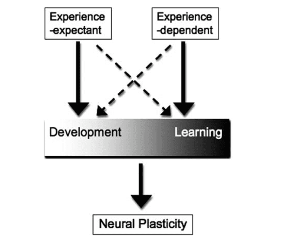
Figure 1: This working model illustrates that development and learning exist on a continuum, as each independently and simultaneously influence neural plasticity. While development is largely guided by experience-expectant mechanisms, it also receives input from experience-dependent mechanisms. Similarly, learning is mostly guided by experience-dependent mechanisms, but also receives experience-expectant input (72 × 72 DPI).

The notion that development and learning are complementary processes is not new. Karmiloff-Smith [ 1994] introduced the idea that together, they involve the gradual process of making behaviors more habitual while simultaneously, increasing explicit accessibility (as when learning a new task or skill). Another reason to believe that these constructs are not entirely separable, but rather that neural mechanisms are shared between the two, is that evolution tends to modify and borrow from existing systems, as opposed to creating entirely new ones to serve a similar purpose [Greenough et al., 1987]. As Karmiloff-Smith has argued, “A specifically human way to gain knowledge is for the mind to exploit internally the information that it has already stored, by redescribing its representations or, more precisely, by iteratively rerepresenting in different representational formats what its internal representations represent” [Karmiloff-Smith, 1994]. What will of course differ between the immature and adult system are the internal representations already available to them when new experiences are introduced into the system; the mature system will have a broader repertoire of previous experience. For instance, an axon that is growing toward a target has a very different landscape to navigate in the infant than in the adult brain. In this review, the terms learning and development are not used interchangeably but their linked effects on the developing system are implicitly acknowledged and appreciated.

Neural Plasticity

Neural plasticity is one of the most fascinating and challenging questions in neuroscience. Almost five decades ago, Hebb established a theoretical framework describing the phenomenon that the brain adapts to its environment based on experience and development [Hebb, 1949]. According to the theories of neuroplasticity, thinking and learning change both the brain's physical structure and functional organization. Basic mechanisms that are involved in plasticity include neurogenesis, programmed cell death, and activity-dependent synaptic plasticity. Repetitive stimulation of synapses can cause long-term potentiation or long-term depression of neurotransmission. Together, these changes are associated with physical changes in dendritic spines and neuronal circuits that eventually influence behavior. These same mechanisms stand out as important contributors to the developing brain's ability to acquire new information, adapt to the rapidly changing environment and recover from injury [Johnston, 2009]. As reviewed in several other articles in this issue, neuroimaging has significantly contributed to the study of neural development. It will undoubtedly serve important roles in disentangling neural substrates of development and learning.

Neuroimaging tools to study human neural plasticity

In previous studies of plasticity using neuroimaging, two main experimental approaches have been employed. While structural and functional approaches measure distinct neural attributes (delineated below), examples from studies which measure each are intermingled here to highlight examples of the two experimental approaches, cross-sectional and longitudinal, most commonly used to examine neural plasticity.

The first approach is a cross-sectional approach, in which individuals with varying levels of a given skill are compared and differences in neural structure or function related to their skill level are identified. For instance, in a study measuring structural change, Elbert et al. [ 1995] examined the relation between increased cortical representation of the fingers of musicians relative to those of nonmusicians. They found that musicians showed larger cortical representations and that the extent of this representation was correlated with the age at which the person had begun to play. Similar findings have been reported in the auditory cortex of musicians relative to nonmusicians [Pantev et al., 1998], suggesting that neural plasticity of relevant cortices depends on use and changes to accommodate the needs and experiences of the individual. The cross-sectional approach is often used in developmental and/or clinical developmental studies. There are numerous examples of this approach in the functional imaging literature, showing distinct neural activation patterns across age that correspond to differences in cognitive ability [Bunge et al. 2002; Casey et al., 1997; Durston et al., 2002; Luna et al., 2001; van Leijenhorst et al., 2007]. While their statistical power limitations are often acknowledged, it is argued that the major savings in time and cost of cross-sectional studies commonly used with fMRI, relative to longitudinal studies, make them an attractive alternative [Kraemer et al., 2000]. However, cross-sectional analyses may falsely suggest changes over time through confounds accidentally introduced into the study design, such as a time scale related to the variables of interest [Kraemer et al., 2000]. In addition, cross-sectional studies may suffer from cohort effects, in which different groups (e.g. children and adults; musicians; and nonmusicians) differ from each other by factors other than the factor of interest. For example, children growing up in the last 10 years will have received much more experience with computers than children growing up 30 years ago [Poldrack, 2000].

The second strategy to study neural plasticity is the longitudinal method. In this approach, participants are examined multiple times over the course of learning or development. In essence, training is a way to flood the organism with experience-dependent processes by saturating it with one particular experience. In the first experiment of this sort, structural changes were examined in rodents. Rats were trained on a changing series of patters in the Hebb-Williams maze during a period of 25 days [Greenough et al., 1979]. They found that in the visual cortex of trained animals, two types of neurons had more dendrites than in nontrained animals, while a third neuron type was unaffected. Thus, these findings demonstrated the specific and robust effects of training on synaptic connectivity. In humans, functional neural activation is assessed in a cognitive task before and after (and sometimes during) training on a task, in comparison with a baseline task that is not practiced [e.g., Karni et al., 1995; Karni and Sagi, 1993]. Then it is determined whether brain activity has changed in association with training on the task. The main advantage to this approach is that it provides optimal power to identify neural changes associated with the experience of interest because it is within-subject. However, in addition to the logistically challenging and expensive nature of longitudinal work, practice effects could potentially introduce confounds, as participants may acquire skills related to participation in the study.

Recent advances in the use of transcranial magnetic stimulation (TMS) have demonstrated the utility of this tool in the study of neural plasticity as well. Since its introduction, it has been known that repeated TMS (rTMS) of the motor cortex in healthy adult human participants can lead to relatively lasting effects (usually of the order of 30–60 minutes) on the excitability of the corticospinal output [Rothwell, 2007; Siebner and Rothwell, 2003]. For instance, a recent study showed that rTMS delivered to the superior temporal cortex causes macroscopic cortical changes in gray matter in the auditory cortex as early as within 5 days of continuous intervention [May et al., 2007] and work in patient populations has also uncovered the utility of this tool in investigating plasticity in the diseased brain [Rothwell, 2007]. Collectively, these approaches highlight the utility of neuroimaging tools in studying human neural plasticity.

The distinction between structural and functional changes

While experience can influence both structural and functional changes, it is important to note that there are clear distinctions between the two. Structural changes, as measured with MRI, typically refer to how experience influences anatomical changes. This change is indexed as volumetric differences in morphometry of particular brain region. Functional changes instead refer to differences in neural activation patterns following a particular experience. The functional methodology measures changes in blood oxygenation in the brain that are assumed to reflect changes in brain activity [Logothetis et al., 2001]. Structural and functional changes are not synonymous with each other, as different mechanisms likely underlie the two. Further, each can and do occur without the other. For instance, it is quite possible to observe functional remapping without any significant structural changes. Similarly, morphometric changes in a particular brain region do not always confer functional and/or behavioral changes.

Progressive and Regressive Changes With Learning

Most studies on neural plasticity have been conducted in adults and have yielded important insights that help inform developmental work. At the structural level, one of the first studies demonstrating the effects of experience on the brain showed that London taxi drivers had posterior hippocampi that were larger than controls in proportion to the length of driving experience [Maguire et al., 2000]. This study showed that the hippocampus, which is critical for spatial representation, is structurally altered by increased navigational experience. Subsequent studies showed structural changes using a variety of training paradigms, including complex visuomotor tasks such as juggling, [Draganski and May, 2008; Draganski et al., 2004] and music training [Zatorre et al., 2007], which each yielded changes in grey matter in motor cortex and regions in the parietal sulci. Together, these studies provide evidence that regions relevant for the task at hand are directly influenced by experience.

Using functional MRI, several groups have shown that training or experience is related to functional neural changes. However, these studies have yielded mixed results and interpretations. Numerous articles have addressed this topic [e.g. Draganski and May, 2008; Poldrack, 2000], so only a brief review of these findings will be provided here [for review, see Kelly and Garavan, 2005]. While some studies have shown general decreases in neural activity following training [e.g. Chein and Schneider, 2005], others have shown general increases with training or experience [e.g. Karni et al., 1995; Westerberg and Klingberg, 2007]. One explanation for the observed decreases in cortical activity with training or learning is based on the dual-processing framework of learning, which posits that a set of central resources mediating controlled processing is assumed to play a critical role in scaffolding novice performance, but becomes less essential as skilled, automatic processing emerges [Chein and Schneider, 2005]. With training, therefore, changes in fMRI signal are generalized as decreases in the extent and/or magnitude of activity, which may reflect local changes in synaptic efficacy [Haier et al., 1992]. Further, synaptic changes are assumed to affect the strength of local associations by tightening connections between neurons that contribute effectively toward task processing and weakening connections between those that do not. Across development, this phenomenon results in selective pruning of synapses that are overproduced early in development [e.g. Boothe et al., 1979; Huttenlocher, 1979]. As development collides with experience, extra synapses are lost, such that the final system consists of synapses that were selectively retained [Changeux and Danchin, 1977].

In contrast, several reports have shown general increases in activation with learning in both developmental and adult studies. For instance, Klingberg et al. reported that increased prefrontal and parietal activation was related to working memory capacity in children [Klingberg et al., 2002]. In adults, several groups have reported increased activation following skill learning [Karni et al., 1995] and other interventions such as meditation [Davidson et al., 2003], suggesting that these findings reflect the recruitment of additional cortical units with practice. This view is in agreement with the constructivist manifesto proposed by Quartz and Sejnowski [ 1997], which posits that there is a ‘progressive increase in the representational properties of cortex’ and that connectivity progresses from fewer to greater connections.

While strong evidence exists for both progressive and regressive changes (i.e. increasing and decreasing neural activation involved in cognitive tasks), change in a system from an immature to mature state is a product of both. This dual influence directly stems from the contribution of experience-expectant and experience-dependent mechanisms. While experience-expectant mechanisms encourage the elimination of unnecessary synapses or neural units (regressive change, presumably indexed by decreases in fMRI signal activity), experience-dependent mechanisms will guide activity-dependent creation and strengthening of synapses based on the individual organism's experience and needs (progressive changes, presumably indexed by increases in fMRI signal). Experience-expectant processes found in early development seem to assemble an excess of synapses, which are then selectively pruned back by experience to a functional subset. In later development and learning in adulthood, synapses appear to be generated in response to events that provide (learned) information to be encoded in the nervous system. At the synaptic level, this phenomenon is called metaplasticity, and refers to the notion that properties of synaptic plasticity can change as a function of previous plasticity and previous activation of synapses [Kalantzis and Shouval, 2009]. In other words, plasticity itself is plastic and the way the brain changes in response to its environment is contingent on the existing neural environment, the cause of (i.e. learning or development or both) and the behavioral consequences of the change.

What are the mechanisms of structural and functional neural changes?

The pronounced contributions of neuroimaging have been documented extensively. However, the conflicting findings reviewed above demonstrate how limited resolution continues to constrain interpretations of structural and functional neuroimaging data. This restriction is further complicated when examining developmental data, as described in more detail in this issue [Poldrack, 2010]. Our interpretations of increases and/or decreases in neural activity and grey matter are merely informed speculations of how they relate to neural plasticity, most of which are deduced from insights in molecular and cellular investigations.

Animal studies suggest that increases in cortical grey matter are the result of a complex array of morphological changes including synaptic events such as the formation of new connections by dendritic spine growth and alterations in the strength of existing connections [Chklovskii et al., 2004; Draganski and May, 2008; Hirata et al., 2004; Holtmaat et al., 2006; Trachtenberg et al., 2002]. In animals exposed to enriched environments, increased size of the soma and nucleus of neurons, glia and capillary dimensions have also been shown to influence cortical morphology [Kozorovitskiy et al., 2005; Muotri and Gage, 2006]. In addition, further mechanisms linked to training and experience-related plasticity include changes in the synaptic contacts known to be the morphological substrates of long-term potentiation and long-term depression [Draganski and May, 2008], synaptic pruning [Huttenlocher, 1979], changes in gene expression [Kleim et al., 1996], protein synthesis [e.g. McAllister et al., 1995], and dendritic density [Comery et al., 1995].

The Plasticity of Developmental Timing

Understanding whether the same neural mechanisms underlie both development and learning will address larger questions about developmental timing and experience-expectant processes. Are there certain cognitive processes that can be “sped up” with training or developmentally prolonged with experience? Animal work has suggested that the length of time that the developing nervous system remains sensitive to experience-expectant events can be manipulated. Cynader and Mitchell [ 1980] found that kittens reared in the dark until 6, 8, or 10 months of age remained highly sensitive to monocular deprivation effects. In contrast, kittens reared normally (i.e. reared in the light), show peak sensitivity to monocular deprivation within the first 2 months of life [Hubel and Wiesel, 1970]. In humans, the most insightful experiments to address these questions have been conducted in infants. In these experiments, researchers introduce tools that facilitate motor skills early in development, before the age at which these behaviors are typically observed. Although most of these studies do not have associated measures of neural activity, they provide considerable insight into how experience-expectant processes can be manipulated to occur earlier that is developmentally expected.

Needam et al. [ 2002] have shown that early, simulated experience serves to jump-start processes that are considered to be developmentally constrained. That is, by providing infants with scaffolding tools that they are not typically exposed to before a certain age, they can be trained to exhibit certain characteristics earlier than normal. In general, infants do not systematically reach for objects until ∼ 5 months of age [Butterworth and Hopkins, 1988; Rochat, 1993]; this is likely a reflection of their relatively immature gross motor skills (e.g. arm and hand strength, fine motor control) before this time [Halverson, 1933; Jeannerod, 1984]. Needham et al. [ 2002] tested 3-month-old infants (before spontaneous effective reaching and grasping) using an “enrichment experience.” The enrichment experience consisted of 12 to 14 brief parent-led object play sessions held at the infant's home. During the play sessions, the infant sat on a parent's lap at a table and wore mittens with the soft side of Velcro covering the palms. On the table in front of the infant were small, lightweight objects with edges covered in the corresponding side of the Velcro. With a quick swipe of the hand, the infant could easily “pick up” an object as it stuck to the mitten. After the enrichment phase for the infants in the experimental condition, these infants as well as the infants in the control condition (who did not play with the “sticky mittens”) were taken to the lab for an assessment of object exploration skills. Infants who had had the enrichment experience showed accelerated reaching behavior toward the new objects compared with control infants, even when not wearing the sticky mittens. These data suggest that experience may be a critical factor in manipulating processes considered to be under developmental constraint.

Rovee-Collier's conjugate reinforcement paradigm [see Rovee-Collier and Hayne, 2000] also produced observable actions on objects by infants before they typically do so on their own. In this paradigm, a ribbon is tied to an infant's ankle and the other end is tied to a mobile stand; once an infant makes the association between their ankle and the rewarding mobile movement, the infant's rate of leg kicks increases sharply. This is an additional evidence that producing actions on objects with observable effects is highly reinforcing for young infants. It remains an open question whether this experience with leg-kicking and mobile-moving would generalize to other abilities. These experiments suggest that behaviors that seem developmentally constrained can, in fact, be manipulated in developmental time. That is, processes that presumably rely on experience-expectant mechanisms (e.g. motor skill) show experience-related plasticity in their developmental onset. An ERP experiment in conjunction with these types of enrichment manipulations would help the field examine neural mechanisms that support these accelerated learning experiences. For instance, one could imagine that these accelerated behavioral experiences are subserved by precocial activity in motor cortices. As neural development is hierarchical and highly linked across the brain, these early enriched experiences likely have subsequent effects on downstream neural and behavioral development. Conducting such studies in infants is particularly useful as it would help avoid some of the potential confounds of domain-general experience that older children are already equipped with.

Do Development and Learning Processes Share the Same Neural Mechanisms?

Without delving too deeply into the different types of learning processes that occur, both in development and beyond, there are undoubtedly some types of learning that are highly similar in both. For instance, learning by trial and error is one common and lifelong way that organisms, from rodents to primates, master their environment. Given their limited speaking and language comprehension abilities, infants constantly learn through trial and error; as such, they are problem-solvers who are constantly faced with a problem and the challenge of solving it. A common dilemma a young infant encounters is how to balance and sit upright. After repeated collapses and attempts at a solution, the infant eventually learns to use an arm in a so-called “tripod stance” to support him/herself. In adults, the neural activation that accompanies learning by trial and error, particularly through unexpected outcomes, is referred to as the “prediction error signal” [e.g. Hollerman and Schultz, 1998; Schultz et al., 1997], thought to be mediated primarily through the neurotransmitter dopamine. Briefly, there is an increased dopamine firing rate in nonhuman primates and increased activation in dopamine-rich region in humans when the organism receives an unexpected event [Fiorillo et al., 2003]. Eventually, the dopamine signal decreases as the organism learns to expect the event [Fiorillo et al., 2003; 2008]. These findings have implicated dopamine as a learning signal. While methodological constraints have precluded examination of the neural basis of prediction error learning in the young infant, I would argue that the dopaminergic neural mechanisms are the same. That is, dopamine neurons respond to expected and unexpected events similarly in the infant as they do in the adult. However, I would also assert that, because the system in which dopamine is acting is very different across development, that prediction error cannot be exactly the same in the infant as it is in the adult. While prediction error learning is a form of environmental adaptation across development, neural plasticity that arises from it differs. In the young infant, this plasticity will influence the basic architecture of the neural system (i.e. how the brain is going to be organized) while in the adult, this plasticity is modifying the existing architecture of the brain (i.e. reorganizing and modifying but not laying the groundwork). This dichotomy is analogous to building a house, where building a brand new house represents developmental plasticity and a house remodel represents plasticity in the mature system. The tools and mechanisms are identical, but the environment in which the change is occurring is vastly different.

The utility of “noise”

One of the greatest confounds in developmental work is the significant difference in ability or performance between children and adults. As compared with adults, who can and do implicitly draw on domain-general neural resources, the child's ability to perform any given cognitive operation is inefficient at best, as they require additional effortful, explicit, and implicit requirements to perform complex cognitive demands (e.g. response inhibition) as well as adults. For example, numerous studies have shown that even when children perform a given task as well as, or without any differences in observable behavior, as adults, they recruit distinct neural strategies. Tamm et al. [ 2002] compared the changing performance of children, adolescents, and young adults on the Go/No-Go task, a measure of inhibitory control. Despite an overall reduction in reaction time with age, younger subjects showed the same level of accuracy as adults. However, the fMRI data collected alongside the performance measures revealed that younger children demonstrated a greater level of activity within left superior and middle frontal gyri than did older children and that, conversely, older participants demonstrated an increased focal activation in the left inferior frontal gyrus relative to their younger counterparts. In a separate study of cognitive control, effective interference suppression in children was associated with prefrontal activation in the opposite hemisphere as adults while effective response inhibition was associated with activation in posterior, but not prefrontal, regions activated by adults [Bunge et al., 2002]. The authors also reported that children failed to activate a region in right ventrolateral prefrontal cortex that was recruited for overall cognitive control by adults. Similarly, a more recent study showed that children recruit distinct activation profiles from adults also differ temporally (i.e. show different time-courses of activation) across relational reasoning tasks [Crone et al., 2009]. Together, these studies provide evidence for the alternative neural strategies that immature systems often engage to support more mature behavioral demands. Despite the apparent nuisance that such extreme behavioral and neural variability introduces into the study of development, dynamic systems theory celebrates this variability [Smith and Thelan, 2003]. This noise allows investigators to examine developmental trajectories of change over the short timescales of problem-solving (i.e. because of intersubject individual differences) and/or over a longer developmental span (i.e. as when comparing children with adults).

Schlaggar et al. have elegantly demonstrated how variability can be used to gather insight into developmental versus performance-related neural activity. Using a single-word processing task, they compared neural activity in a performance-matched subgroup of children and adults taken from a larger sample [Schlaggar et al., 2002]. That is, the children and adults in the matched group did not differ in behavioral performance, making it possible to determine whether any functional activation differences were due to developmental stage or performance. They found distinct patterns of activation that were age-related, performance-related, or independent from either. As such, their data shed new light on age-related regions (regions that were more/less active in children regardless of performance) that most likely reflect effects of brain development. In a follow-up study [Brown et al., 2005], the same group reported a more thorough examination of progressive and regressive neural changes across development. Using lexical association tasks, the authors identified increases and decreases in different brain regions that varied by age, performance ability, or neither. Seventy-five percent of the regions identified as showing age-related changes (i.e. independent of performance) showed decreases in activity over age. These regions were most prominently located in medial frontal and anterior cingulate cortex, right frontal cortex medial parietal, and posterior cingulate cortex. The remaining 25% of regions that showed increases in brain activity with age, were primarily later-stage processing regions, including lateral and medial dorsal frontal cortex and left parietal cortex. This strategy, of taking advantage of developmental and behavioral variability post hoc, is precisely the approach that needs to be adopted to disentangle neural mechanisms of development- and learning-related plasticity.

The approach by Schlaggar et al. described above can easily be modified to examine training-related plasticity. By substituting a training component for the “performance” group (i.e. the group who had naturally occurring variability in performance ability), one could separate neural activation changes related to age, training, or neither. For instance, a group of individuals ranging in age from childhood to adulthood could be trained on a motor task, such as juggling, that all were naive to. Participants would be scanned before and after training. Post hoc, participants would be divided into groups based on their level of juggling skill. In this manner, neural regions would be divided in those that are age-related and training-related, thereby allowing insight into neural regions more susceptible to experience and those with greater developmental constraints.

Longitudinal training studies across development

Despite the inherent challenges, the only way to identify the root of neural plasticity as either developmental or experiential is to conduct a longitudinal training study. There is no question that the incredibly challenging, logistically difficult, and expensive nature of this type of work is what has precluded the field from embarking more vigorously on this type of research. However, a few recent studies have proven its feasibility. For instance, Durston et al. [ 2006] used a combined longitudinal and cross-sectional study to examine shifts in cortical activity during a response inhibition task. The longitudinal findings, relative to the cross-sectional data, showed attenuated activity in dorsolateral prefrontal cortical areas with age. In parallel, there was increased focal activation in ventral prefrontal cortex that was related to improvements in task performance [Durston et al., 2006]. A more recent study by Hyde et al. [ 2009] implemented a training component. Their study builds on previous studies in adult musicians and matched nonmuscians that have revealed structural and functional differences in brain regions relevant to music production [Bermudez and Zatorre, 2005; Elbert et al., 1995; Gaser and Schlaug, 2003; Pantev et al., 1998; Zatorre et al., 2007]. The authors were motivated by the question begged of this type of research: Do musicians (or others who show skill-related neural plasticity) do so because of biological predispositions to music or because of intensive music training? Hyde et al. compared structural changes in relation to behavioral changes in young children who received 15 months of instrumental musical training relative to a group of children who did not. The children who received private keyboard lessons showed greater behavioral improvements on music discrimination and related tasks than the nontrained children; neither group showed differences between baseline and testing on nonmusical tasks. In addition, the musically trained children showed greater structural changes in right precentral gyrus, corpus callosum, and the primary auditory region [Hyde et al., 2009], consistent with findings in adults [Zatorre et al., 2007 for review]. Their data provide new evidence for training-induced structural brain plasticity in early childhood. Using structural MRI, Schlaug et al. [ 2005, 2009] also identified structural differences in the corpus callosum in young musicians. Based on total weekly practice time, they divided a sample of 5- to 7-year-old children into three groups: high-practicing, low-practicing, and controls. There were no differences in corpus callosum size at baseline, but differences emerged after approximately 29 months, with the greatest increased change in the high-practicing group of children [Schlaug et al., 2009]. Further, total weekly music exposure predicted degree of change in the corpus callosum as well as improvement on a nonmusic related motor-sequencing task.

In addition, training interventions have been implemented in clinical populations and have similarly shown robust plasticity. In a group of children with ADHD, training significantly improved performance on a nontrained visuospatial working memory tasks. In addition, motor activity, as measured by the number of head movements during a computerized task, was significantly reduced in the treatment group [Klingberg et al., 2002]. In a separate study, strong improvement in attention was found after only 5 days of attention training in a group of 4- and 6-year-old children. This change was paralleled by changes in EEG patterns that resembled a more mature pattern of activation, such that training had specific effects on the scalp distribution of the ERPs that was similar to the influence of development [Rueda et al., 2005].

Several studies have provided strong support for the claim that children with reading disabilities can benefit significantly from intervention techniques; the impact of such interventions on neural plasticity has been assessed using fMRI [McCandliss and Noble, 2003]. In one study, dyslexic children received an intervention after an initial baseline scan showing the typical reduced activation of the left posterior superior temporal gyrus (STG) during a phonologically challenging task [Simos et al., 2002]. Following the 80-hour intervention, all dyslexic children showed significant increases in reading skill, as well as increased activation in the left posterior STG [Simos et al., 2002]. Similar changes in neural activation were reported in separate intervention training in children with other dyslexia [McCandliss et al., 2001; Temple, 2002]. In a recent report, Keller and Just showed that reading remediation induces changes in white matter of poor readers [Keller and Just, 2009], such that fractional anisotrophy (FA) was significantly increased following remedial instruction. This FA increase was correlated with improvement in phonological decoding ability, which demonstrates how behavioral intervention can influence neural plasticity.

A recent study was able to examine the effects of reading on structural neural change without the influence of development [Carreiras et al., 2009]. Structrual MRI scans were obtained from adult participants who had recently completed a literacy program in adulthood (before the program, they were illiterate) and matched illiterates who had not yet started the literacy program. Their findings suggest that learning to read strengthens the coupling between left and right angular gyri and between the left dorsal occipital gyrus and left supramarginal gyrus [Carreiras et al., 2009].

Collectively, these studies have suggested that plasticity within the immature brain shows similarities to plasticity in the adult system. First behavioral improvements related to intensive training or experience are associated with neural plasticity specific to the task at hand (e.g. increased activation in the STG following reading intervention). What this suggests is that experience-dependent mechanisms do not differ greatly across the lifespan. Second, neural regions previously associated with experience-expectant mechanisms, such as motor abilities and language, show a high degree of plasticity across development, suggesting that perhaps there is plasticity in processes that are initially precipitated by expectant interactions with the environment.

These studies have also led to more questions that will undoubtedly be addressed in the next generation of research on this topic. First, which neural systems show greater or less training-related plasticity earlier in development? For example, there is significantly greater plasticity in receiving and learning from language input during infancy than in any other point in life. As infants receive increasing exposure to their native language, neural systems sensitive to language lose plasticity, which is translated into more difficulty discriminating speech sounds of foreign languages and learning new languages [Doupe and Kuhl, 2008]. Are there other examples of such extreme behavioral and neural loss of plasticity across the lifespan, whereby learning itself imposes constraints on plasticity? Second, how do the timescales of neural plasticity change across development? That is, do observed behavioral and neural changes occur more or less quickly in the developing brain? Again, to borrow from the language literature, young children learn to discriminate foreign languages more quickly and more proficiently than adults [Snow, 1987]; does this accelerated timescale hold for all cognition? Last, which behaviors cannot be “sped up” by exposure earlier in development because of time-locked experience-expectant mechanisms? Certainly, pubertal constraints (as described in more detail in Blakemore et al. [2010], this issue) will impose at least some limits on plasticity associated with this maturational change.

Methodological Considerations and Potential Confounds

The dearth of studies that examine learning versus development is a consequence of the potential confounds and fairly prohibitive methodological factors inherent in this type of work. In this closing section I outline the most common issues.

Resources and attrition

Cost and attrition are two significant reasons that training and development studies are not more prevalent in the literature, despite calls for the need of this kind of work [Casey et al., 2005]. In addition to steep scanner costs at many institutions and scanning facilities, a healthy sample size of both children and adults are necessary for sufficient statistical power to detect learning versus developmental differences. Also, additional staff is needed to conduct the training portion of the study, as participants will need to visit and the lab and receive training at least weekly and in some cases daily, depending on the study. If the training aspect involves a particular still, such as music training, the staff will need to be proficient in this as well. Together, these requirements lead to a very expensive experiment.

Subject attrition also poses a potential hurdle in this type of work. Subject burden for a training and/or longitudinal study is relatively high, as participants visit the lab for multiple sessions and are required to maintain cognitive and training engagement. However, several groups have successfully conducted longitudinal studies [e.g. Durston et al., 2006, Giedd et al., 2009, and an ongoing study at UCLA] by increased attention and sensitivity to families' needs. Some useful strategies for successful subject retention are listed in Table I.

Table I. Strategies to increase subject retention

1. During the first study visit, ask parents to give the names and phone numbers of three people who know how to contact them.

2. Contact parents by phone every 2 months for a longitudinal study that spans 1 year or longer.

3. Send children birthday and holiday cards as well as a small gifts or gift cards for these occasions.

4. Provide snacks and breaks at each visit.

5. Reinforce children at each visit and evaluation with small age-appropriate toys, shirts, hats, and school supplies.

6. Compensate parent for their time and effort at each visit.

7. Send manuscripts and copies of posters/presentations that describe relevant findings to demonstrate the importance of the work they are participating in.

Scanner-related anxiety

Subjects, particularly children, may exhibit scanner-related anxiety (e.g. Eatough et al., 2009]. Initial anxiety might lead to increased head motion, increased vigilance, or attention, all of which can skew the observed results and/or interpretations. As such, investigators should implement techniques to reduce the possibility of this potential confound. In addition to acclimation to the scanning environment with a mock scanner, as many groups do, it would be prudent to conduct two baseline scans in training studies. The first baseline scan, which could be conducted using an unrelated task, would capture the novelty-related anxiety while the second baseline scan would serve as the “true” baseline of the training session.

Performance differences

As elegantly reviewed in Schlaggar (this issue) and stated above, performance differences between developmental and adult populations can introduce significant confounds and make difficult the accurate interpretation of neuroimaging data. Without addressing performance differences, the observed differences in brain activity between children and adults could be misinterpreted as maturational differences in functional neuroanatomy. Successful ways to address this issue include the use passive tasks in the scanner, the use of tasks that are equated for difficulty, and performance-matching, as reviewed earlier. In addition, the dependent measure should test for accuracy (as equated task difficulty), rather than reaction time, as the latter is well known to lag in developmental populations.

A problem in the interpretation of training studies is the possibility that brain effects might arise from stress, sensory stimulation, motor activity, or other nonspecific consequences of the training procedure, rather than information acquired through training [Greenough et al., 1987]. As such, it is critical that appropriate control groups are studied in tandem.

Last, choosing the optimal time between scans is a potential dilemma, as the field simply does not have enough knowledge of the precise nature of the underlying neural events, how quickly they are affected by experience, and their impact on the imaging signal [Draganski and May, 2008]. MRI voxel-based morphometry has revealed changes within 1 week after training [Driemeyer et al., 2008]. Other groups [Draganski et al., 2006] have waited 3 months between scans, as this is the length of time needed for newly generated stem cells to differentiate into neurons [Cummings et al., 2005].

CONCLUSIONS

Neural plasticity is the brain's solution to the challenge of integrating new information into its repertoire of neural networks. Three main points have been outlined in this review. First, the reader is asked to consider how and if development differs from learning and, subsequently, how this distinction translates to differences or similarities in neural mechanisms. I have proposed that rather than struggling to parse out distinctions between two constructs that are nearly inextricable, it is important to recognize how their effects bleed into one another, and how experience-expectant and experience-dependent mechanisms simultaneously and independently influence each one.

Second, that the local neural environment in which plasticity occurs is a critical component of change. While the actual neural mechanisms (e.g. synaptic mechanisms) that underlie plasticity probably do not differ significantly in the immature and mature system, the local environment in which that plasticity is occurring is undoubtedly different. This difference exists not only in terms of the cellular, anatomical, and metabolic environment, but in the much larger contextual environment of the individual. For instance, modifying synapses that are already committed (e.g. learning a motor skill such as juggling) is very different than committing the synapse for the first time (e.g. learning the motor coordination necessary for the first time a baby holds himself up).

Last, the challenges of this type of work have precluded significant advances in addressing plasticity-related questions. Despite these hurdles, it is critical that the field more vigorously embrace longitudinal training studies in developmental groups if we are truly committed to uncovering the mechanisms underlying plasticity across development. The greatest utility of cross-sectional studies of development is the insight into developmental differences. However, to understand developmental changes in the brain, a longitudinal approach is critical. Eventually, answers to these questions will be useful in identifying strategies and developmental timing for optimal learning, remediation, and rescue from brain injury.

Link to Article

Abstract

Development and learning are powerful agents of change across the lifespan that induce robust structural and functional plasticity in neural systems. An unresolved question in developmental cognitive neuroscience is whether development and learning share the same neural mechanisms associated with experience-related neural plasticity. In this article, I outline the conceptual and practical challenges of this question, review insights gleaned from adult studies, and describe recent strides toward examining this topic across development using neuroimaging methods. I suggest that development and learning are not two completely separate constructs and instead, that they exist on a continuum. While progressive and regressive changes are central to both, the behavioral consequences associated with these changes are closely tied to the existing neural architecture of maturity of the system. Eventually, a deeper, more mechanistic understanding of neural plasticity will shed light on behavioral changes across development and, more broadly, about the underlying neural basis of cognition.

Development and Learning: Shared Mechanisms of Neural Plasticity

Introduction

Early life stages witness profound brain transformations intertwined with normative development and learning experiences. Both agents of change rely on emerging neural processes that mirror and facilitate behavioral adaptations. Consequently, developmental cognitive neuroscientists have long sought to determine if the biological underpinnings of learning and development are congruent. While a conclusive answer to this multifaceted question remains elusive, advancements in cognitive neuroscience methodologies, particularly magnetic resonance imaging (MRI), functional MRI (fMRI), and electroencephalogram (EEG), have provided an unparalleled opportunity to investigate neural and behavioral alterations in concert.

Development and Learning: The Problem of Definition

Development and learning represent intricately intertwined constructs. While a definitive and mutually exclusive definition for each remains challenging, for clarity, this article proposes that development encompasses changes in an organism resulting from growth, maturation, and/or experience. In contrast, learning refers to acquiring skills or knowledge through study, instruction, and/or experience. The shared influence of experience complicates disentangling these two.

Some developmentalists argue that development and learning are inseparable, as learning invariably occurs within a developmental context [Casey et al., 2006]. Others differentiate them based on timescales: learning transpires over hours, days, or months, while developmental change unfolds over weeks, months, or years, with evolution spanning even longer periods [Smith and Thelan, 2003]. This article posits that development and learning exist on a continuum, with each endpoint influenced by both experience-expectant and experience-dependent processes (Figure 1).

Figure 1. This working model illustrates that development and learning exist on a continuum, as each independently and simultaneously influence neural plasticity. While development is largely guided by experience-expectant mechanisms, it also receives input from experience-dependent mechanisms. Similarly, learning is mostly guided by experience-dependent mechanisms, but also receives experience-expectant input (72 × 72 DPI).

This model suggests that while both development and learning induce neural and behavioral plasticity, development emerges primarily through experience-expectant mechanisms, while learning is more heavily shaped by experience-dependent ones. As defined by Greenough et al. [1987], experience-expectant mechanisms utilize environmental information consistently encountered by a species throughout its evolutionary history. Essentially, the neural system "anticipates" these experiences, such as visual contrast or language input, to shape sensory and motor systems. Conversely, experience-dependent mechanisms are attuned to an individual's unique encounters. While experience-expectant mechanisms operate on relatively consistent timelines across individuals, experience-dependent ones are more fluid, reflecting the diverse timing of individual experiences and learning opportunities.

Consequently, plasticity arising from typical development signifies neural change aligning with the phylogenetic norm. In contrast, plasticity stemming from learning represents neural adaptations specific to individual experiences. Though the relative influence of these mechanisms may shift throughout life, an organism ultimately represents the culmination of its developmental and experiential environment, both molded by experience-expectant and experience-dependent mechanisms.

Language acquisition exemplifies the transition from experience-expectant to experience-dependent plasticity [Doupe and Kuhl, 2008]. Kuhl et al. have demonstrated that infants initially discriminate phonetic sounds from all languages [Eimas et al., 1976; Kuhl and Meltzoff, 1982]. However, with language exposure, infants gradually lose this universal discrimination ability while becoming increasingly adept in their native tongue [Kuhl et al., 1997]. This elegantly illustrates how biological predispositions (experience-expectant development) are subsequently refined by individual experiences (experience-dependent learning).

The notion of complementary development and learning processes is not novel. Karmiloff-Smith [1994] proposed that both involve gradually making behaviors more habitual while simultaneously enhancing explicit accessibility. This is exemplified when mastering a new skill: initial explicit efforts transition into more automatic, implicit processing with practice. The evolutionary tendency to adapt and build upon existing systems rather than create entirely new ones for similar functions [Greenough et al., 1987] further supports shared neural mechanisms. Karmiloff-Smith [1994] argues that a uniquely human approach to knowledge acquisition involves the mind internally leveraging stored information by "redescribing its representations" or "iteratively rerepresenting in different representational formats what its internal representations represent."

While both immature and mature systems engage in this process, they differ in the internal representations available for incorporating new experiences. The mature system possesses a richer repertoire built upon more extensive prior experiences. For instance, an axon navigating towards its target encounters a vastly different landscape in the developing infant brain compared to the adult brain.

It is important to clarify that while this review acknowledges the intertwined effects of learning and development on the developing system, the terms are not used interchangeably.

Neural Plasticity

Neural plasticity stands as a captivating and intricate puzzle in neuroscience. Hebb [1949] laid the foundation by proposing that the brain adapts to its environment based on experience and development. Neuroplasticity theories posit that thinking and learning modify both the brain's physical structure and functional organization. Fundamental mechanisms underlying plasticity include neurogenesis, programmed cell death, and activity-dependent synaptic plasticity. Repetitive synaptic stimulation can induce long-term potentiation or depression of neurotransmission. These changes are linked to physical alterations in dendritic spines and neuronal circuits, ultimately influencing behavior. These mechanisms are crucial for the developing brain's capacity to acquire information, adapt to its dynamic environment, and recover from injury [Johnston, 2009]. As highlighted in this issue, neuroimaging has significantly advanced our understanding of neural development and will undoubtedly prove invaluable in unraveling the neural substrates of both development and learning.

Neuroimaging Tools to Study Human Neural Plasticity

Two primary experimental approaches utilizing neuroimaging have been employed to investigate plasticity. While structural and functional approaches assess distinct neural attributes (detailed below), examples from studies employing both are interwoven here to illustrate the two most common experimental designs: cross-sectional and longitudinal.

The cross-sectional approach compares individuals with varying levels of a specific skill, aiming to identify structural or functional neural differences associated with skill proficiency. For instance, Elbert et al. [1995] investigated structural changes by examining the cortical representation of fingers in musicians versus non-musicians. Musicians exhibited larger cortical representations, with the extent of this representation correlating with the age at which they began playing. Similar findings in the auditory cortex of musicians [Pantev et al., 1998] suggest that neural plasticity in relevant cortices is experience-dependent, adapting to individual needs and experiences.

The cross-sectional approach frequently features in developmental and clinical developmental research. Functional imaging studies utilizing this approach have revealed distinct age-related neural activation patterns corresponding to cognitive ability differences [Bunge et al., 2002; Casey et al., 1997; Durston et al., 2002; Luna et al., 2001; van Leijenhorst et al., 2007]. While acknowledging their statistical power limitations, the time and cost-effectiveness of cross-sectional studies, particularly with fMRI, make them an attractive alternative to longitudinal designs [Kraemer et al., 2000]. However, cross-sectional analyses risk falsely inferring changes over time due to confounds like time-related variables [Kraemer et al., 2000] or cohort effects, where distinct groups differ in factors beyond the variable of interest. For example, children raised in the past decade likely have greater computer experience than those raised 30 years ago [Poldrack, 2000].

The longitudinal method, in contrast, examines participants repeatedly throughout learning or development. This approach essentially "floods" the organism with a specific experience, amplifying experience-dependent processes. Greenough et al. [1979] conducted one of the first such studies, investigating structural changes in rodents trained on a series of patterns in the Hebb-Williams maze. Trained animals exhibited more dendrites in two neuron types within the visual cortex compared to untrained animals, highlighting the specific and robust effects of training on synaptic connectivity.

In human studies, functional neural activation is assessed before and after (and sometimes during) training on a cognitive task, compared to a non-practiced baseline task [e.g., Karni et al., 1995; Karni and Sagi, 1993]. This approach offers greater power to detect experience-related neural changes due to its within-subject design. However, besides logistical challenges and expense, practice effects (e.g., skill acquisition related to study participation) can confound interpretations.

Recent advancements in transcranial magnetic stimulation (TMS) have highlighted its potential in studying neural plasticity. Repeated TMS (rTMS) of the motor cortex in healthy adults can induce relatively lasting effects (30-60 minutes) on corticospinal excitability [Rothwell, 2007; Siebner and Rothwell, 2003]. May et al. [2007] demonstrated that rTMS applied to the superior temporal cortex can induce macroscopic gray matter changes in the auditory cortex within five days. Studies in patient populations further underscore the utility of TMS in investigating plasticity in diseased brains [Rothwell, 2007]. Collectively, these approaches demonstrate the value of neuroimaging tools in studying human neural plasticity.

The Distinction Between Structural and Functional Changes

While experience can influence both structural and functional changes, it's crucial to recognize their distinct nature. Structural changes, measured via MRI, typically refer to experience-induced anatomical alterations, indexed as volumetric differences in specific brain regions. Conversely, functional changes pertain to differences in neural activation patterns following an experience. Functional methodologies measure alterations in blood oxygenation, assumed to reflect changes in brain activity [Logothetis et al., 2001].

It is important to note that structural and functional changes are not interchangeable, likely arising from distinct mechanisms. Moreover, they can occur independently. For instance, functional remapping can occur without significant structural modifications, and conversely, morphometric changes in a brain region may not always translate into functional or behavioral differences.

Progressive and Regressive Changes With Learning

Most neural plasticity studies, primarily conducted in adults, offer valuable insights applicable to developmental research. One of the earliest studies demonstrating experience-dependent structural changes revealed that London taxi drivers had larger posterior hippocampi compared to controls, with size correlating with driving experience [Maguire et al., 2000]. This finding suggested that the hippocampus, crucial for spatial representation, undergoes structural modifications with increased navigational experience. Subsequent studies employing diverse training paradigms, including complex visuomotor tasks like juggling [Draganski and May, 2008; Draganski et al., 2004] and music training [Zatorre et al., 2007], have reported training-induced gray matter changes in relevant motor and parietal cortices, further supporting the idea that experience directly shapes task-relevant regions.

Functional MRI studies have also demonstrated experience-dependent functional neural changes, albeit with mixed findings and interpretations [for review, see Kelly and Garavan, 2005]. While some studies report general decreases in neural activity following training [e.g., Chein and Schneider, 2005], others observe increases with training or experience [e.g., Karni et al., 1995; Westerberg and Klingberg, 2007].

The observed decreases in cortical activity with training or learning are often explained through the dual-processing framework of learning. This framework posits that controlled processing, mediated by central resources, is crucial for novice performance but becomes less essential as skilled, automatic processing emerges [Chein and Schneider, 2005]. Thus, training-induced fMRI signal changes often manifest as reduced activity extent or magnitude, potentially reflecting local synaptic efficacy changes [Haier et al., 1992]. Synaptic changes are thought to refine local associations by strengthening connections between neurons contributing effectively to task processing and weakening those that do not. Throughout development, this translates into selective pruning of initially overproduced synapses [e.g., Boothe et al., 1979; Huttenlocher, 1979]. As development intersects with experience, superfluous synapses are eliminated, leaving a functionally refined subset [Changeux and Danchin, 1977].

Conversely, several studies report increased activation with learning in both developmental and adult populations. Klingberg et al. [2002] found that increased prefrontal and parietal activation correlated with working memory capacity in children. In adults, heightened activation has been observed following skill learning [Karni et al., 1995] and interventions like meditation [Davidson et al., 2003], suggesting recruitment of additional cortical units with practice. This aligns with the constructivist manifesto proposed by Quartz and Sejnowski [1997], which posits a "progressive increase in the representational properties of cortex" and a developmental trajectory from fewer to greater connections.

While evidence exists for both progressive and regressive changes, the transition from an immature to a mature system likely involves both. This dual influence stems from the interplay between experience-expectant and experience-dependent mechanisms. While experience-expectant mechanisms promote the elimination of unnecessary synapses or neural units (regressive change, reflected in decreased fMRI signal), experience-dependent mechanisms guide activity-dependent synapse creation and strengthening based on individual experiences and needs (progressive change, reflected in increased fMRI signal). Early development's experience-expectant processes appear to initially generate an overabundance of synapses, subsequently pruned by experience into a functional subset. Later development and adult learning, however, involve synapse generation in response to events encoding learned information in the nervous system.

At the synaptic level, this phenomenon, termed metaplasticity, describes the property of synaptic plasticity being modifiable by prior plasticity and synaptic activation history [Kalantzis and Shouval, 2009]. In essence, plasticity itself is plastic, with the brain's response to its environment depending on the existing neural architecture, the source of change (learning, development, or both), and the behavioral consequences of that change.

What are the Mechanisms of Structural and Functional Neural Changes?

Neuroimaging has significantly advanced our understanding of neural plasticity. However, the conflicting findings discussed above highlight how limited resolution continues to constrain interpretations of structural and functional neuroimaging data. This limitation is further compounded when examining developmental data [Poldrack, 2010]. Our interpretations of increased or decreased neural activity and gray matter remain informed speculations guided by insights from molecular and cellular investigations.

Animal studies suggest that increased cortical gray matter arises from a complex interplay of morphological changes, including synaptic events like new connection formation through dendritic spine growth and alterations in existing connection strength [Chklovskii et al., 2004; Draganski and May, 2008; Hirata et al., 2004; Holtmaat et al., 2006; Trachtenberg et al., 2002]. Enriched environments have been linked to increased soma and nucleus size in neurons, as well as changes in glia and capillary dimensions, all influencing cortical morphology [Kozorovitskiy et al., 2005; Muotri and Gage, 2006].

Further mechanisms implicated in training and experience-related plasticity include:

  • Changes in synaptic contacts, the morphological substrates of long-term potentiation and depression [Draganski and May, 2008].

  • Synaptic pruning [Huttenlocher, 1979].

  • Altered gene expression [Kleim et al., 1996].

  • Modifications in protein synthesis [e.g., McAllister et al., 1995].

  • Shifts in dendritic density [Comery et al., 1995].

The Plasticity of Developmental Timing

Understanding whether development and learning share neural mechanisms is crucial for addressing broader questions regarding developmental timing and experience-expectant processes. Can specific cognitive processes be accelerated through training or prolonged through experience? Animal studies suggest that the duration of the developing nervous system's sensitivity to experience-expectant events can be manipulated. Cynader and Mitchell [1980] found that kittens reared in darkness until 6, 8, or 10 months remained sensitive to monocular deprivation effects. In contrast, normally reared kittens exhibit peak sensitivity to monocular deprivation within the first two months of life [Hubel and Wiesel, 1970].

Human infant studies offer compelling insights into manipulating experience-expectant processes. By introducing tools that facilitate motor skills earlier than typically observed, researchers can investigate how experience-expectant processes can be accelerated. While most of these studies lack corresponding neural activity measures, they provide valuable information on manipulating developmentally constrained processes.

Needham et al. [2002] demonstrated that early simulated experience can "jump-start" developmentally constrained processes. Infants typically don't exhibit systematic reaching for objects until around five months of age [Butterworth and Hopkins, 1988; Rochat, 1993], likely due to immature gross motor skills [Halverson, 1933; Jeannerod, 1984]. Needham et al. [2002] tested three-month-old infants (before independent reaching and grasping) using an "enrichment experience" involving 12-14 brief parent-led object play sessions. During these sessions, infants wore mittens with Velcro on the palms, interacting with objects covered in corresponding Velcro. This allowed infants to easily "pick up" objects by brushing their hands against them. After the enrichment phase, both experimental and control infants (who did not experience the "sticky mittens") underwent object exploration assessments. Infants exposed to the enrichment experience displayed accelerated reaching towards novel objects compared to controls, even without the mittens. This suggests that experience can influence the timing of processes previously considered developmentally constrained.

Rovee-Collier's conjugate reinforcement paradigm [Rovee-Collier and Hayne, 2000] further demonstrates the ability to elicit object-directed actions in infants earlier than typically observed. In this paradigm, a ribbon connects an infant's ankle to a mobile. As infants learn the association between their ankle movement and the rewarding mobile movement, their leg-kicking rate increases drastically. This reinforces the notion that actions producing observable effects on objects are highly reinforcing for young infants. Whether this experience generalizes to other abilities remains an open question.

These experiments collectively suggest that developmental timing for behaviors previously considered fixed can be influenced by experience. Processes seemingly reliant on experience-expectant mechanisms, such as motor skills, demonstrate experience-dependent plasticity in their developmental onset.

Integrating ERP experiments with such enrichment manipulations could illuminate the neural mechanisms underlying accelerated learning. For instance, one might hypothesize that precocious motor cortex activity supports these accelerated behavioral experiences. Given the hierarchical and interconnected nature of neural development, such early enriched experiences likely have cascading effects on downstream neural and behavioral development. Conducting such studies in infants is particularly valuable as it minimizes confounds arising from domain-general experiences accumulated in older children.

Do Development and Learning Processes Share the Same Neural Mechanisms?

While acknowledging the diverse learning processes occurring throughout development and beyond, some learning types remain remarkably consistent. Trial-and-error learning, for instance, is a ubiquitous strategy employed by organisms across species to master their environments. Infants, limited in their linguistic abilities, rely heavily on trial and error, constantly encountering and needing to solve problems. A classic example is learning to balance and sit upright. Through repeated attempts and adjustments, infants eventually discover the "tripod stance," utilizing an arm for support.

In adults, the neural correlate of trial-and-error learning, particularly through unexpected outcomes, is the "prediction error signal" [e.g., Hollerman and Schultz, 1998; Schultz et al., 1997], primarily attributed to dopaminergic activity. Studies in non-human primates and humans have revealed increased dopamine firing rates and activation in dopamine-rich regions, respectively, following unexpected events [Fiorillo et al., 2003]. This dopaminergic response diminishes as the organism learns to anticipate the event [Fiorillo et al., 2003; 2008], implicating dopamine as a learning signal.

While methodological constraints hinder the investigation of prediction error learning in infants, it is plausible that the underlying dopaminergic mechanisms are consistent across development. That is, dopamine neurons likely respond similarly to expected and unexpected events in both infants and adults. However, the system in which dopamine operates differs significantly across development, suggesting that prediction error may not be identical. In infants, plasticity shapes the fundamental neural architecture, while in adults, it modifies the existing architecture – reorganizing and refining rather than laying the foundation.

This distinction can be likened to building versus remodeling a house. While the tools and mechanisms are similar, the context of change differs significantly. Building a new house represents developmental plasticity, while remodeling represents plasticity in the mature system.

The Utility of "Noise"

Developmental research grapples with the substantial performance gap between children and adults. Unlike adults, who implicitly draw upon domain-general neural resources, children operate less efficiently, requiring greater effortful, explicit, and implicit processing to manage complex cognitive demands [e.g., response inhibition].

Numerous studies have demonstrated that even when children match adult performance on a task, their neural strategies differ. Tamm et al. [2002] compared performance on the Go/No-Go task (inhibitory control) across children, adolescents, and young adults. Despite age-related reductions in reaction time, younger subjects achieved similar accuracy levels as adults. However, fMRI data revealed that younger children exhibited greater activity in the left superior and middle frontal gyri compared to older children, while older participants showed increased focal activation in the left inferior frontal gyrus.

Similarly, Bunge et al. [2002] found that in a cognitive control task, effective interference suppression in children was associated with prefrontal activation in the opposite hemisphere compared to adults, while effective response inhibition involved posterior, but not prefrontal, regions activated in adults. Additionally, children did not activate a region in the right ventrolateral prefrontal cortex recruited for overall cognitive control by adults. Crone et al. [2009] reported that children exhibit distinct activation profiles and temporal dynamics compared to adults during relational reasoning tasks.

These studies collectively suggest that immature systems often employ alternative neural strategies to support mature behavioral demands. While such variability poses challenges for developmental research, dynamic systems theory embraces this "noise" [Smith and Thelan, 2003], allowing researchers to examine developmental trajectories over short timescales (problem-solving) and longer developmental spans (comparing children to adults).

Schlaggar et al. have elegantly demonstrated how variability can be leveraged to disentangle developmental and performance-related neural activity. Using a single-word processing task, they compared neural activity in performance-matched subgroups of children and adults [Schlaggar et al., 2002]. By ensuring comparable behavioral performance, they could isolate functional activation differences attributable to developmental stage. Their findings revealed distinct activation patterns related to age, performance, or independent of both, highlighting age-related regions (more/less active in children regardless of performance) likely reflecting developmental influences.

In a follow-up study [Brown et al., 2005], the same group conducted a more comprehensive examination of progressive and regressive neural changes across development. Using lexical association tasks, they identified age-related, performance-related, or independent increases and decreases in brain activity across various regions. Notably, 75% of age-related changes (independent of performance) involved decreased activity with age, primarily in the medial frontal and anterior cingulate cortex, right frontal cortex, medial parietal cortex, and posterior cingulate cortex. The remaining 25%, showing increased activity with age, were primarily later-stage processing regions, including the lateral and medial dorsal frontal cortex and left parietal cortex.

This strategy of leveraging developmental and behavioral variability post hoc is crucial for disentangling the neural mechanisms underlying development- and learning-related plasticity.

Schlaggar et al.'s approach can be readily adapted to examine training-related plasticity. By substituting a training component for the "performance" group, one could isolate neural activation changes associated with age, training, or neither. For example, individuals across a developmental spectrum could be trained on a novel motor skill like juggling, undergoing pre- and post-training scans. Participants could then be grouped based on skill level, allowing for the identification of age-related and training-related neural regions. This would provide insights into experience-sensitive regions and those subject to greater developmental constraints.

Longitudinal Training Studies Across Development

Despite inherent challenges, longitudinal training studies are essential for determining whether plasticity stems from developmental or experiential factors. The logistical complexities, cost, and subject burden associated with such research have hindered its widespread adoption. However, recent studies demonstrate its feasibility.

Durston et al. [2006] combined longitudinal and cross-sectional approaches to investigate cortical activity shifts during a response inhibition task. Longitudinal findings revealed attenuated activity in dorsolateral prefrontal areas with age, accompanied by increased focal activation in the ventral prefrontal cortex related to task performance improvements.

Hyde et al. [2009] extended this approach by incorporating a training component. Building upon previous research revealing structural and functional differences in music-related brain regions between adult musicians and non-musicians [Bermudez and Zatorre, 2005; Elbert et al., 1995; Gaser and Schlaug, 2003; Pantev et al., 1998; Zatorre et al., 2007], they investigated whether such differences stem from biological predispositions or intensive training. They compared structural changes and behavioral improvements in young children receiving 15 months of instrumental musical training to untrained controls. Musically trained children exhibited greater improvements in music discrimination and related tasks, with no between-group differences on non-musical tasks. Moreover, trained children showed greater structural changes in the right precentral gyrus, corpus callosum, and primary auditory region [Hyde et al., 2009], consistent with adult findings [Zatorre et al., 2007]. These results provide further evidence for training-induced structural brain plasticity in early childhood.

Using structural MRI, Schlaug et al. [2005, 2009] also identified structural differences in the corpus callosum of young musicians. Five- to seven-year-old children were categorized into high-practicing, low-practicing, and control groups based on weekly practice time. While no corpus callosum size differences existed at baseline, differences emerged after approximately 29 months, with the greatest increase in the high-practicing group [Schlaug et al., 2009]. Notably, total weekly music exposure predicted corpus callosum changes and improvements on a non-musical motor sequencing task.

Training interventions have also demonstrated robust plasticity in clinical populations. Klingberg et al. [2002] found that training significantly improved performance on untrained visuospatial working memory tasks in children with ADHD, accompanied by reduced motor activity during a computerized task. Similarly, Rueda et al. [2005] reported significant attention improvements after only five days of training in 4- and 6-year-old children. This coincided with EEG pattern changes resembling a more mature activation pattern, suggesting that training specifically influenced the scalp distribution of ERPs.

Numerous studies have established the efficacy of intervention techniques for children with reading disabilities. fMRI studies have further assessed the impact of such interventions on neural plasticity [McCandliss and Noble, 2003]. Simos et al. [2002] observed that dyslexic children, initially exhibiting reduced left posterior superior temporal gyrus (STG) activation during a phonologically demanding task, demonstrated significant reading skill improvements and increased left posterior STG activation following an 80-hour intervention. Similar neural activation changes have been reported in other dyslexia intervention studies [McCandliss et al., 2001; Temple, 2002].

Keller and Just [2009] recently demonstrated that reading remediation can induce white matter changes in poor readers, with fractional anisotropy (FA) increasing significantly following remedial instruction. This FA increase correlated with improved phonological decoding ability, illustrating how behavioral interventions can influence neural plasticity.

A recent study by Carreiras et al. [2009] examined the effects of reading on structural neural changes in adults without the influence of typical development. Structural MRI scans were obtained from adults who had recently learned to read through a literacy program and compared to matched illiterate controls. Their findings suggest that learning to read strengthens the coupling between the left and right angular gyri and between the left dorsal occipital gyrus and left supramarginal gyrus.

These studies collectively suggest that plasticity in the developing brain shares similarities with adult plasticity. First, behavioral improvements arising from intensive training or experience are associated with task-specific neural plasticity (e.g., increased STG activation after reading interventions), suggesting that experience-dependent mechanisms remain relatively consistent across the lifespan. Second, neural regions typically associated with experience-expectant mechanisms, like motor and language areas, exhibit significant plasticity throughout development. This implies that even processes initially driven by experience-expectant interactions with the environment retain a degree of plasticity.

These findings raise further questions for future research:

  1. Which neural systems demonstrate greater or lesser training-related plasticity early in development? For instance, language learning exhibits heightened plasticity during infancy compared to other life stages. As infants gain exposure to their native language, neural systems involved in language processing become less plastic, making foreign language discrimination and acquisition more challenging [Doupe and Kuhl, 2008]. Do other cognitive domains exhibit such dramatic experience-dependent plasticity loss across the lifespan?

  2. How do the timescales of neural plasticity change developmentally? Do behavioral and neural changes occur more rapidly in the developing brain? For example, young children learn foreign languages more quickly and proficiently than adults [Snow, 1987]. Does this accelerated timescale apply to other cognitive domains?

  3. Which behaviors cannot be accelerated by early exposure due to time-locked experience-expectant mechanisms? Pubertal constraints [Blakemore et al., 2010] likely impose some limits on plasticity associated with this maturational stage.

Methodological Considerations and Potential Confounds

The scarcity of studies directly comparing learning and development stems partly from the potential confounds and methodological challenges inherent in such research. This section outlines some common issues:

Resources and Attrition

Cost and attrition significantly contribute to the underrepresentation of training and developmental studies, despite calls for such research [Casey et al., 2005]. Besides the high cost of scanner time and the need for large, well-powered samples of both children and adults, these studies require additional personnel to administer training, often involving weekly or even daily lab visits. If specialized skills are involved, such as music training, finding qualified staff adds another layer of complexity.

Subject attrition poses another obstacle. Longitudinal training studies place a higher burden on participants, demanding multiple lab visits and sustained cognitive engagement. However, several groups have successfully conducted longitudinal studies [e.g., Durston et al., 2006; Giedd et al., 2009; ongoing study at UCLA] by prioritizing participant needs and implementing strategies to enhance retention (Table 1).

Table 1. Strategies to Increase Subject Retention

Strategy

Description

Flexible scheduling

Accommodate families' busy schedules by offering appointments on weekends, evenings, and at multiple locations.

Transportation assistance

Provide transportation reimbursement or arrange for transportation to and from appointments.

Child-friendly environment

Create a welcoming and comfortable environment for children, including a designated play area and age-appropriate toys and activities.

Regular communication

Maintain consistent contact with families, providing updates on the study's progress and addressing any concerns they may have.

Incentives for participation and retention

Offer appropriate compensation for participants' time and effort, as well as bonuses for completing all study components.

Scanner-Related Anxiety

Scanner anxiety, particularly in children [e.g., Eatough et al., 2009], can manifest as increased head motion, heightened vigilance, or altered attention, potentially skewing results and interpretations. Researchers should implement strategies to mitigate this confound. Beyond acclimation procedures like mock scanner sessions, incorporating two baseline scans in training studies could be beneficial. The first, using an unrelated task, would capture novelty-related anxiety, while the second would serve as the "true" baseline for the training session.

Performance Differences

As discussed earlier and reviewed by Schlaggar (this issue), performance disparities between children and adults can confound interpretations of neuroimaging data. Without accounting for such differences, observed brain activity variations between age groups could be misconstrued as maturational differences in functional neuroanatomy. Employing passive tasks, equating task difficulty, and using performance-matching strategies can help address this issue. Additionally, focusing on accuracy-based dependent measures rather than reaction time, which often lags in children, can mitigate performance-related confounds.

Another challenge in interpreting training studies is discerning brain effects specifically attributable to information acquired through training versus those arising from stress, sensory stimulation, motor activity, or other nonspecific consequences of the training procedure [Greenough et al., 1987]. Employing appropriate control groups is paramount for addressing this issue.

Finally, determining the optimal interval between scans presents a challenge. Our understanding of the precise timing of underlying neural events, their susceptibility to experience-dependent modulation, and their impact on the imaging signal remains limited [Draganski and May, 2008]. While MRI voxel-based morphometry has revealed changes within a week after training [Driemeyer et al., 2008], other studies [Draganski et al., 2006] have opted for 3-month intervals, aligning with the time needed for newly generated stem cells to differentiate into neurons [Cummings et al., 2005].

Conclusions

Neural plasticity allows the brain to dynamically incorporate new information into its intricate network of neural circuitry. This review has highlighted three key points:

  1. Development and Learning: A Continuum: Rather than striving for a strict dichotomy, recognizing the interplay between development and learning is crucial. Experience-expectant and experience-dependent mechanisms exert simultaneous and independent influences on both processes.

  2. The Importance of Context: While the neural mechanisms underlying plasticity might be relatively consistent across development, the environment in which they operate differs significantly. This includes the cellular, anatomical, and metabolic environment, as well as the broader individual context. Modifying existing synapses (e.g., learning to juggle) differs vastly from establishing those connections for the first time (e.g., an infant learning to support their own weight).

  3. The Need for Longitudinal Training Studies: Despite methodological hurdles, longitudinal training studies in developmental populations are essential for unraveling the mechanisms underlying plasticity across the lifespan. While cross-sectional designs offer valuable insights into developmental differences, understanding developmental changes necessitates a longitudinal approach. Uncovering the intricacies of neural plasticity will ultimately inform strategies and optimize the timing of interventions for learning, remediation, and recovery from brain injury.

Link to Article

Abstract

Development and learning are powerful agents of change across the lifespan that induce robust structural and functional plasticity in neural systems. An unresolved question in developmental cognitive neuroscience is whether development and learning share the same neural mechanisms associated with experience-related neural plasticity. In this article, I outline the conceptual and practical challenges of this question, review insights gleaned from adult studies, and describe recent strides toward examining this topic across development using neuroimaging methods. I suggest that development and learning are not two completely separate constructs and instead, that they exist on a continuum. While progressive and regressive changes are central to both, the behavioral consequences associated with these changes are closely tied to the existing neural architecture of maturity of the system. Eventually, a deeper, more mechanistic understanding of neural plasticity will shed light on behavioral changes across development and, more broadly, about the underlying neural basis of cognition.

Development and Learning: How Experience Shapes the Brain

Our brains change dramatically throughout our lives, especially during childhood. This transformation is driven by natural development and our experiences, both of which modify our brains and behavior. Scientists who study how our brains and minds develop are fascinated by the connection between learning and development, specifically whether they rely on the same biological processes. While there isn’t a clear answer yet, advanced brain imaging techniques like MRI, fMRI, and EEG allow us to observe these changes in detail.

Defining Development and Learning

Clearly defining "development" and "learning" is surprisingly tricky. For clarity, let's consider development as changes due to growth, maturation, and experience, while learning is gaining skills or knowledge through study, instruction, or experience. The overlap of "experience" makes separating the two difficult.

Some argue development and learning are intertwined, as learning always happens within a developmental context [Casey et al., 2006]. Others differentiate them by timescale: learning happens over hours, days, or months, while development occurs over weeks, months, years, and even across generations [Smith and Thelan, 2003].

Another perspective is that development and learning exist on a continuum (Figure 1). Both drive brain changes, but development is mostly influenced by experience-expectant mechanisms, relying on environmental input common to everyone (like seeing contrast or hearing language). Learning, however, leans more on experience-dependent mechanisms, shaped by our unique experiences. Think of it like this: experience-expectant processes lay the foundation, while experience-dependent processes customize it based on individual experiences.

Figure 1: This model illustrates how development and learning, each influenced by experience-expectant and experience-dependent mechanisms, interact to shape brain plasticity.

Language acquisition illustrates this shift. Babies initially distinguish sounds from all languages [Eimas et al., 1976; Kuhl and Meltzoff, 1982]. Exposure to their native language refines this ability, leading to mastery while losing the ability to differentiate sounds from other languages [Kuhl et al., 1997]. This demonstrates how biology (experience-expectant) interacts with experience (experience-dependent).

Karmiloff-Smith [1994] suggests both processes gradually make behaviors more automatic while making knowledge more accessible. Our brains likely adapt existing systems for new purposes [Greenough et al., 1987], explaining why learning and development might share mechanisms. The difference lies in the "building blocks" available: a mature brain has more experience to draw from.

Neural Plasticity: The Brain's Ability to Change

Neural plasticity, the brain's remarkable ability to adapt based on experience and development, is a cornerstone of neuroscience [Hebb, 1949]. Learning and thinking physically and functionally change our brains. Key players in plasticity include:

  • Neurogenesis: The birth of new brain cells.

  • Programmed Cell Death: The controlled elimination of unnecessary cells.

  • Synaptic Plasticity: Strengthening or weakening connections between brain cells based on their activity.

These processes allow our brains to absorb new information, adapt, and recover from injury [Johnston, 2009]. Brain imaging helps us understand how these changes happen.

Studying Brain Plasticity with Imaging

Two main approaches are used:

1. Cross-sectional Studies: Compare people with different skill levels to identify brain structure or function differences. For example, musicians have larger cortical representations of their fingers compared to non-musicians [Elbert et al., 1995], demonstrating how experience shapes the brain.

2. Longitudinal Studies: Examine the same individuals over time during learning or development. An early example showed that rats trained on a maze had increased dendrites (connections between brain cells) in their visual cortex [Greenough et al., 1979]. In humans, fMRI can track how brain activity changes before, during, and after learning a new skill [e.g., Karni et al., 1995; Karni and Sagi, 1993].

While both approaches have limitations, they provide valuable insights into how the brain changes with experience.

Structural vs. Functional Changes

While experience influences both, they are distinct:

  • Structural Changes: Measured by MRI, reflect physical changes in brain anatomy, like the size of specific regions.

  • Functional Changes: Measured by fMRI, show differences in brain activity patterns.

Importantly, they can occur independently. You can have functional changes (reorganization of activity) without noticeable structural changes.

Brain Changes with Learning: Increasing and Decreasing Activity

Studies in adults show that experience can both increase and decrease brain activity. Some studies show decreases in activity after training [e.g., Chein and Schneider, 2005], suggesting the brain becomes more efficient. This could be due to "synaptic pruning," where unnecessary connections are eliminated [e.g., Boothe et al., 1979; Huttenlocher, 1979].

Conversely, other studies report increased activity after training [e.g., Karni et al., 1995; Westerberg and Klingberg, 2007]. This could reflect the recruitment of additional brain areas or the strengthening of existing connections [Quartz and Sejnowski, 1997].

These seemingly contradictory findings might reflect the interplay between experience-expectant and experience-dependent mechanisms. Early development might involve eliminating excess connections (decreased activity), while later learning involves strengthening and creating new connections (increased activity).

Understanding the Underlying Mechanisms

Neuroimaging provides valuable insights, but limitations in resolution mean we are still figuring out exactly how structural and functional changes relate to neural plasticity. Animal studies suggest several mechanisms are at play, including:

  • Dendritic Spine Growth: New connections between brain cells.

  • Synaptic Pruning: Elimination of unused connections.

  • Changes in Gene Expression and Protein Synthesis: Altering how cells function.

The Plasticity of Developmental Timing: Can We Speed Up Development?

A key question is whether experience can alter the timing of developmental milestones. Animal studies show that the sensitive periods for experience-expectant processes can be manipulated [Cynader and Mitchell, 1980]. In humans, studies using "enrichment experiences" have shown that exposing infants to specific experiences earlier than usual can accelerate the development of related skills [Needam et al., 2002].

For example, by providing 3-month-old infants with mittens that allowed them to easily pick up objects, researchers found that these infants showed earlier and more advanced reaching behavior compared to those without this experience. This suggests that experience can influence the timing of developmental milestones, even those thought to be heavily influenced by biological maturation.

Shared Mechanisms: Do Development and Learning Use the Same Tools?

While different types of learning exist, some are common across the lifespan. For example, learning by trial and error is something we do throughout life. The "prediction error signal," thought to be mediated by the neurotransmitter dopamine, is a neural signature of this type of learning. It involves increased activity when we experience something unexpected, which then decreases as we learn to predict the event [e.g., Hollerman and Schultz, 1998; Schultz et al., 1997].

This mechanism likely operates similarly in infants and adults, but its impact might differ due to the developmental stage of the brain. Early on, it shapes the fundamental organization of the brain, while later it modifies existing structures.

Embracing Variability in Development

Studying brain development is challenging because children's brains and behavior are constantly changing. They often use different brain regions and strategies compared to adults, even when performing the same task [e.g., Bunge et al., 2002; Tamm et al., 2002]. However, this variability is valuable, as it allows researchers to pinpoint brain activity related to development versus performance.

By comparing brain activity in children and adults who perform a task equally well, researchers can identify brain regions where activity changes are specifically related to age [Schlaggar et al., 2002; Brown et al., 2005]. This approach helps disentangle the influence of development and learning on brain function.

Longitudinal Training Studies: The Gold Standard

To truly understand the interplay between learning and development, longitudinal training studies are essential. These studies, while challenging to conduct, involve training individuals on a specific skill and tracking brain changes over time. A few studies have demonstrated the feasibility and value of this approach, showing that training can lead to structural and functional changes in the developing brain, similar to those observed in adults [Durston et al., 2006; Hyde et al., 2009; Schlaug et al., 2005, 2009].

For example, children who received musical training showed changes in brain structure and function related to music processing, as well as improvements in unrelated motor skills [Hyde et al., 2009; Schlaug et al., 2009]. This suggests that training can have a broader impact on brain development, potentially influencing the maturation of related skills.

Conclusion: Embracing the Complexity of Brain Development

Neural plasticity allows our brains to continuously adapt and learn throughout life. Understanding how development and learning interact is crucial for optimizing learning environments, developing interventions for learning disabilities, and promoting healthy brain development.

Future research needs to address several key questions:

  • Which brain systems are more or less plastic at different ages?

  • How does the speed of brain changes vary across development?

  • Which behaviors are more influenced by experience-expectant mechanisms and thus less amenable to acceleration?

Answering these questions will require overcoming methodological challenges and embracing the inherent variability in brain development. By combining advanced neuroimaging techniques, well-designed training studies, and a deep appreciation for the complexities of the developing brain, we can unravel the fascinating interplay between nature and nurture in shaping who we become.

Link to Article

Abstract

Development and learning are powerful agents of change across the lifespan that induce robust structural and functional plasticity in neural systems. An unresolved question in developmental cognitive neuroscience is whether development and learning share the same neural mechanisms associated with experience-related neural plasticity. In this article, I outline the conceptual and practical challenges of this question, review insights gleaned from adult studies, and describe recent strides toward examining this topic across development using neuroimaging methods. I suggest that development and learning are not two completely separate constructs and instead, that they exist on a continuum. While progressive and regressive changes are central to both, the behavioral consequences associated with these changes are closely tied to the existing neural architecture of maturity of the system. Eventually, a deeper, more mechanistic understanding of neural plasticity will shed light on behavioral changes across development and, more broadly, about the underlying neural basis of cognition.

How Our Brains Change: Development vs. Learning

Our brains change a lot, especially when we're young. This happens as we grow and mature, but also because of our experiences and what we learn. Scientists who study the brain want to understand if these changes use the same "tools" in our brains. This is a tough question, but new brain imaging technology like MRI and EEG give scientists a peek into how our brains change as we learn and grow.

Development and Learning: Two Sides of the Same Coin?

It’s really hard to separate "development" from "learning." For now, let's say development is about changes as we grow up, and learning is about picking up new skills and knowledge. Since both involve experience, telling them apart is tricky.

Think about it like this: learning to ride a bike takes practice over days or weeks, while developmental changes, like learning language, happen over months and years. Maybe development and learning exist on a spectrum, both shaping our brains (See Figure 1).

[Image of a spectrum with "Development" on one end and "Learning" on the other. Experience-expectant is under Development and Experience-dependent is under learning. In the middle where they overlap it says "Neural and Behavioral Plasticity"]

Figure 1: Development and learning work together to change our brains.

Our brains are "plastic," meaning they can change and adapt. This plasticity is driven by two things:

  • Experience-expectant: Our brains are pre-wired to expect certain experiences, like seeing and hearing, which help them develop normally.

  • Experience-dependent: These are changes based on our unique experiences, like learning to play a specific sport or instrument.

Development relies heavily on experience-expectant processes, while learning leans more on experience-dependent ones. Think of it like a road trip: the route is planned (experience-expectant), but detours and stops (experience-dependent) make each trip unique.

Our Ever-Changing Brains

The brain's ability to change is called "neural plasticity." Like playdough, our brains are moldable, especially at a young age. Learning and new experiences actually change the physical structure and function of our brains!

How Do We Study Brain Changes?

Scientists use brain imaging to study plasticity in two main ways:

  1. Cross-sectional: Compare brain structure or function in people with different skill levels. For example, musicians have larger brain areas related to music processing than non-musicians.

  2. Longitudinal: Track brain changes in the same individuals over time as they learn a new skill. For example, scientists might scan someone's brain before, during, and after learning to juggle.

Each method has pros and cons. Cross-sectional studies are faster and cheaper, but longitudinal studies give us a more accurate picture of how each individual's brain changes.

Structure vs. Function: What's the Difference?

When we talk about brain changes, we can look at:

  • Structural changes: Physical differences in brain areas, like size or shape.

  • Functional changes: Differences in brain activity patterns.

It's important to remember that these changes are linked but not the same. You can have functional changes without structural ones, and vice-versa.

The Brain: Growing and Shrinking at the Same Time?

Studies show that learning can lead to both increases and decreases in brain activity. This might seem contradictory, but it makes sense if we think about how the brain becomes more efficient with practice:

  • Decreases: As we become skilled at something, our brains streamline the process, requiring less effort and activity in certain areas. It's like taking a shortcut.

  • Increases: Learning also strengthens connections in brain areas crucial for a specific skill, leading to increased activity. It's like adding lanes to a highway to handle more traffic.

Think about learning to read. Initially, it requires a lot of effort, but as we improve, our brains become more efficient, showing less activity. However, areas related to language comprehension might show increased activity as our vocabulary expands.

Can We Speed Up Development?

A fascinating question is whether we can accelerate brain development through experience. Studies with babies suggest it might be possible.

For example, babies typically don't reach for objects until around five months old. But when researchers gave three-month-old babies special mittens that allowed them to easily pick up Velcro toys, these babies started reaching for objects sooner than those who didn't have the mittens.

This suggests that providing the right experiences can jumpstart developmental milestones.

Development and Learning: A Shared Toolbox?

So, do development and learning use the same mechanisms in the brain? While more research is needed, evidence suggests there are similarities.

For example, both children and adults learn through trial and error. This type of learning involves a brain chemical called dopamine, which acts as a "reward signal." When something unexpected happens, our brains release dopamine, helping us learn from that experience.

However, even if the mechanisms are similar, the context is different. Imagine learning to play the piano as a child versus as an adult. The adult brain already has a foundation of knowledge and skills that the child's brain is still building.

Challenges and Future Directions

Studying brain plasticity in development is super complex! We need more long-term studies that follow individuals over time to truly understand how our brains change as we grow and learn.

Despite the challenges, this research is incredibly valuable. By understanding how our brains change, we can develop better strategies for learning, education, and even treating brain injuries. The brain is an amazing organ with a remarkable capacity for change, and unraveling its secrets is a journey full of exciting discoveries!

Link to Article

Abstract

Development and learning are powerful agents of change across the lifespan that induce robust structural and functional plasticity in neural systems. An unresolved question in developmental cognitive neuroscience is whether development and learning share the same neural mechanisms associated with experience-related neural plasticity. In this article, I outline the conceptual and practical challenges of this question, review insights gleaned from adult studies, and describe recent strides toward examining this topic across development using neuroimaging methods. I suggest that development and learning are not two completely separate constructs and instead, that they exist on a continuum. While progressive and regressive changes are central to both, the behavioral consequences associated with these changes are closely tied to the existing neural architecture of maturity of the system. Eventually, a deeper, more mechanistic understanding of neural plasticity will shed light on behavioral changes across development and, more broadly, about the underlying neural basis of cognition.

How Our Brains Change: Is It Growing Up or Learning New Things?

Growing Up and Learning: What's the Difference?

Growing up and learning are connected. Development is how we change as we get older and experience new things. Learning is when we gain new skills or knowledge through studying or practicing. Both involve experiencing things, so it's hard to separate the two. Some scientists think of them as points on a line, with development on one end and learning on the other.

Imagine your brain is like a garden. Experience-expectant things are like sunshine and water that all brains need to grow, like learning to see or talk. Experience-dependent things are like the different plants you choose to grow in your garden, depending on what you like.

Learning a language is a good example. Babies can hear the differences between sounds in any language, but as they grow and hear only one language, their brains focus on those sounds. That's why it's harder to learn a new language when you're older!

Our Flexible Brains

Our brains are amazingly flexible! They can change and adapt based on what we experience. This flexibility is called neural plasticity. Think of it like Play-Doh - you can mold and reshape it. Our brains can change in two ways:

  • Structural changes are like changing the shape of the Play-Doh. These changes happen when we learn new things and certain parts of our brain grow bigger.

  • Functional changes are like making the Play-Doh work differently. These changes happen when different parts of our brain start talking to each other in new ways.

Scientists use special tools like MRI scans to study these changes. They can see which parts of the brain are working harder when we're learning something new.

Can We Learn Faster?

Sometimes, we learn things gradually. Other times, it seems like our brains change suddenly! Scientists are trying to understand if we can learn faster by practicing more. They've done experiments with babies to see if they can learn to do things earlier than usual.

For example, babies don't usually start reaching for objects until they are about 5 months old. But scientists gave some babies special mittens that helped them pick things up. These babies started reaching for objects sooner than babies who didn't have the mittens! This makes scientists wonder if practice can help our brains learn faster.

Do Our Brains Learn the Same Way Throughout Life?

When we're learning, our brains try to figure out what to expect. If something unexpected happens, our brains take notice and learn from it. This is called the "prediction error signal." Scientists think this signal helps us learn throughout our lives.

However, even if the way our brains learn stays the same, what we learn changes how our brains grow. Imagine you're building a house. Building the house for the first time is like when our brains are young and developing. Later, if you want to remodel the house, you're changing things that are already built. That's like learning new things when we're older.

Studying Brains is Tricky!

Studying how brains change is really hard! Kids and adults learn and behave differently. Kids' brains are still developing and they haven't had as much experience as adults. This makes it difficult for scientists to compare them.

Scientists have to be really clever in how they design their experiments. They have to find ways to make sure that kids and adults are doing the same task with the same level of effort.

Also, studying brains takes a long time and costs a lot of money! Scientists need to scan people's brains multiple times to see how they change, and they need to pay for the special equipment and the people who run it.

What Does it All Mean?

Our brains are always changing! They change as we grow, and they change as we learn. Scientists are working hard to understand how these changes happen.

  • They want to know if we can make our brains learn faster by practicing more.

  • They want to know if there are things we can only learn at certain ages.

By studying brains, scientists hope to help people learn better and overcome learning challenges. Maybe one day, you'll be the one studying brains and making new discoveries!

Link to Article

Footnotes and Citation

Cite

Galván, A. (2010). Neural plasticity of development and learning. Human Brain Mapping, 31(6), 879-890. https://doi.org/10.1016/j.metabol.2011.10.005

    Highlights