Internal capsule microstructure mediates the relationship between childhood maltreatment and PTSD following adulthood trauma exposure
SimpleOriginal

Summary

Study of trauma survivors shows childhood maltreatment predicts later PTSD symptoms by reducing internal capsule white matter integrity, which mediates risk after new trauma and may serve as a biomarker of vulnerability.

2023

Internal capsule microstructure mediates the relationship between childhood maltreatment and PTSD following adulthood trauma exposure

Keywords Childhood trauma; PTSD; White matter microstructure; Fractional Anisotropy; Internal Capsule; DTI; Posttraumatic symptoms; Neural pathway; Maltreatment; Stress reactions

Abstract

Childhood trauma is a known risk factor for trauma and stress-related disorders in adulthood. However, limited research has investigated the impact of childhood trauma on brain structure linked to later posttraumatic dysfunction. We investigated the effect of childhood trauma on white matter microstructure after recent trauma and its relationship with future posttraumatic dysfunction among trauma-exposed adult participants (n = 202) recruited from emergency departments as part of the AURORA Study. Participants completed self-report scales assessing prior childhood maltreatment within 2-weeks in addition to assessments of PTSD, depression, anxiety, and dissociation symptoms within 6-months of their traumatic event. Fractional anisotropy (FA) obtained from diffusion tensor imaging (DTI) collected at 2-weeks and 6-months was used to index white matter microstructure. Childhood maltreatment load predicted 6-month PTSD symptoms (b = 1.75, SE = 0.78, 95% CI = [0.20, 3.29]) and inversely varied with FA in the bilateral internal capsule (IC) at 2-weeks (p = 0.0294, FDR corrected) and 6-months (p = 0.0238, FDR corrected). We observed a significant indirect effect of childhood maltreatment load on 6-month PTSD symptoms through 2-week IC microstructure (b = 0.37, Boot SE = 0.18, 95% CI = [0.05, 0.76]) that fully mediated the effect of childhood maltreatment load on PCL-5 scores (b = 1.37, SE = 0.79, 95% CI = [−0.18, 2.93]). IC microstructure did not mediate relationships between childhood maltreatment and depressive, anxiety, or dissociative symptomatology. Our findings suggest a unique role for IC microstructure as a stable neural pathway between childhood trauma and future PTSD symptoms following recent trauma. Notably, our work did not support roles of white matter tracts previously found to vary with PTSD symptoms and childhood trauma exposure, including the cingulum bundle, uncinate fasciculus, and corpus callosum. Given the IC contains sensory fibers linked to perception and motor control, childhood maltreatment might impact the neural circuits that relay and process threat-related inputs and responses to trauma.

Introduction

Childhood trauma is a well-established risk factor for development of trauma and stress-related disorders in adulthood. Early life stress may interact with stressors in adulthood to increase an individual’s risk for posttraumatic stress disorder (PTSD), major depression, substance use, or behavioral disorders. Furthermore, childhood trauma is associated with variability in brain circuits known to play a role in PTSD, which could represent potential neural signatures of PTSD susceptibility. However, limited work to date has investigated neural correlates of how earlier childhood trauma augments posttraumatic reactions after a trauma sustained as an adult. Identifying the neurobiological correlates of childhood trauma related risk for acute stress reactions in adulthood may advance neuroscience-based approaches for prediction and prevention of PTSD development.

PTSD is thought to be partially driven by dysfunction of threat learning neurocircuitry – particularly the prefrontal cortex, hippocampus, and amygdala – as a result of a traumatic experience. White matter tracts such as the cingulum bundle, uncinate fasciculus, and fornix/stria terminalis interconnect threat neurocircuitry regions and are thought to be involved in PTSD-related dysfunction, potentially due to experience-dependent changes in tract microstructure. In line with this reasoning, previous PTSD research has investigated Fractional Anisotropy (FA) as one of several measures to index white matter microstructure derived from Diffusion Tensor Imaging (DTI). Greater FA indicates greater linearity in the flow of water molecules due to constraint by myelinated tracts. Individuals with PTSD show reduced FA of the cingulum bundle and uncinate fasciculus, which interconnects the prefrontal cortex, amygdala, and hippocampus, although there is some heterogeneity in findings. Successful psychotherapy for PTSD appears to lead to increased FA in tracts such as the cingulum and fornix. Further, studies of recent trauma exposure suggest variability in these same tracts are related to future development of PTSD such that lower FA is generally related to greater PTSD symptom severity. Taken together, the previous work suggests white matter tracts of core threat neurocircuitry are related to the development and expression of PTSD symptoms.

Despite the importance of threat neurocircuitry white matter tracts, emergent research in childhood and adult trauma suggests that PTSD-related white matter alterations may additionally occur within other tracts. Previous retrospective and meta-analytic DTI studies demonstrate that childhood trauma exposure is associated with alterations in FA both within threat neurocircuitry tracts and sensory integration tracts such as the anterior thalamic radiation, superior longitudinal fasciculus, inferior fronto-occipital fasciculus, optic radiations, and arcuate fasciculus. Further, recent meta-analyses from the PGC-PTSD and ENIGMA groups found that the largest reduction in FA for individuals with PTSD was not within threat neurocircuitry tracts, but instead within the tapetum of the corpus callosum. Perception and integration of sensory stimuli is necessary for appropriate threat learning. The prior findings thus suggest trauma and PTSD-related FA reductions may extend outside threat neurocircuitry and encompass regions necessary for stimulus perception.

Limited research exists on the interrelationship between childhood trauma, white matter microstructure, and posttraumatic outcomes following a more recent trauma, though it may improve our understanding of the biological basis of PTSD. However, previous studies have found relationships between childhood trauma, brain structure, and stressors in adulthood. In one study, total childhood trauma exposure moderated the effect of later combat exposure on FA within the hippocampal component of the cingulum, with greater childhood trauma and combat exposure related to decreased FA. In a longitudinal study of young adults, uncinate fasciculus FA values at baseline moderated the relationship between recent stressors (e.g., break up with romantic partner, failing a course, or financial problems) and mood and anxiety symptoms at follow up among those with higher reported childhood maltreatment. Limited work, however, has considered potential associations with white matter tracts outside threat neurocircuitry, which may be important in light of recent findings of PTSD-related FA reductions.

The present study investigated whether, among recent trauma survivors, brain white matter microstructure mediated the effect of childhood maltreatment exposure on posttraumatic dysfunction. Given prior findings in both studies on threat neurocircuitry of PTSD and emergent work implicating sensory and other white matter tracts, we assessed FA across white matter tracts using a whole-brain approach following previous work by the PGC-ENIGMA consortium. We hypothesized that white matter FA at 2 weeks post-trauma, in general, would be negatively associated with childhood maltreatment load. We further hypothesized that white matter FA associated with childhood maltreatment would mediate associations between childhood maltreatment and posttraumatic outcomes after a recent trauma. Our findings highlight a neural pathway through which childhood trauma may confer risk for acute stress reactions in adulthood and shed light on white matter markers of susceptibility for PTSD.

Materials and methods

Participants

Participants were recruited as part of the AURORA study, a longitudinal multisite investigation of adverse neuropsychiatric sequalae. Participants included in this investigation have been reported on in previous work. However, the investigation described here is the first to consider the relationships of childhood maltreatment exposure, white matter microstructure, and later posttraumatic outcomes. As detailed in our prior reports, enrollment occurred at emergency departments (ED) and focused on those presenting within the 72 h following exposure to a qualifying trauma (physical or sexual assault, motor vehicle accident, fall >10 feet, mass casualty incident, or other life-threatening traumatic event reported on a screener question and agreed upon as a plausible qualifying event by the study staff). Participants were included if they were English-speaking, between 18 and 75 years-old, and able to consent and follow study procedures. Participants were recruited regardless of prior PTSD symptoms or diagnosis and were asked to report retrospectively on prior PTSD (and other disorders) symptoms in the emergency department. General exclusion criteria for the AURORA study have been described previously. MRI collection exclusion criteria were having metal or ferromagnetic implants, history of seizure or epilepsy, history of Parkinson’s disease, dementia, or Alzheimer’s disease, current pregnancy, and/or declining to complete the MRI. From the beginning of study enrollment in September 2017 to July 2020, MRI data were collected within ~2 weeks of trauma exposure for 439 participants and DTI data were available from 353 participants. Participants were excluded for MRI quality issues (n = 37) (e.g., motion artefact, anatomical barriers, or low-quality data). The present analyses focused on participants who completed both DTI and the abbreviated Childhood Trauma Questionnaire (described below) at 2-weeks and posttraumatic outcome measures at 6-months post qualifying trauma and excluded participants missing a required questionnaire (n = 153). A total of 202 participants were retained for final analyses. Further analyses of DTI data from a subset of 85 participants (n = 111 collected, n = 26 excluded) collected at a 6-month follow-up imaging session also were completed. All participants provided informed consent as approved by the Biomedical IRB at UNC Chapel Hill through the office of Human Research Ethics, the central IRB for all study sites.

Baseline surveys and socio-demographics

Participants completed a baseline assessment in the ED that included self-reported trauma characteristics and demographic characteristic. Age, sex assigned at birth, race/ethnicity, highest education level, marital status, employment status, and total household income were obtained in the ED baseline surveys. Patients were also asked if they hit their head or experienced a head injury during the event that brought them to the ED (n = 86 endorsed).

Childhood maltreatment load

An abbreviated 11-item version of the Childhood Trauma Questionnaire—Short Form (CTQ-SF) was used to index childhood maltreatment. Items were selected from the CTQ-SF to capture maltreatment subscales while minimizing participant burden (individual questions selected provided in the supplementary information). Items selected to capture childhood maltreatment showed high internal reliability (Cronbach’s a = 0.92). The questionnaire was administered two weeks after the qualifying trauma. Items were self-reported on a 5-point Likert scale (0: never, 1: rarely, 2: sometimes, 3: often, 4: very often). The maltreatment subtypes evaluated include emotional abuse (sub-score range: 0 to 8), physical abuse (sub-score range: 0 to 8), sexual abuse (sub-score range: 0 to 12), emotional neglect (sub-score range: 0 to 8), and physical neglect (sub-score range: 0 to 8). Total possible summed scores ranged from 0 to 44. We indexed childhood maltreatment load as the endorsements of moderate to extreme levels of each maltreatment subtype. Moderate to extreme abuse or neglect for other maltreatment subtypes were defined as a subtype score of 4 or above. Moderate to extreme sexual abuse was defined as a sub-score of 3 or above on sexual abuse items. These cutoffs were modified for the abbreviated assessment from clinical cutoffs previously suggested. The sum of moderate to extreme maltreatment types was used to index total childhood maltreatment load.

Lifetime trauma

Lifetime trauma exposure was assessed with the Life Events Checklist (LEC-5), an established 17-item instrument assessing exposure to 17 traumatic life events. Participants completed the LEC-5 at 8-weeks after their qualifying exposure. Participants indicated if a selection of traumatic experiences happened to them personally, if they witnessed it happen to someone else, learned about it happening to someone close to them, or was exposed to details about it as part of their job. A modified total LEC score (mLEC-5, range: 0 to 17) was calculated by summing the types of traumatic events endorsed, regardless of exposure modality. Although participants could endorse experiencing a life event in multiple ways (e.g., “happened to me,” “witnessed it”), any exposure to a given traumatic life event resulted in the maximal score of one for the modified total LEC score.

Posttraumatic outcomes

Posttraumatic dysfunction was assessed in terms of PTSD, depression, anxiety, and dissociation symptoms at 6-months following the index trauma. PTSD symptoms were assessed using the Posttraumatic Stress Disorder (PTSD) checklist for DSM-5 (PCL-5), a psychometrically rigorous 20-item questionnaire on symptom presence and severity. Participants rated symptom severity on a scale of 0 (not at all) to 4 (extremely). Depression symptoms were assessed with the 8-item Patient-Reported Outcomes Measurement Information System (PROMIS) Depression instrument, short form 8b. A total raw score was computed from summing the individual items and then converted to a T-score. Anxiety symptoms were assessed with 4-items from the PROMIS Anxiety bank. Participants rated how often they felt tense, worried about things, had trouble relaxing, or felt anxious on a scale of 1 (none of the time) to 5 (all or almost all of the time), and item scores were summed to create a total anxiety score. Dissociation was assessed using a modified 2-item Brief Dissociative Experiences Scale (DES-B-Modified). Participants rated how often they felt people, objects, or the world around them seemed unreal, and how often they felt they were looking through a fog so that people and things seemed unclear on a scale from 1 (none of the time) to 5 (all or almost all of the time). A sum of the two questions was used as an index of dissociation severity.

Diffusion tensor imaging

Diffusion weighted imaging (DWI) data were collected across five sites (Table S1). Data processing was similar to prior reports, following the recommendations of the ENIGMA consortium (http://enigma.ini.usc.edu/protocols/dti-protocols/). To ensure quality data, raw data were visually inspected, and we calculated metrics of temporal signal-to-noise ratio and outlier maximum voxel intensity as in a prior report. Participants who demonstrated both: (a) TSNR values lower than 4.88 and (b) maximum voxel intensities greater than 5000 were removed from analyses to retain the maximum number of participants while removing low-quality data. Briefly, motion and eddy current effects in the DWI data were reduced using the ‘eddy’ subroutine in FSL and susceptibility effects were corrected for using nonlinear warping of the DWI data to the participant’s T1-weighted anatomical scan, Tract-Based Spatial Statistics (TBSS) processing was used as implemented in the ENIGMA-DTI working group processing standards to extract FA values across white matter regions. First, FA maps were non-linearly registered to the standard ENIGMA FA map in Montreal Neurological Institute (MNI) standard space. The ENIGMA FA skeleton map was then projected onto each subjects FA maps in standard space. Finally, regional FA values were extracted from the John’s Hopkins University (JHU) White matter atlas and used in group level analyses. We also extracted axial diffusivity (AD), radial diffusivity (RD) and mean diffusivity (MD) for exploratory follow-up analyses (see Supplementary Information).

Statistical analysis

Statistical analyses were performed with IBM SPSS Statistics for Macintosh, Version 28. Participant demographics, trauma histories, and symptoms were evaluated with chi-square tests, Pearson’s correlations, and independent sample t-tests for differences across imaging sites. Linear regressions covarying for MRI scanner site, age, and sex at birth assessed effects of childhood maltreatment load and posttraumatic outcomes on FA in bilateral white matter tracts. These tests were conducted for the 18 individual white matter tracts included in the JHU atlas. FA was examined due to its predominance in the literature. Relationships with AD, RD, and MD were examined in exploratory follow-up analyses for significant tracts in the FA analysis (see Supplementary information). Identical follow-up tests evaluated the contribution of the subcomponents of tracts significantly associated with childhood maltreatment load. A nominal significance threshold was set at p < 0.05, 2-tailed. False discovery rate (FDR) correction using the Benjamini–Hochberg method was used to control for multiple comparisons and maintain α = 0.05. For statistically significant models where subcomponent data was available (e.g., the anterior limb of the internal capsule), identical follow-up models were completed with separate FDR correction using the Benjamini–Hochberg method. Linear models covarying for MRI scanner site, age, and sex at birth evaluated effects of summed exposure to moderate to extreme threat (physical, emotional, and sexual abuse) and deprivation (emotional and physical neglect) components of childhood maltreatment load, as well as their interaction, on major bilateral white matter tracts significantly associated with childhood maltreatment load. Tracts that showed a significant association with childhood trauma were also included in subsequent mediation analyses, conducted using the PROCESS macro version 4, including childhood trauma load, posttraumatic outcomes at 6-months, and a mediator of white matter microstructure. For mediation analyses, we completed bootstrapping with 5000 permutations to obtain 95% bias-corrected confidence intervals as an inferential test of direct and indirect effects. Lastly, univariate effects of childhood maltreatment load on 6-month bilateral white matter tracts significantly associated with childhood maltreatment at 2-weeks were evaluated with ANOVA, in models covarying for scanner site, age, and sex assigned at birth.

Results

Participant characteristics

Participant demographics and trauma characteristics are detailed in Table 1. Samples from the imaging sites were well matched across sex assigned at birth, age, educational attainment, employment, total family income, and marital status (Table S2). Further, each MRI scanning site sample had similar distributions of participants’ qualifying traumas and proportions of individuals who hit their head as part of the trauma (Supplementary Information). Participant racial/ethnic identity significantly differed by site (p < 0.001).

Table 1 Demographics and trauma characteristics.

Table 1

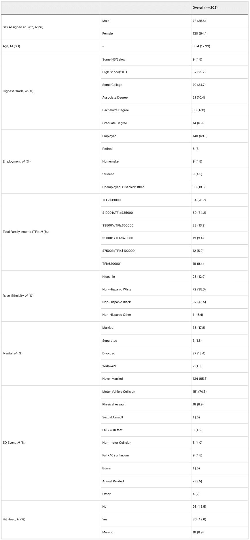
ED Emergency Department.

Childhood and lifetime trauma load among participants are detailed in Table 2 and distribution of childhood maltreatment load scores in Table S3. On average, participants endorsed greater than one moderate to extreme childhood maltreatment type, with emotional abuse (32.7%) being the most frequently endorsed followed by sexual abuse (24.8%) and emotional neglect (24.8%). There were no significant site differences in the prevalence of any maltreatment subtype or the average number of moderate to extreme maltreatment subtypes endorsed (Table S4). However, there were significant site differences in the modified total LEC score (p = 0.002). Associations between childhood maltreatment load, modified total LEC score, and 6-month symptom scores at each site are shown in Table S5. Participants did not significantly differ in 6-month PTSD, depression, anxiety, or dissociation symptoms across sites (Table S6). Of note, participants included in the present analyses reported significantly lower 6-month PTSD symptoms and total childhood trauma scores than those excluded due to MRI issues but did not differ in childhood maltreatment load (Supplementary Information).

Table 2 Childhood and lifetime trauma load.

Table 2

Note: Mlx Maltreatment; CTQ Childhood Trauma Questionnaire (11 item); mLEC-5 Score modified total Life Events Checklist Score.

Childhood maltreatment and white matter

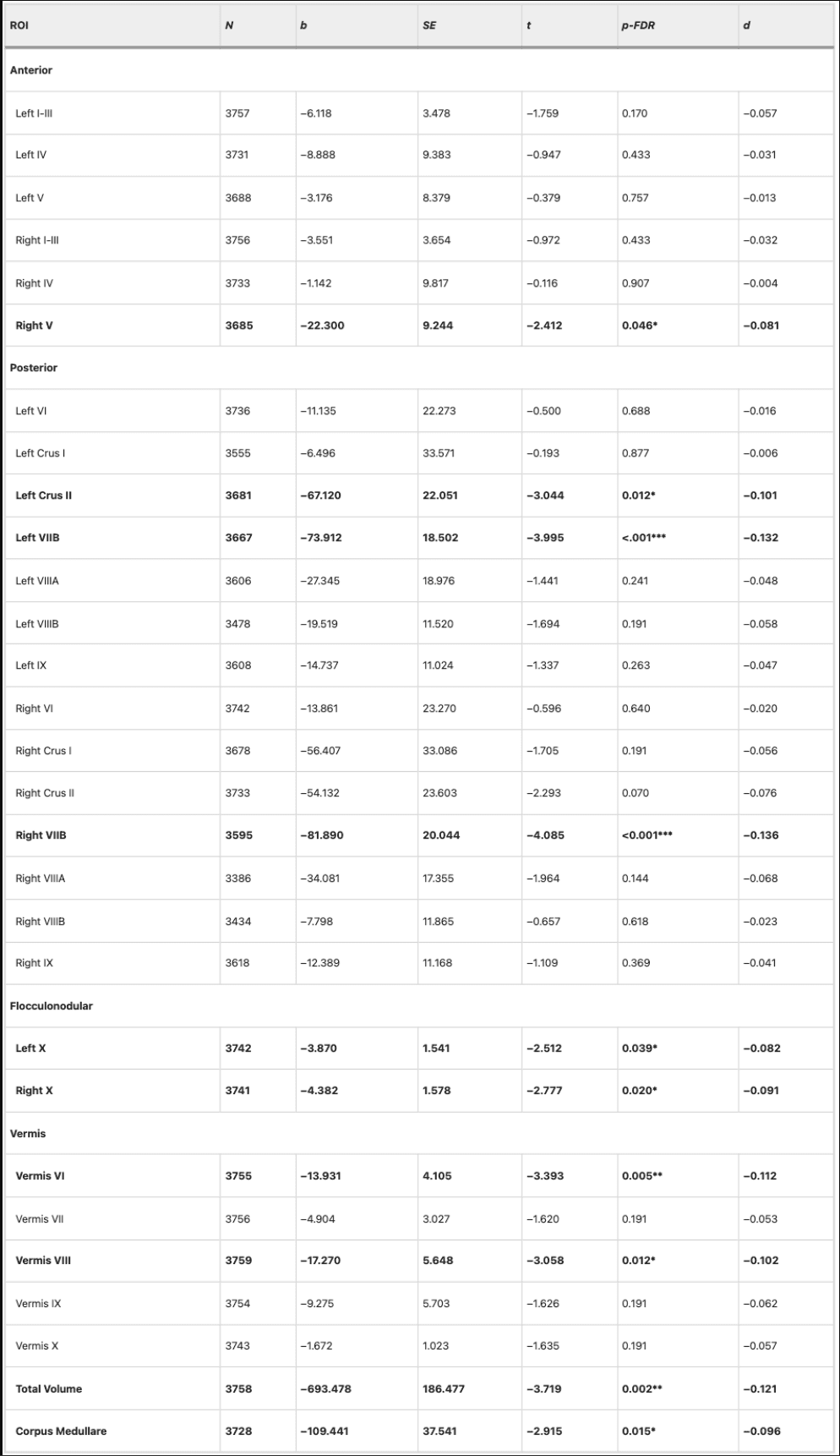
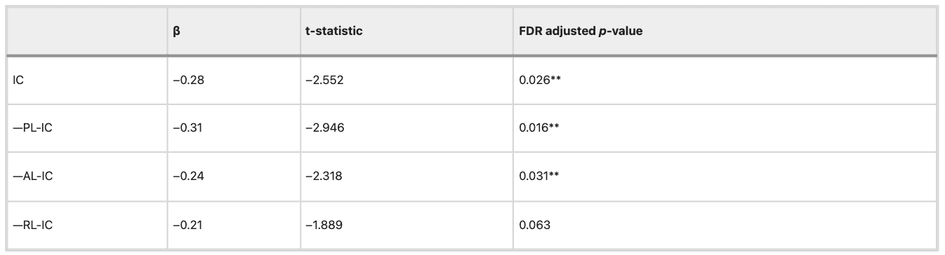
Childhood maltreatment load was associated with FA of several white matter tracts (Table 3). Following FDR correction, childhood maltreatment load negatively varied with FA in bilateral internal capsule (IC) at 2-weeks post-trauma, after covarying for sex assigned at birth, scanner site, and age (Table 3). Given the significant relationship between the IC and childhood maltreatment load, we considered the contribution of the IC subcomponents including the Posterior Limb of the IC (PL-IC), the Retrolenticular Part of the IC (RL-IC), and the Anterior Limb of the IC (AL-IC) in identical models and found childhood maltreatment load significantly negatively varied with all 3 IC subcomponents after FDR correction, though the PL-IC was the strongest contributor to the effect (Table 3; Fig. 1). Exploratory follow-up analyses with other diffusivity metrics revealed childhood maltreatment load was also associated with RD in the PL-IC (Table S7). Additional statistical analyses found only the threat (physical, emotional, and sexual abuse; β = −0.19, p = 0.01), not deprivation (emotional and physical neglect; β = −0.05, p = 0.53), component of childhood maltreatment load significantly contributed to the observed effect on the IC when both dimensions were included in an identical model as described above. The interaction between threat and deprivation was not significant. We further conducted sensitivity analyses to determine if associations between IC FA and childhood maltreatment remained while controlling for prior (i.e., endorsed pre-trauma) PCL-5 scores or mLEC-5 scores. Inclusion of either covariate did not impact the relationship between IC FA and childhood maltreatment load (see Supplementary information).

Table 3 Significant univariate effects in childhood maltreatment load model (2-Week).

Table 3

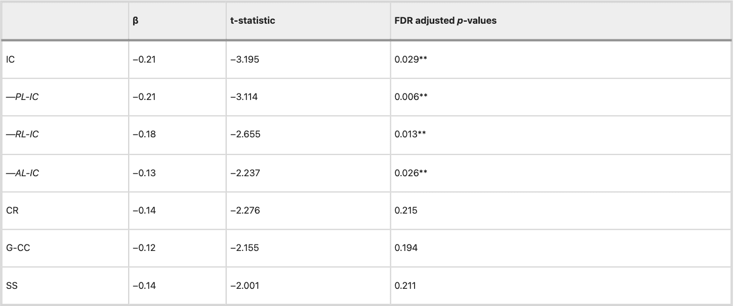
Reported tracts were significant at a nominal p < 0.05. Benjamini–Hochberg adjusted p-values were calculated by multiplying raw p-values by m/i (i = rank, m = total number of tests). Internal Capsule subcomponents were included in a follow-up, identical model, and FDR corrected with Benjamini–Hochberg. The PL-IC, RL-IC, and AL-IC were tested in follow-up models given the significant IC effect and separately FDR corrected. **Significant After Benjamini–Hochberg (p < 0.05) Adjustment. IC Internal Capsule, CR Corona Radiata, G-CC Genu of the Corpus Callosum, SS Sagittal Stratum, PL-IC Posterior Limb of the Internal Capsule, RL-IC Retrolenticular Part of Internal Capsule, AL-IC Anterior Limb of Internal Capsule.

Fig. 1: The relationship between childhood maltreatment load and internal capsule FA values at 2-weeks and 6-months.

Fig 1

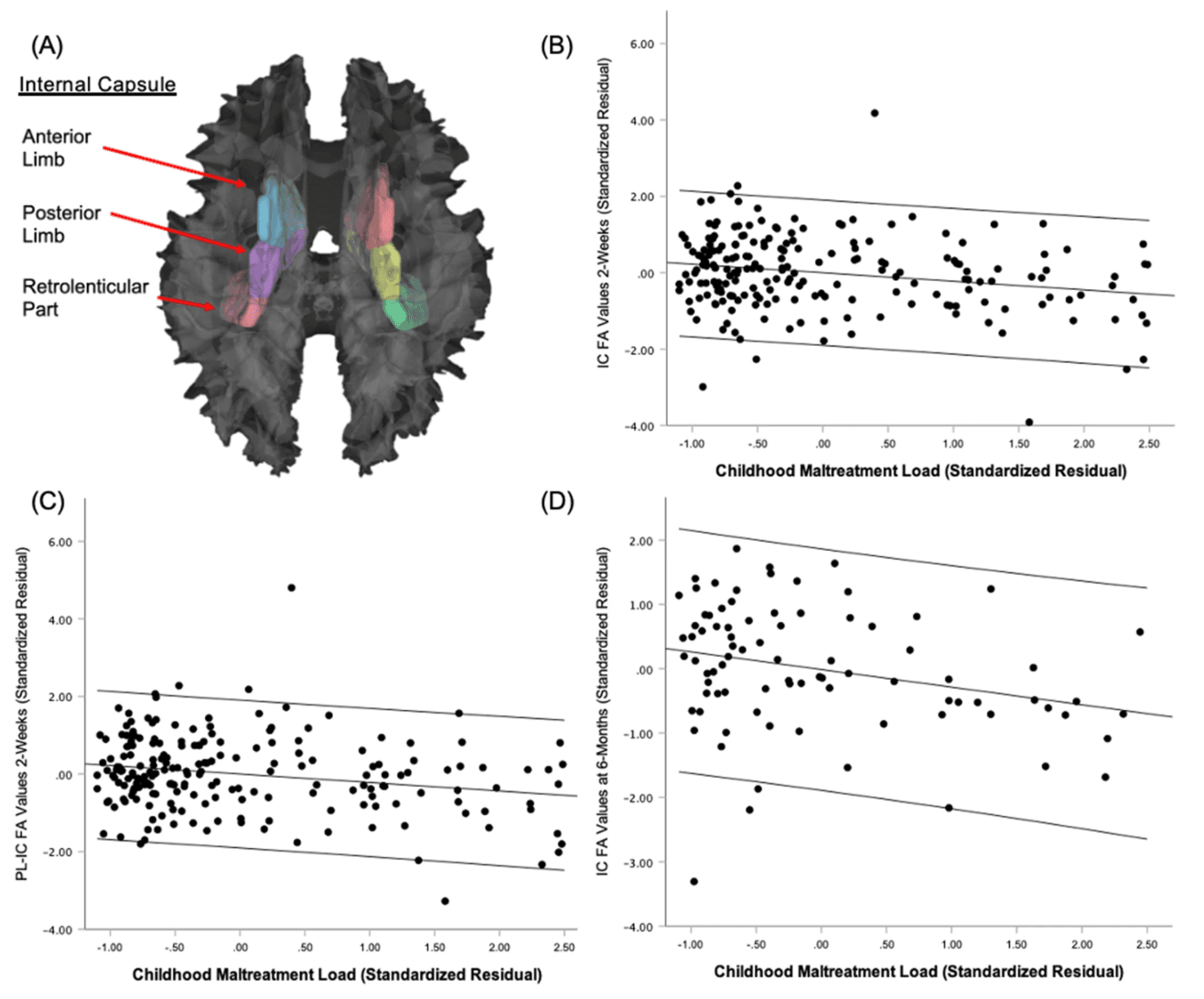
The internal capsule and its subcomponents are displayed on 3D rendering of human white matter tracts (A) Standardized residual plot of the regression of Childhood Maltreatment Load on 2-Week Internal Capsule FA Values depicts the significant negative effect (B). Standardized residual plot of Childhood Maltreatment Load on 2-Week Posterior-Limb of the Internal Capsule FA Values depicts the significant negative effect (C). Standardized residual plot of the regression of Childhood Maltreatment Load on Internal Capsule FA Values indexed 6-months post-trauma depicts the significant negative effect (D).

We performed follow-up analyses to test whether associations between childhood maltreatment and FA of the IC were also observed at 6-months post-trauma. Childhood maltreatment load negatively predicted bilateral IC FA indexed 6-months after trauma (Table 4; Fig. 1). In further analyses of IC subparts, negative predictive relationships of childhood maltreatment load with PL-IC and AL-IC microstructure were significant following Benjamini–Hochberg FDR correction (Table 4).

Table 4 Univariate effects in childhood maltreatment load model (6-Month).

Table 4

Benjamini–Hochberg adjusted p-values were calculated by multiplying raw p-values by m/i (i = rank, m = total number of tests). Internal Capsule subcomponents were included in a follow-up, identical model. The Benjamini–Hochberg FDR-correction included all 6-month tests. **Significant After Benjamini–Hochberg (p < 0.05) Adjustment. PL-IC Posterior Limb of the Internal Capsule, IC Internal Capsule, AL-IC Anterior Limb of Internal Capsule, RL-IC Retrolenticular Part of Internal Capsule.

Mediation analyses: childhood maltreatment, IC microstructure, and 6-month PTSD symptoms

Mediation analyses revealed a total effect of childhood maltreatment load on PCL-5 scores at 6-months (b = 1.75, SE = 0.78. 95% CI = [0.20, 3.29]). We found a significant indirect effect of childhood maltreatment load on 6-month PCL-5 scores through IC microstructure (b = 0.37, Boot SE = 0.18, 95% CI = [0.05, 0.76]) that completely mediated the effect of childhood maltreatment load on PCL-5 scores (b = 1.37, SE = 0.79, 95% CI = [−0.18, 2.93]) (Fig. 2). Similar analyses were performed with the total childhood maltreatment score, and we observed similar results (see Supplementary information).

Fig. 2: Mediation model of the effect of childhood maltreatment load on 6-month PCL-5 through Internal Capsule FA values indexed 2-weeks post-trauma.

Fig 2

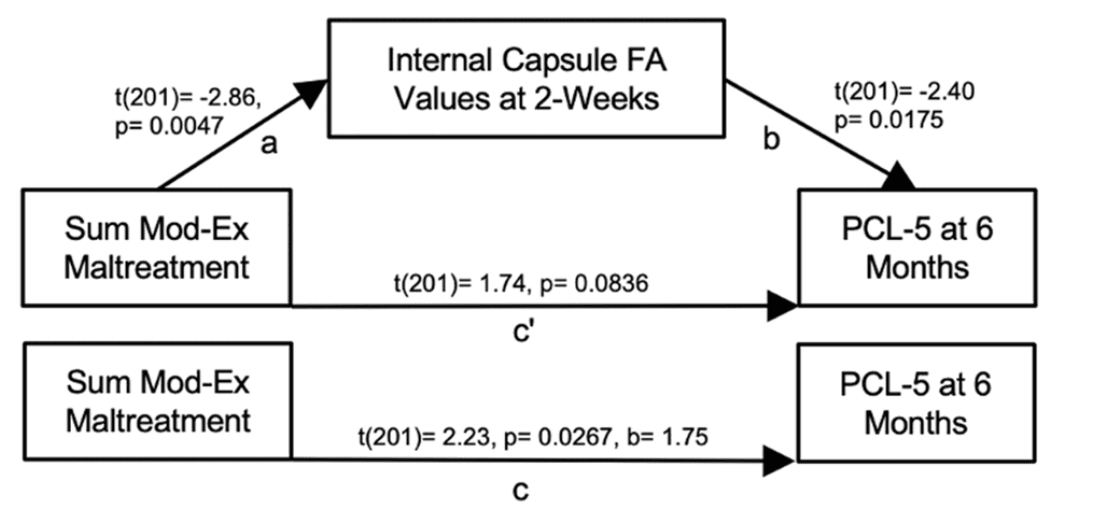
The indirect effect is significant based on a 5000 permutation, bootstrapped 95% confidence interval (i.e., path ab; b = 0.37, Boot SE = 0.18, 95% CI = [0.05, 0.76]), completely mediating the effect of childhood maltreatment load on PCL-5 scores (i.e., path c'; b = 1.37, SE = 0.79, 95% CI = [−0.18, 2.93]).

Exploratory mediation analyses assessed if findings were specific to future PTSD symptoms or if similar relationships were observed with other posttraumatic outcomes including depression, anxiety, and dissociation (Supplementary Information; Figure S1). Although there was a total effect of childhood maltreatment load on 6-month PROMIS-Depression (b = 1.29, SE = 0.48, 95% CI = [0.34, 2.24]), the indirect effect of childhood maltreatment load on 6-month PROMIS-Depression through IC microstructure (b = 0.15, Boot SE = 0.12, 95% CI = [−0.07, 0.39]) was not significant and did not mediate the effect of childhood maltreatment load on 6-month PROMIS-Depression scores (b = 1.14, SE = 0.49, 95% CI = [0.17, 2.11]). There was a total effect of childhood maltreatment load on 6-month PROMIS-Anxiety (b = 0.43, SE = 0.20, 95% CI = [0.05, 0.82]); however, neither the indirect effect of childhood maltreatment load on 6-month PROMIS-Anxiety through IC microstructure (b = 0.05, Boot SE = 0.05, 95% CI = [−0.04, 0.14]) nor the direct effect of childhood maltreatment load on 6-month PROMIS-Anxiety (b = 0.39, SE = 0.20, 95% CI = [−0.01, 0.78]) met statistical significance. No total effect of childhood maltreatment load on dissociation emerged (b = 0.14, SE = 0.08, 95% CI = [−0.01, 0.29]).

Discussion

To our knowledge, the present study is the first to investigate the relationship between childhood maltreatment load and white matter microstructure with posttraumatic symptoms in the early aftermath of trauma. We observed robust relationships between childhood maltreatment load and fractional anisotropy (FA) in the internal capsule (IC) in the early aftermath of an acute trauma event (2-weeks) and 6-months later. Furthermore, variations in IC FA values at 2-weeks fully mediated the relationship between childhood maltreatment load and later posttraumatic symptoms at 6-months. The mediation was specific to posttraumatic symptoms and not observed for depressive, anxiety, or dissociative symptomatology. Given that childhood maltreatment was related to IC microstructure at 2-weeks and 6-months following the adulthood traumatic event, our findings may point to IC FA values as a stable biomarker of later posttraumatic dysfunction and suggest a potential neurobiological pathway through which childhood trauma could confer risk for acute stress reactions in adulthood. Additionally, this study did not reproduce effects of white matter tracts previously found to vary with PTSD symptoms and childhood trauma exposure, including the cingulum bundle, uncinate fasciculus, and corpus callosum.

Our findings implicate the IC as a critical substrate for the effects of childhood trauma on PTSD development. The IC is a dense fiber bundle that contains several projections including the corticospinal tract, frontopontine and corticofugal fibers, the anterior and superior thalamic radiation, the optic radiation, and the auditory radiation. Anatomically, the IC is limited laterally by the pallidum and medially by the thalamus, the head of the caudate nucleus, and the corticospinal tract. The IC further appears to contain fibers for both medial (hippocampal formation, mammillary bodies, anterior thalamic nuclei, and cingulate gyrus) and basolateral (orbitofrontal cortex, dorsomedial thalamic nucleus, amygdala, and anterior temporal cortex) limbic circuits.

The present findings may be related to dysfunctional stimulus processing during PTSD. Although threat processing and its neural substrates are commonly dysregulated in PTSD, these components are dependent on the ability to perceive and integrate sensory stimuli. Recent work suggests variability in structure of visual processing regions, such as the ventral visual stream, is associated with susceptibility to PTSD symptom development. This pathway supports important processes, such as object recognition, integral to threat learning and includes core threat-related regions such as the amygdala and medial PFC. In the current study, we found that higher childhood maltreatment load was associated with lower FA of the IC and its subcomponents. The IC encompasses occipital connections between the higher order visual cortex and temporal lobe, as well as components of major motor tracts and somatosensory relays from the thalamus to the cortex. Prior work found trauma-exposed children and adults with childhood maltreatment histories had reduced FA of the IC and its component tracts, including the optic radiations and left anterior thalamic radiation. Speculatively, reduced FA of the IC may reflect disrupted white matter myelination and membrane integrity in fibers that transmit visual sensory information and contribute to altered perception and processing of threat-related information, which, in turn, may contribute to PTSD-related disruptions. Disruption of the IC could further be related to altered ability to consolidate, encode, or retrieve sensory components of trauma memories leading emotion dysregulation. In line with such reasoning, ischemic damage to the IC can lead to cognitive and behavioral alterations such as agitation and impaired attention, and deep brain stimulation of the ventral IC/ventral striatum enhances prefrontal cortex driven cognitive control. However, corresponding data on visual processing was not collected in the present study, and specific interpretations of IC function should thus be tempered. Taken together with prior literature, the present results suggest childhood maltreatment has a pronounced effect on IC microstructure which may confer risk for PTSD-related dysregulation following subsequent trauma.

Of note, we did not observe effects in canonical threat circuitry often associated with PTSD. Past studies have not typically considered childhood maltreatment when evaluating white matter markers of PTSD susceptibility, and it is possible that reduced IC FA may be a sequela of childhood maltreatment exposure. Moreover, although we analyzed imaging data from over 200 participants, we could have been underpowered to detect all effects with our unbiased whole-brain analytic approach. There are likely different biological subtypes of PTSD that are not accounted for here and such heterogeneity may have decreased our ability to detect associations in other tracts. For example, subtypes that show stronger intrusive symptoms or disruptions in emotional memory may be more associated with canonical threat neurocircuitry. Notably, varied white matter microstructure in the IC and its component tracts has been previously implicated in PTSD, with recent works suggesting a role in predicting PTSD in the acute aftermath of trauma exposure and in treatment response. Further investigation of the role of childhood maltreatment load in the relationship between white matter microstructure and PTSD development might assist in developing robust predictive models.

The findings of the current work should be interpreted with several considerations. First, we assessed childhood maltreatment load with a retrospective self-assessment. Since we did not query the age participants experienced childhood trauma, we could not assess the role of the developmental timing of trauma on the observed effects. Future longitudinal work is needed to investigate white matter microstructural variability in children and recently traumatized adults with more granular information on childhood trauma exposure and timing. Further, although we used items from the childhood trauma questionnaire, which is itself a validated and broadly used tool, and prospective research suggests the reliability of such retrospective reporting, we were unable to administer the full questionnaire as to minimize participant burden within the parent study. It would also be beneficial to investigate these associations in longitudinal studies of childhood trauma as opposed to using purely retrospective reports. Secondly, data that could be related to hypothesized contributions of the IC to sensory processes were not available, and thus the specific functional role of variability within IC microstructure in relation to PTSD is unclear. Future research considering the specific targets and functional outcomes of variable IC microstructure among those with childhood trauma would further clarify the present findings. Lastly, our analyses do not consider potential protective or socioeconomic factors that may contribute to early life stress load or resiliency. We did, however, consider the site at which MRI scanning occurred, which largely accounted for participant race and ethnicity. Given the relationships between racial discrimination, neighborhood disadvantage, and socioeconomic status with white matter microstructure, a critical next step will be to understand how these factors and protective agents impact white matter markers of PTSD susceptibility. As participants in this work reported substantial childhood maltreatment and low levels of PTSD symptoms prior to the presenting trauma, resiliency factors may be especially critical to decipher. Relatedly, it will also be important to consider potentially salient factors such as prenatal exposures and genetics.

The present study of recent trauma survivors examined the relationship of childhood maltreatment load with white matter microstructure and posttraumatic symptoms in the early aftermath of trauma. Childhood maltreatment load stably, inversely varied with the FA of the IC following the acute trauma event. Further, the FA of the IC in the early aftermath of an acute trauma event mediated the relationship between childhood maltreatment load and PTSD symptoms 6-months following the adulthood trauma exposure. These findings suggest a unique role for IC microstructure as a neural pathway between childhood trauma and future PTSD symptoms following a recent trauma. Furthermore, these data suggest DTI imaging may assist in revealing neural signatures of risk for later stress-related dysfunction in those with earlier childhood trauma.

Open Article as PDF

Abstract

Childhood trauma is a known risk factor for trauma and stress-related disorders in adulthood. However, limited research has investigated the impact of childhood trauma on brain structure linked to later posttraumatic dysfunction. We investigated the effect of childhood trauma on white matter microstructure after recent trauma and its relationship with future posttraumatic dysfunction among trauma-exposed adult participants (n = 202) recruited from emergency departments as part of the AURORA Study. Participants completed self-report scales assessing prior childhood maltreatment within 2-weeks in addition to assessments of PTSD, depression, anxiety, and dissociation symptoms within 6-months of their traumatic event. Fractional anisotropy (FA) obtained from diffusion tensor imaging (DTI) collected at 2-weeks and 6-months was used to index white matter microstructure. Childhood maltreatment load predicted 6-month PTSD symptoms (b = 1.75, SE = 0.78, 95% CI = [0.20, 3.29]) and inversely varied with FA in the bilateral internal capsule (IC) at 2-weeks (p = 0.0294, FDR corrected) and 6-months (p = 0.0238, FDR corrected). We observed a significant indirect effect of childhood maltreatment load on 6-month PTSD symptoms through 2-week IC microstructure (b = 0.37, Boot SE = 0.18, 95% CI = [0.05, 0.76]) that fully mediated the effect of childhood maltreatment load on PCL-5 scores (b = 1.37, SE = 0.79, 95% CI = [−0.18, 2.93]). IC microstructure did not mediate relationships between childhood maltreatment and depressive, anxiety, or dissociative symptomatology. Our findings suggest a unique role for IC microstructure as a stable neural pathway between childhood trauma and future PTSD symptoms following recent trauma. Notably, our work did not support roles of white matter tracts previously found to vary with PTSD symptoms and childhood trauma exposure, including the cingulum bundle, uncinate fasciculus, and corpus callosum. Given the IC contains sensory fibers linked to perception and motor control, childhood maltreatment might impact the neural circuits that relay and process threat-related inputs and responses to trauma.

Introduction

Childhood trauma increases the risk of developing trauma- and stress-related disorders in adulthood. Stressful experiences in early life can combine with adult stressors, making an individual more likely to experience conditions such as post-traumatic stress disorder (PTSD), severe depression, substance use, or behavioral issues. Additionally, childhood trauma is linked to changes in brain areas known to be involved in PTSD, which could indicate a tendency toward developing the disorder. However, limited research has explored how childhood trauma affects brain activity after an adult experiences a new trauma. Understanding these brain links between childhood trauma and acute stress reactions in adulthood could help predict and prevent PTSD.

PTSD is thought to stem partly from issues with brain circuits involved in learning about threats, specifically the prefrontal cortex, hippocampus, and amygdala, after a traumatic event. Connections between these brain regions, called white matter tracts (like the cingulum bundle, uncinate fasciculus, and fornix/stria terminalis), are also believed to play a role in PTSD-related problems, possibly due to changes in their structure from past experiences. Researchers have studied Fractional Anisotropy (FA), a measure derived from Diffusion Tensor Imaging (DTI), to assess white matter structure. A higher FA indicates more organized water movement in these tracts. Individuals with PTSD often show reduced FA in the cingulum bundle and uncinate fasciculus, although findings can vary. Successful therapy for PTSD seems to increase FA in tracts like the cingulum and fornix. Studies of recent trauma also suggest that changes in these tracts are linked to future PTSD development, with lower FA generally correlating with more severe PTSD symptoms. This past research suggests that white matter tracts in key threat-processing brain circuits are connected to the development and expression of PTSD symptoms.

Beyond these core threat circuits, new research on childhood and adult trauma suggests that PTSD-related white matter changes might also occur in other tracts. Previous DTI studies and meta-analyses show that childhood trauma is linked to FA changes in both threat-related and sensory integration tracts, such as the anterior thalamic radiation and superior longitudinal fasciculus. Recent meta-analyses from research groups found that the biggest FA reduction in individuals with PTSD was in the tapetum of the corpus callosum, not within traditional threat circuits. Proper perception and integration of sensory information are crucial for learning about threats. These findings suggest that trauma and PTSD-related FA reductions might extend beyond threat-related circuits to include areas essential for processing sensory input.

There is limited research on how childhood trauma, white matter structure, and post-traumatic outcomes after a recent trauma are related, but understanding this could improve our knowledge of PTSD's biological basis. Some studies have found links between childhood trauma, brain structure, and adult stressors. For example, the total amount of childhood trauma experienced changed how later combat exposure affected FA in the hippocampal part of the cingulum; more childhood trauma and combat exposure were linked to lower FA. In a long-term study of young adults, baseline uncinate fasciculus FA values influenced the connection between recent stressors (like a breakup or financial problems) and later mood and anxiety symptoms in those who reported more childhood maltreatment. However, limited work has considered potential links with white matter tracts outside the threat circuits, which might be important given recent findings of PTSD-related FA reductions.

This study investigated whether white matter structure in recent trauma survivors explained how childhood maltreatment led to post-traumatic difficulties. Considering previous findings on both PTSD threat circuits and newer work involving sensory and other white matter tracts, FA was assessed across all white matter tracts using a whole-brain approach. It was hypothesized that white matter FA at two weeks post-trauma would generally be negatively associated with the extent of childhood maltreatment. Furthermore, it was hypothesized that white matter FA linked to childhood maltreatment would explain the connections between childhood maltreatment and post-traumatic outcomes after a recent trauma. The findings highlight a brain pathway through which childhood trauma may increase the risk for acute stress reactions in adulthood and identify white matter markers that indicate a susceptibility to PTSD.

Materials and Methods

Participants

Participants were recruited as part of the AURORA study, a long-term, multi-site study of negative mental health consequences. Participants included in this investigation have been reported on previously. However, this is the first investigation to examine the relationships between childhood maltreatment, white matter structure, and later post-traumatic outcomes. As detailed in prior reports, enrollment occurred in emergency departments (EDs) and focused on individuals presenting within 72 hours of a qualifying trauma (physical or sexual assault, motor vehicle accident, fall over 10 feet, mass casualty incident, or other life-threatening traumatic event reported on a screener question and agreed upon by study staff). Participants were included if they were English-speaking, 18 to 75 years old, and able to consent and follow study procedures. Participants were recruited regardless of previous PTSD symptoms or diagnosis and were asked to report retrospectively on prior PTSD (and other disorders) symptoms in the emergency department. General exclusion criteria for the AURORA study have been described previously. MRI collection exclusion criteria included having metal implants, a history of seizure or epilepsy, a history of Parkinson’s disease, dementia, or Alzheimer’s disease, current pregnancy, and/or declining to complete the MRI. From September 2017 to July 2020, MRI data were collected within approximately two weeks of trauma exposure for 439 participants, and DTI data were available from 353 participants. Participants were excluded for MRI quality issues (n = 37), such as motion artifacts or low-quality data. The current analyses focused on participants who completed both DTI and the abbreviated Childhood Trauma Questionnaire at two weeks and post-traumatic outcome measures at six months post-trauma; participants missing a required questionnaire were excluded (n = 153). A total of 202 participants were included in the final analyses. Further DTI data analyses from a subset of 85 participants (111 collected, 26 excluded) collected at a six-month follow-up imaging session were also completed. All participants provided informed consent as approved by the Biomedical IRB at UNC Chapel Hill, the central IRB for all study sites.

Baseline Surveys and Socio-Demographics

Participants completed a baseline assessment in the emergency department that included self-reported trauma characteristics and demographic information. Age, sex assigned at birth, race/ethnicity, highest education level, marital status, employment status, and total household income were collected at the ED baseline surveys. Participants were also asked if they hit their head or experienced a head injury during the event that brought them to the ED (n = 86 endorsed).

Childhood Maltreatment Load

An abbreviated 11-item version of the Childhood Trauma Questionnaire—Short Form (CTQ-SF) was used to measure childhood maltreatment. Items were selected from the CTQ-SF to cover maltreatment subtypes while minimizing the burden on participants. The selected items showed strong internal consistency (Cronbach’s a = 0.92). The questionnaire was given two weeks after the trauma. Participants self-reported on a 5-point Likert scale (0: never, 1: rarely, 2: sometimes, 3: often, 4: very often). The maltreatment subtypes assessed included emotional abuse (score range: 0 to 8), physical abuse (score range: 0 to 8), sexual abuse (score range: 0 to 12), emotional neglect (score range: 0 to 8), and physical neglect (score range: 0 to 8). Total possible scores ranged from 0 to 44. Childhood maltreatment load was measured by how often participants reported moderate to extreme levels of each maltreatment subtype. Moderate to extreme abuse or neglect for most subtypes was defined as a subtype score of 4 or above. Moderate to extreme sexual abuse was defined as a sub-score of 3 or above. These cutoffs were adjusted for the abbreviated assessment from previously suggested clinical cutoffs. The sum of moderate to extreme maltreatment types was used to calculate the total childhood maltreatment load.

Lifetime Trauma

Lifetime trauma exposure was assessed using the Life Events Checklist (LEC-5), a standard 17-item tool that evaluates exposure to 17 traumatic life events. Participants completed the LEC-5 eight weeks after their trauma. They indicated if a traumatic experience happened to them personally, if they witnessed it, learned about it happening to someone close to them, or were exposed to details about it through their job. A modified total LEC score (mLEC-5, range: 0 to 17) was calculated by adding up the types of traumatic events reported, regardless of how they were experienced. Even if participants reported experiencing an event in multiple ways (e.g., "happened to me," "witnessed it"), any exposure to a given traumatic event resulted in the maximum score of one for the modified total LEC score.

Posttraumatic Outcomes

Posttraumatic dysfunction was assessed by examining symptoms of PTSD, depression, anxiety, and dissociation six months after the initial trauma. PTSD symptoms were measured using the Posttraumatic Stress Disorder (PTSD) checklist for DSM-5 (PCL-5), a reliable 20-item questionnaire on symptom presence and severity. Participants rated symptom severity on a scale from 0 (not at all) to 4 (extremely). Depression symptoms were assessed with the 8-item Patient-Reported Outcomes Measurement Information System (PROMIS) Depression instrument, short form 8b. A total raw score was calculated by summing individual items and then converted to a T-score. Anxiety symptoms were assessed with four items from the PROMIS Anxiety bank. Participants rated how often they felt tense, worried, had trouble relaxing, or felt anxious on a scale from 1 (none of the time) to 5 (all or almost all of the time), and item scores were summed to create a total anxiety score. Dissociation was assessed using a modified 2-item Brief Dissociative Experiences Scale (DES-B-Modified). Participants rated how often they felt people, objects, or the world around them seemed unreal, and how often they felt they were looking through a fog so that people and things seemed unclear, on a scale from 1 (none of the time) to 5 (all or almost all of the time). A sum of the two questions served as an index of dissociation severity.

Diffusion Tensor Imaging

Diffusion weighted imaging (DWI) data were collected across five sites. Data processing followed recommendations from the ENIGMA consortium. To ensure data quality, raw data were visually inspected, and metrics of temporal signal-to-noise ratio and outlier maximum voxel intensity were calculated. Participants with both (a) TSNR values lower than 4.88 and (b) maximum voxel intensities greater than 5000 were removed to retain the maximum number of participants while removing low-quality data. Briefly, motion and eddy current effects in the DWI data were reduced using FSL’s ‘eddy’ subroutine, and susceptibility effects were corrected using nonlinear warping of the DWI data to the participant’s T1-weighted anatomical scan. Tract-Based Spatial Statistics (TBSS) processing, as implemented in the ENIGMA-DTI working group standards, was used to extract FA values across white matter regions. First, FA maps were non-linearly registered to the standard ENIGMA FA map in Montreal Neurological Institute (MNI) standard space. The ENIGMA FA skeleton map was then projected onto each subject's FA maps in standard space. Finally, regional FA values were extracted from the John’s Hopkins University (JHU) White matter atlas and used in group-level analyses. Axial diffusivity (AD), radial diffusivity (RD), and mean diffusivity (MD) were also extracted for exploratory follow-up analyses.

Statistical Analysis

Statistical analyses were performed using IBM SPSS Statistics for Macintosh, Version 28. Participant demographics, trauma histories, and symptoms were evaluated with chi-square tests, Pearson’s correlations, and independent sample t-tests for differences across imaging sites. Linear regressions, controlling for MRI scanner site, age, and sex at birth, assessed the effects of childhood maltreatment load and post-traumatic outcomes on FA in bilateral white matter tracts. These tests were conducted for the 18 individual white matter tracts included in the JHU atlas. FA was examined due to its prevalence in the literature. Relationships with AD, RD, and MD were explored in follow-up analyses for significant tracts in the FA analysis. Identical follow-up tests evaluated the contribution of the subcomponents of tracts significantly associated with childhood maltreatment load. A nominal significance threshold was set at p < 0.05, two-tailed. False discovery rate (FDR) correction using the Benjamini–Hochberg method was used to control for multiple comparisons and maintain α = 0.05. For statistically significant models where subcomponent data were available (e.g., the anterior limb of the internal capsule), identical follow-up models were completed with separate FDR correction using the Benjamini–Hochberg method. Linear models, controlling for MRI scanner site, age, and sex at birth, evaluated the effects of summed exposure to moderate to extreme threat (physical, emotional, and sexual abuse) and deprivation (emotional and physical neglect) components of childhood maltreatment load, as well as their interaction, on major bilateral white matter tracts significantly associated with childhood maltreatment load. Tracts that showed a significant association with childhood trauma were also included in subsequent mediation analyses, conducted using the PROCESS macro version 4, including childhood trauma load, post-traumatic outcomes at six months, and a mediator of white matter microstructure. For mediation analyses, bootstrapping with 5000 permutations was completed to obtain 95% bias-corrected confidence intervals as an inferential test of direct and indirect effects. Lastly, univariate effects of childhood maltreatment load on six-month bilateral white matter tracts significantly associated with childhood maltreatment at two weeks were evaluated with ANOVA, in models controlling for scanner site, age, and sex assigned at birth.

Results

Participant Characteristics

Participant demographics and trauma characteristics are detailed in Table 1. Samples from the imaging sites were well-matched across sex assigned at birth, age, educational attainment, employment, total family income, and marital status. Furthermore, each MRI scanning site sample had similar distributions of participants’ qualifying traumas and proportions of individuals who hit their head during the trauma. Participant racial/ethnic identity differed significantly by site (p < 0.001).

Childhood and lifetime trauma load among participants are detailed in Table 2, and the distribution of childhood maltreatment load scores in Table S3. On average, participants reported more than one moderate to extreme childhood maltreatment type, with emotional abuse (32.7%) being the most frequently reported, followed by sexual abuse (24.8%) and emotional neglect (24.8%). There were no significant site differences in the prevalence of any maltreatment subtype or the average number of moderate to extreme maltreatment subtypes reported. However, there were significant site differences in the modified total LEC score (p = 0.002). Associations between childhood maltreatment load, modified total LEC score, and six-month symptom scores at each site are shown in Table S5. Participants did not significantly differ in six-month PTSD, depression, anxiety, or dissociation symptoms across sites. Notably, participants included in the current analyses reported significantly lower six-month PTSD symptoms and total childhood trauma scores than those excluded due to MRI issues but did not differ in childhood maltreatment load.

Childhood Maltreatment and White Matter

Childhood maltreatment load was associated with FA in several white matter tracts (Table 3). After correcting for false discovery rate (FDR), childhood maltreatment load was negatively related to FA in the bilateral internal capsule (IC) at two weeks post-trauma, after adjusting for sex assigned at birth, scanner site, and age (Table 3). Given this significant relationship, the contributions of the IC subcomponents—including the Posterior Limb of the IC (PL-IC), the Retrolenticular Part of the IC (RL-IC), and the Anterior Limb of the IC (AL-IC)—were examined in similar models. Childhood maltreatment load was found to be significantly negatively related to all three IC subcomponents after FDR correction, with the PL-IC being the strongest contributor to the effect (Table 3; Fig. 1). Exploratory follow-up analyses with other diffusivity metrics showed that childhood maltreatment load was also associated with RD in the PL-IC (Table S7). Additional statistical analyses found that only the threat component of childhood maltreatment load (physical, emotional, and sexual abuse; β = −0.19, p = 0.01), not the deprivation component (emotional and physical neglect; β = −0.05, p = 0.53), significantly contributed to the observed effect on the IC when both dimensions were included in the same model. The interaction between threat and deprivation was not significant. Sensitivity analyses were also conducted to determine if the associations between IC FA and childhood maltreatment remained when controlling for prior (i.e., reported pre-trauma) PCL-5 scores or mLEC-5 scores. Including either covariate did not affect the relationship between IC FA and childhood maltreatment load.

Follow-up analyses were performed to test whether associations between childhood maltreatment and FA of the IC were also observed at six months post-trauma. Childhood maltreatment load negatively predicted bilateral IC FA indexed six months after trauma (Table 4; Fig. 1). In further analyses of IC subparts, negative predictive relationships of childhood maltreatment load with PL-IC and AL-IC microstructure were significant following Benjamini–Hochberg FDR correction (Table 4).

Mediation Analyses: Childhood Maltreatment, IC Microstructure, and 6-Month PTSD Symptoms

Mediation analyses revealed a total effect of childhood maltreatment load on PCL-5 scores at six months (b = 1.75, SE = 0.78, 95% CI = [0.20, 3.29]). A significant indirect effect of childhood maltreatment load on six-month PCL-5 scores through IC microstructure was found (b = 0.37, Boot SE = 0.18, 95% CI = [0.05, 0.76]). This effect fully explained the relationship between childhood maltreatment load and PCL-5 scores (b = 1.37, SE = 0.79, 95% CI = [−0.18, 2.93]) (Fig. 2). Similar results were observed when analyses were performed with the total childhood maltreatment score.

Exploratory mediation analyses assessed whether these findings were specific to future PTSD symptoms or if similar relationships were observed with other post-traumatic outcomes, including depression, anxiety, and dissociation. Although there was a total effect of childhood maltreatment load on six-month PROMIS-Depression (b = 1.29, SE = 0.48, 95% CI = [0.34, 2.24]), the indirect effect of childhood maltreatment load on six-month PROMIS-Depression through IC microstructure (b = 0.15, Boot SE = 0.12, 95% CI = [−0.07, 0.39]) was not significant and did not explain the effect of childhood maltreatment load on six-month PROMIS-Depression scores (b = 1.14, SE = 0.49, 95% CI = [0.17, 2.11]). There was a total effect of childhood maltreatment load on six-month PROMIS-Anxiety (b = 0.43, SE = 0.20, 95% CI = [0.05, 0.82]); however, neither the indirect effect of childhood maltreatment load on six-month PROMIS-Anxiety through IC microstructure (b = 0.05, Boot SE = 0.05, 95% CI = [−0.04, 0.14]) nor the direct effect of childhood maltreatment load on six-month PROMIS-Anxiety (b = 0.39, SE = 0.20, 95% CI = [−0.01, 0.78]) reached statistical significance. No total effect of childhood maltreatment load on dissociation emerged (b = 0.14, SE = 0.08, 95% CI = [−0.01, 0.29]).

Discussion

This study is the first to investigate the relationship between childhood maltreatment load, white matter structure, and post-traumatic symptoms soon after a trauma. Strong relationships were observed between childhood maltreatment load and fractional anisotropy (FA) in the internal capsule (IC) at two weeks and six months following an acute traumatic event. Furthermore, variations in IC FA values at two weeks fully explained the relationship between childhood maltreatment load and later post-traumatic symptoms at six months. This mediating effect was specific to post-traumatic symptoms and was not observed for depressive, anxiety, or dissociative symptoms. Given that childhood maltreatment was related to IC microstructure both at two weeks and six months after the adult traumatic event, these findings suggest that IC FA values might serve as a stable marker for later post-traumatic dysfunction, pointing to a potential brain pathway through which childhood trauma could increase the risk for acute stress reactions in adulthood. Additionally, this study did not replicate previously found effects in white matter tracts often associated with PTSD symptoms and childhood trauma exposure, such as the cingulum bundle, uncinate fasciculus, and corpus callosum.

The findings indicate that the IC plays a crucial role in how childhood trauma affects the development of PTSD. The IC is a dense bundle of nerve fibers containing various connections, including those for movement, sensation, and vision. Anatomically, the IC is bordered by the pallidum laterally and the thalamus medially, along with the head of the caudate nucleus and the corticospinal tract. It also appears to contain fibers for both medial (hippocampal formation, mammillary bodies, anterior thalamic nuclei, and cingulate gyrus) and basolateral (orbitofrontal cortex, dorsomedial thalamic nucleus, amygdala, and anterior temporal cortex) limbic circuits.

These findings might relate to problems with how stimuli are processed in PTSD. While threat processing and its brain networks are often impaired in PTSD, these functions depend on the ability to perceive and integrate sensory information. Recent research suggests that variations in the structure of visual processing regions, such as the ventral visual stream, are linked to a susceptibility to developing PTSD symptoms. This pathway supports important processes like object recognition, which are vital for learning about threats and include key threat-related regions such as the amygdala and medial prefrontal cortex. In the current study, higher childhood maltreatment load was associated with lower FA of the IC and its subcomponents. The IC includes connections between the higher-order visual cortex and the temporal lobe, as well as parts of major motor tracts and sensory relays from the thalamus to the cortex. Previous work found that trauma-exposed children and adults with histories of childhood maltreatment had reduced FA in the IC and its component tracts, including the optic radiations and left anterior thalamic radiation. It is possible that reduced FA of the IC reflects disrupted white matter insulation and membrane integrity in fibers that transmit visual sensory information, contributing to altered perception and processing of threat-related information, which, in turn, may contribute to PTSD-related disruptions. Disruption of the IC could also be related to an altered ability to consolidate, encode, or retrieve sensory components of trauma memories, leading to emotion dysregulation. In line with this, ischemic damage to the IC can lead to cognitive and behavioral changes such as agitation and impaired attention, and deep brain stimulation of the ventral IC/ventral striatum enhances cognitive control driven by the prefrontal cortex. However, corresponding data on visual processing were not collected in the present study, so specific interpretations of IC function should be cautious. Taken with prior literature, these results suggest that childhood maltreatment has a significant effect on IC microstructure, which may increase the risk for PTSD-related problems after subsequent trauma.

Notably, effects were not observed in the canonical threat circuitry often associated with PTSD. Past studies have not typically considered childhood maltreatment when evaluating white matter markers of PTSD susceptibility, and it is possible that reduced IC FA may be a consequence of childhood maltreatment exposure. Moreover, although imaging data from over 200 participants were analyzed, the study might have been underpowered to detect all effects with the unbiased whole-brain analytic approach. There are likely different biological subtypes of PTSD that are not accounted for here, and such variability may have decreased the ability to detect associations in other tracts. For example, subtypes that show stronger intrusive symptoms or disruptions in emotional memory may be more associated with canonical threat neurocircuitry. Notably, varied white matter microstructure in the IC and its component tracts has been previously implicated in PTSD, with recent works suggesting a role in predicting PTSD soon after trauma exposure and in treatment response. Further investigation of the role of childhood maltreatment load in the relationship between white matter microstructure and PTSD development might help in developing robust predictive models.

The findings of this work should be interpreted with several considerations. First, childhood maltreatment load was assessed using a retrospective self-assessment. Since the age at which participants experienced childhood trauma was not inquired, the role of the developmental timing of trauma on the observed effects could not be assessed. Future long-term research is needed to investigate white matter microstructural variability in children and recently traumatized adults with more detailed information on childhood trauma exposure and timing. Furthermore, although items from the Childhood Trauma Questionnaire, a validated and widely used tool, were used, the full questionnaire could not be administered to minimize participant burden within the parent study, despite prospective research suggesting the reliability of such retrospective reporting. It would also be beneficial to investigate these associations in longitudinal studies of childhood trauma rather than relying purely on retrospective reports. Secondly, data related to hypothesized contributions of the IC to sensory processes were not available, thus the specific functional role of variability within IC microstructure in relation to PTSD remains unclear. Future research considering the specific targets and functional outcomes of variable IC microstructure among individuals with childhood trauma would further clarify the present findings. Lastly, the analyses do not consider potential protective or socioeconomic factors that may contribute to early life stress or resilience. However, the site at which MRI scanning occurred, which largely accounted for participant race and ethnicity, was considered. Given the relationships between racial discrimination, neighborhood disadvantage, and socioeconomic status with white matter microstructure, a critical next step will be to understand how these factors and protective agents impact white matter markers of PTSD susceptibility. As participants in this work reported substantial childhood maltreatment and low levels of PTSD symptoms prior to the presenting trauma, resilience factors may be especially critical to understand. Relatedly, it will also be important to consider potentially relevant factors such as prenatal exposures and genetics.

This study of recent trauma survivors examined the relationship between childhood maltreatment load, white matter microstructure, and post-traumatic symptoms soon after trauma. Childhood maltreatment load was consistently and inversely related to the FA of the IC following the acute traumatic event. Furthermore, the FA of the IC soon after an acute traumatic event explained the relationship between childhood maltreatment load and PTSD symptoms six months after the adult trauma exposure. These findings suggest a unique role for IC microstructure as a neural pathway between childhood trauma and future PTSD symptoms following a recent trauma. Additionally, these data suggest that DTI imaging may help reveal brain markers of risk for later stress-related dysfunction in those with earlier childhood trauma.

Open Article as PDF

Abstract

Childhood trauma is a known risk factor for trauma and stress-related disorders in adulthood. However, limited research has investigated the impact of childhood trauma on brain structure linked to later posttraumatic dysfunction. We investigated the effect of childhood trauma on white matter microstructure after recent trauma and its relationship with future posttraumatic dysfunction among trauma-exposed adult participants (n = 202) recruited from emergency departments as part of the AURORA Study. Participants completed self-report scales assessing prior childhood maltreatment within 2-weeks in addition to assessments of PTSD, depression, anxiety, and dissociation symptoms within 6-months of their traumatic event. Fractional anisotropy (FA) obtained from diffusion tensor imaging (DTI) collected at 2-weeks and 6-months was used to index white matter microstructure. Childhood maltreatment load predicted 6-month PTSD symptoms (b = 1.75, SE = 0.78, 95% CI = [0.20, 3.29]) and inversely varied with FA in the bilateral internal capsule (IC) at 2-weeks (p = 0.0294, FDR corrected) and 6-months (p = 0.0238, FDR corrected). We observed a significant indirect effect of childhood maltreatment load on 6-month PTSD symptoms through 2-week IC microstructure (b = 0.37, Boot SE = 0.18, 95% CI = [0.05, 0.76]) that fully mediated the effect of childhood maltreatment load on PCL-5 scores (b = 1.37, SE = 0.79, 95% CI = [−0.18, 2.93]). IC microstructure did not mediate relationships between childhood maltreatment and depressive, anxiety, or dissociative symptomatology. Our findings suggest a unique role for IC microstructure as a stable neural pathway between childhood trauma and future PTSD symptoms following recent trauma. Notably, our work did not support roles of white matter tracts previously found to vary with PTSD symptoms and childhood trauma exposure, including the cingulum bundle, uncinate fasciculus, and corpus callosum. Given the IC contains sensory fibers linked to perception and motor control, childhood maltreatment might impact the neural circuits that relay and process threat-related inputs and responses to trauma.

Summary

Childhood trauma is a known risk factor for developing trauma and stress-related disorders in adulthood. This early life stress can combine with adult stressors, increasing the likelihood of conditions such as post-traumatic stress disorder (PTSD), depression, substance use, or behavioral issues. Research indicates that childhood trauma is linked to differences in brain circuits involved in PTSD, which might be indicators of how susceptible someone is to the disorder. However, few studies have explored the specific brain changes that connect childhood trauma to stronger reactions after an adult trauma. Identifying these brain-based links could help predict and prevent PTSD.

PTSD is partly thought to stem from problems in brain circuits involved in learning about threats, especially the prefrontal cortex, hippocampus, and amygdala, after a traumatic event. White matter tracts like the cingulum bundle, uncinate fasciculus, and fornix/stria terminalis connect these threat-related brain regions. These tracts are believed to be involved in PTSD dysfunction, possibly due to changes in their structure resulting from experiences. Studies using Diffusion Tensor Imaging (DTI) have measured Fractional Anisotropy (FA) to understand white matter structure. Higher FA suggests more organized water flow in nerve fibers. Individuals with PTSD often show reduced FA in certain tracts, though results can vary. Successful therapy for PTSD seems to increase FA in some tracts. Research on recent trauma also suggests that differences in these tracts relate to future PTSD symptoms, with lower FA generally linked to more severe symptoms. This past work indicates that white matter tracts in core threat circuits are involved in how PTSD symptoms develop and appear.

Beyond the threat-related brain circuits, newer research on childhood and adult trauma suggests that PTSD-related white matter changes might also occur in other tracts. DTI studies have shown that childhood trauma is linked to FA changes in both threat-related and sensory integration tracts. Recent large-scale analyses also found that the biggest reduction in FA for people with PTSD was not in threat-related tracts, but in the tapetum of the corpus callosum. The brain needs to correctly perceive and integrate sensory information for proper threat learning. These findings suggest that trauma and PTSD-related FA reductions might extend beyond threat circuits to areas crucial for sensing stimuli.

Limited research exists on how childhood trauma, white matter structure, and post-traumatic outcomes after a recent trauma are interconnected, though understanding this could improve our knowledge of PTSD's biological basis. However, studies have found links between childhood trauma, brain structure, and adult stressors. For instance, the amount of childhood trauma experienced influenced how later combat exposure affected FA in the hippocampus's cingulum, with more childhood trauma and combat linked to decreased FA. Another study found that uncinate fasciculus FA at the start of the study influenced the relationship between recent stressors and mood/anxiety symptoms in young adults who reported more childhood maltreatment. However, few studies have looked at white matter tracts outside the main threat circuits, which might be important based on recent findings about PTSD-related FA reductions.

The current study examined whether white matter structure in recent trauma survivors explained how childhood maltreatment affected post-traumatic problems. Considering past findings in PTSD threat circuits and newer work involving sensory and other white matter tracts, the study used a whole-brain approach to assess FA across white matter tracts. Researchers predicted that white matter FA two weeks after trauma would generally be negatively associated with the amount of childhood maltreatment. They also hypothesized that white matter FA linked to childhood maltreatment would explain the connection between childhood maltreatment and post-traumatic outcomes after a recent trauma. The findings aim to highlight a brain pathway through which childhood trauma might increase the risk for acute stress reactions in adulthood, shedding light on white matter indicators of PTSD susceptibility.

Materials and Methods

Participants

Participants were recruited from emergency departments within 72 hours of a qualifying trauma (e.g., assault, car accident). English-speaking adults aged 18 to 75 who could consent were included, regardless of prior PTSD history. Exclusion criteria for MRI included metal implants, seizure history, certain neurological conditions, pregnancy, or refusal to complete the MRI. MRI data were collected approximately two weeks post-trauma for 439 participants, with DTI data available for 353. After removing data with quality issues (37 participants) and those missing required questionnaires (153 participants), 202 participants were included in the final analyses. A subset of 85 participants also had DTI data collected at a six-month follow-up. All participants gave informed consent.

Baseline Surveys and Socio-demographics

Participants completed surveys in the emergency department, collecting self-reported trauma characteristics and demographic information such as age, sex, race/ethnicity, education, marital status, employment, and household income. Participants also reported if they experienced a head injury during the trauma.

Childhood Maltreatment Load

An 11-item version of the Childhood Trauma Questionnaire—Short Form (CTQ-SF) was used to measure childhood maltreatment, administered two weeks after the trauma. Items were selected to cover different types of maltreatment (emotional, physical, sexual abuse; emotional, physical neglect) with high internal reliability. Scores ranged from 0 to 44. Childhood maltreatment load was defined by the number of moderate to extreme levels endorsed for each maltreatment subtype, using adjusted cutoffs. The total sum of these endorsed subtypes represented the overall childhood maltreatment load.

Lifetime Trauma

Lifetime trauma exposure was measured using the 17-item Life Events Checklist (LEC-5) at eight weeks post-trauma. Participants indicated if they personally experienced, witnessed, learned about, or were exposed to details of 17 different traumatic events. A modified total LEC score (0 to 17) was calculated by summing the types of events endorsed, regardless of how they were experienced.

Post-traumatic Outcomes

Post-traumatic dysfunction was assessed at six months post-trauma, including symptoms of PTSD, depression, anxiety, and dissociation. PTSD symptoms were measured with the 20-item PCL-5. Depression symptoms were assessed with the 8-item PROMIS Depression instrument. Anxiety symptoms were measured with 4 items from the PROMIS Anxiety bank. Dissociation was assessed using a modified 2-item Brief Dissociative Experiences Scale (DES-B-Modified).

Diffusion Tensor Imaging

Diffusion-weighted imaging (DWI) data were collected across five sites and processed according to ENIGMA consortium recommendations. Data underwent visual inspection and quality control checks for signal-to-noise ratio and voxel intensity. Motion and eddy current effects were reduced using FSL, and susceptibility effects were corrected. Tract-Based Spatial Statistics (TBSS) was used to extract Fractional Anisotropy (FA) values across white matter regions. FA maps were registered to a standard space, and regional FA values were extracted from the John's Hopkins University (JHU) White matter atlas for analysis. Axial, radial, and mean diffusivity were also extracted for exploratory analyses.

Statistical Analysis

Statistical analyses used IBM SPSS Statistics. Participant demographics, trauma histories, and symptoms were compared across imaging sites using chi-square tests, Pearson’s correlations, and t-tests. Linear regressions, controlling for MRI scanner site, age, and sex, assessed the effects of childhood maltreatment load and post-traumatic outcomes on FA in 18 bilateral white matter tracts from the JHU atlas. A nominal significance threshold of p < 0.05 was used, with False Discovery Rate (FDR) correction applied for multiple comparisons. Follow-up analyses examined significant tracts and their subcomponents, and the contributions of different childhood maltreatment types (threat vs. deprivation). Sensitivity analyses checked if results held when controlling for prior PTSD symptoms or lifetime trauma. Mediation analyses were conducted to see if white matter microstructure explained the link between childhood maltreatment load and six-month post-traumatic outcomes, specifically PTSD symptoms. Bootstrapping was used for confidence intervals in mediation analyses.

Results

Participant Characteristics

Demographic and trauma characteristics of participants are detailed in tables. Imaging sites were largely similar across sex, age, education, employment, income, and marital status. The types of qualifying traumas and head injury rates were also similar across sites. Participant racial/ethnic identity did differ significantly by site.

Childhood and Lifetime Trauma

Tables show childhood and lifetime trauma loads among participants. On average, participants reported more than one moderate to extreme childhood maltreatment type, with emotional abuse being the most common, followed by sexual abuse and emotional neglect. There were no significant site differences in the prevalence of maltreatment types. However, there were significant site differences in the modified total LEC score. There were no significant site differences in 6-month PTSD, depression, anxiety, or dissociation symptoms. Participants included in the final analyses reported lower 6-month PTSD symptoms and total childhood trauma scores than those excluded due to MRI issues, but did not differ in childhood maltreatment load.

Childhood Maltreatment and White Matter

Childhood maltreatment load was associated with FA in several white matter tracts. After correcting for multiple comparisons, childhood maltreatment load showed a negative relationship with FA in the bilateral internal capsule (IC) two weeks post-trauma, even after accounting for sex, scanner site, and age. Given this significant link, the study further examined IC subcomponents: the Posterior Limb (PL-IC), Retrolenticular Part (RL-IC), and Anterior Limb (AL-IC). All three subcomponents also showed a significant negative relationship with childhood maltreatment load after correction, with the PL-IC being the strongest contributor. Exploratory analyses also found a link between childhood maltreatment load and radial diffusivity in the PL-IC. Further statistical analyses showed that only the threat component (physical, emotional, and sexual abuse) of childhood maltreatment load significantly contributed to the effect on the IC, not the deprivation component (emotional and physical neglect). The interaction between threat and deprivation was not significant. Sensitivity analyses confirmed that the associations between IC FA and childhood maltreatment remained even when controlling for prior PTSD scores or lifetime trauma scores.

Follow-up analyses investigated if the associations between childhood maltreatment and IC FA persisted at six months post-trauma. Childhood maltreatment load negatively predicted bilateral IC FA at six months after trauma. In analyses of IC subparts, negative predictive relationships between childhood maltreatment load and PL-IC and AL-IC microstructure remained significant after correction.

Mediation Analyses: Childhood Maltreatment, IC Microstructure, and 6-Month PTSD Symptoms

Mediation analyses indicated that childhood maltreatment load had a total effect on PTSD symptoms (PCL-5 scores) at six months. A significant indirect effect was found where childhood maltreatment load influenced six-month PCL-5 scores through IC microstructure. This indirect effect fully explained the relationship between childhood maltreatment load and PCL-5 scores. Similar results were found when using the total childhood maltreatment score.

Exploratory mediation analyses examined if these findings were specific to PTSD symptoms or also applied to depression, anxiety, and dissociation. While childhood maltreatment load had a total effect on six-month depression symptoms, the indirect effect through IC microstructure was not significant and did not explain the relationship. There was a total effect of childhood maltreatment load on six-month anxiety, but neither the indirect effect through IC microstructure nor the direct effect was statistically significant. No total effect of childhood maltreatment load on dissociation was observed.

Discussion

This study investigated the relationship between childhood maltreatment, white matter microstructure, and post-traumatic symptoms in the early period after an adult trauma. Robust connections were found between childhood maltreatment load and fractional anisotropy (FA) in the internal capsule (IC) at two weeks and six months post-trauma. Critically, variations in IC FA values at two weeks fully explained the relationship between childhood maltreatment load and later PTSD symptoms at six months. This mediation effect was specific to PTSD symptoms and not seen for depression, anxiety, or dissociation. Given that childhood maltreatment was linked to IC microstructure at both two weeks and six months after the adult trauma, these findings suggest that IC FA values might serve as a stable indicator of later post-traumatic problems, pointing to a potential neurobiological pathway through which childhood trauma increases the risk for acute stress reactions in adulthood. This study did not replicate previously reported effects in white matter tracts like the cingulum bundle, uncinate fasciculus, or corpus callosum, which are often associated with PTSD and childhood trauma.

The findings suggest the IC is important in how childhood trauma affects PTSD development. The IC is a dense bundle of nerve fibers containing various projections, including those involved in motor control, sensory pathways from the thalamus to the cortex, and connections for limbic circuits related to emotion and memory.

These results might be connected to problems with processing stimuli in PTSD. While threat processing and its brain regions are often dysregulated in PTSD, these functions rely on the ability to perceive and integrate sensory information. Recent research suggests that differences in visual processing areas, like the ventral visual stream, are linked to susceptibility to PTSD symptoms. This pathway is crucial for processes like object recognition, which are integral to threat learning and include core threat-related regions. In this study, higher childhood maltreatment load was associated with lower FA in the IC and its subcomponents. The IC includes connections between higher-order visual cortex and the temporal lobe, as well as major motor tracts and somatosensory relays. Previous work found that trauma-exposed children and adults with histories of childhood maltreatment had reduced FA in the IC and its parts, including optic radiations. This reduced FA in the IC could mean disrupted white matter structure in fibers that transmit visual sensory information, leading to altered perception and processing of threat-related information, which might contribute to PTSD-related issues. Disruptions in the IC could also relate to problems consolidating, encoding, or retrieving sensory aspects of trauma memories, leading to emotion dysregulation. However, specific data on visual processing were not collected in this study, so interpretations of IC function should be cautious. Overall, these results, combined with previous literature, suggest that childhood maltreatment significantly affects IC microstructure, which might increase the risk for PTSD-related problems after subsequent trauma.

It is notable that this study did not observe effects in the typical threat circuitry often linked to PTSD. Past studies have not usually considered childhood maltreatment when looking for white matter indicators of PTSD susceptibility, and it is possible that reduced IC FA is a direct result of childhood maltreatment exposure. Also, despite analyzing imaging data from over 200 participants, the study might have lacked the power to detect all effects with its whole-brain approach. There are likely different biological subtypes of PTSD that were not accounted for, and this variability might have reduced the ability to detect associations in other tracts. For example, subtypes with stronger intrusive symptoms or emotional memory disruptions might be more linked to typical threat neurocircuitry. It is worth noting that varied white matter microstructure in the IC has been previously implicated in PTSD, with recent work suggesting its role in predicting PTSD in the early aftermath of trauma and in treatment response. Further investigation into the role of childhood maltreatment load in the relationship between white matter microstructure and PTSD development could help create more accurate predictive models.

These findings should be considered with certain limitations. First, childhood maltreatment load was assessed retrospectively through self-report. Since the age at which participants experienced childhood trauma was not collected, the role of developmental timing of trauma on the observed effects could not be assessed. Future longitudinal studies are needed to investigate white matter microstructure in children and recently traumatized adults with more detailed information on childhood trauma exposure and timing. Although a validated and widely used questionnaire was used, the full version was not administered to minimize participant burden. Longitudinal studies on childhood trauma, rather than relying solely on retrospective reports, would also be beneficial. Second, data related to the hypothesized contributions of the IC to sensory processes were not available, making the specific functional role of IC microstructure variability in relation to PTSD unclear. Future research focusing on the specific targets and functional outcomes of IC microstructure variability in those with childhood trauma would help clarify these findings. Lastly, the analyses did not consider potential protective or socioeconomic factors that might influence early life stress or resilience. However, the study did account for the MRI scanning site, which largely covered participant race and ethnicity. Given the links between racial discrimination, neighborhood disadvantage, and socioeconomic status with white matter microstructure, future steps should explore how these factors and protective agents affect white matter indicators of PTSD susceptibility. As participants in this study reported significant childhood maltreatment and low levels of pre-trauma PTSD symptoms, resilience factors may be particularly important to investigate. Similarly, considering prenatal exposures and genetics will also be crucial.

This study of recent trauma survivors examined the link between childhood maltreatment load, white matter microstructure, and post-traumatic symptoms soon after trauma. Childhood maltreatment load was consistently and inversely related to the FA of the IC after the acute trauma event. Furthermore, the FA of the IC in the early aftermath of an acute trauma mediated the relationship between childhood maltreatment load and PTSD symptoms six months after the adult trauma. These findings suggest a unique role for IC microstructure as a neural pathway connecting childhood trauma to future PTSD symptoms after a recent trauma. Additionally, these data indicate that DTI imaging might help identify brain markers of risk for later stress-related problems in individuals with a history of childhood trauma.

Open Article as PDF

Abstract

Childhood trauma is a known risk factor for trauma and stress-related disorders in adulthood. However, limited research has investigated the impact of childhood trauma on brain structure linked to later posttraumatic dysfunction. We investigated the effect of childhood trauma on white matter microstructure after recent trauma and its relationship with future posttraumatic dysfunction among trauma-exposed adult participants (n = 202) recruited from emergency departments as part of the AURORA Study. Participants completed self-report scales assessing prior childhood maltreatment within 2-weeks in addition to assessments of PTSD, depression, anxiety, and dissociation symptoms within 6-months of their traumatic event. Fractional anisotropy (FA) obtained from diffusion tensor imaging (DTI) collected at 2-weeks and 6-months was used to index white matter microstructure. Childhood maltreatment load predicted 6-month PTSD symptoms (b = 1.75, SE = 0.78, 95% CI = [0.20, 3.29]) and inversely varied with FA in the bilateral internal capsule (IC) at 2-weeks (p = 0.0294, FDR corrected) and 6-months (p = 0.0238, FDR corrected). We observed a significant indirect effect of childhood maltreatment load on 6-month PTSD symptoms through 2-week IC microstructure (b = 0.37, Boot SE = 0.18, 95% CI = [0.05, 0.76]) that fully mediated the effect of childhood maltreatment load on PCL-5 scores (b = 1.37, SE = 0.79, 95% CI = [−0.18, 2.93]). IC microstructure did not mediate relationships between childhood maltreatment and depressive, anxiety, or dissociative symptomatology. Our findings suggest a unique role for IC microstructure as a stable neural pathway between childhood trauma and future PTSD symptoms following recent trauma. Notably, our work did not support roles of white matter tracts previously found to vary with PTSD symptoms and childhood trauma exposure, including the cingulum bundle, uncinate fasciculus, and corpus callosum. Given the IC contains sensory fibers linked to perception and motor control, childhood maltreatment might impact the neural circuits that relay and process threat-related inputs and responses to trauma.

Summary

Early childhood trauma can increase a person's risk for developing trauma and stress-related disorders as an adult, such as PTSD, depression, and substance use issues. Childhood trauma is also linked to changes in brain areas important for PTSD. However, there has been limited research on how childhood trauma affects the brain's response to new trauma later in life. Understanding these brain changes could help predict and prevent PTSD.

PTSD is thought to involve problems in brain circuits that handle threats, especially the prefrontal cortex, hippocampus, and amygdala. White matter tracts, like the cingulum bundle and uncinate fasciculus, connect these areas and are believed to be affected in PTSD, possibly due to past experiences. Studies often look at Fractional Anisotropy (FA), a measure of white matter structure. Lower FA in these tracts is often seen in individuals with PTSD and is linked to more severe PTSD symptoms after recent trauma. Successful therapy for PTSD seems to increase FA in some tracts. This suggests that white matter tracts in key threat processing areas are involved in developing and showing PTSD symptoms.

Recent research on childhood and adult trauma suggests that changes in white matter related to PTSD might also occur in other tracts beyond those involved in threat processing. Studies have found that childhood trauma is linked to FA changes in both threat processing tracts and sensory integration tracts. Furthermore, large studies found that the biggest FA reduction in individuals with PTSD was in the tapetum of the corpus callosum, not just in threat processing tracts. Since processing sensory information is crucial for learning about threats, these findings suggest that trauma and PTSD-related FA reductions might extend to brain regions needed for perception.

Limited research has explored the connection between childhood trauma, white matter structure, and outcomes after a recent trauma, but this could improve understanding of PTSD's biological basis. Previous studies have linked childhood trauma, brain structure, and adult stressors. For example, more childhood trauma combined with adult combat exposure was linked to decreased FA in a part of the cingulum. Another study found that FA in the uncinate fasciculus influenced how recent stressors affected mood and anxiety in young adults with a history of childhood mistreatment. However, few studies have looked at white matter tracts outside of threat processing circuits, which may be important given recent findings about PTSD-related FA reductions.

This study examined whether white matter structure in recent trauma survivors explained the link between childhood mistreatment and later post-traumatic problems. Using a whole-brain approach, researchers hypothesized that FA would be negatively linked to childhood mistreatment and that FA changes would explain the connection between childhood mistreatment and post-traumatic outcomes after a recent trauma. The findings aim to highlight a brain pathway through which childhood trauma might increase the risk for acute stress reactions in adulthood, shedding light on brain markers for PTSD susceptibility.

Materials and Methods

Participants

The study included participants from the AURORA study, a large investigation of long-term mental health effects after trauma. While some participants were part of earlier work, this study uniquely examined childhood mistreatment, white matter structure, and post-traumatic outcomes. Participants were recruited from emergency departments within 72 hours of a traumatic event (such as assault, car accidents, or other life-threatening incidents). They had to be English-speaking, aged 18-75, and able to consent. Prior PTSD symptoms or diagnoses did not exclude them. General exclusion criteria included metal implants, a history of seizures, Parkinson’s, dementia, Alzheimer’s, pregnancy, or declining MRI. MRI data was collected about two weeks after trauma for 439 participants, with DTI data available for 353. After excluding 37 participants due to poor MRI quality and 153 for missing questionnaires, 202 participants remained for the main analyses. A smaller group of 85 participants also had DTI data collected at a 6-month follow-up. All participants provided informed consent approved by the relevant ethics boards.

Baseline Surveys and Socio-Demographics

At the emergency department, participants completed a baseline survey. This survey gathered information about the trauma event and personal details like age, sex, race/ethnicity, education, marital status, employment, and household income. Participants also reported if they experienced a head injury during the trauma event.

Childhood Maltreatment Load

An 11-item version of the Childhood Trauma Questionnaire—Short Form (CTQ-SF) was used to measure childhood mistreatment. These items were chosen to cover different types of mistreatment while being quick to complete. The selected items showed strong internal consistency. The questionnaire was given two weeks after the trauma. Participants rated items on a 5-point scale (0: never to 4: very often). The types of mistreatment included emotional abuse, physical abuse, sexual abuse, emotional neglect, and physical neglect, each with a specific score range. The study defined "childhood maltreatment load" by summing how many types of mistreatment participants reported at moderate to extreme levels, using modified cutoffs for the abbreviated assessment.

Lifetime Trauma

Lifetime exposure to traumatic events was measured using the Life Events Checklist (LEC-5), a 17-item survey completed eight weeks after the initial trauma. Participants indicated if they personally experienced, witnessed, learned about, or were exposed to details of 17 different traumatic events through their job. A modified total LEC score (mLEC-5, 0 to 17) was calculated by counting the number of different traumatic events endorsed, regardless of how they were experienced.

Posttraumatic Outcomes

Post-traumatic problems were assessed at six months after the trauma, focusing on symptoms of PTSD, depression, anxiety, and dissociation. PTSD symptoms were measured using the 20-item Posttraumatic Stress Disorder (PTSD) checklist for DSM-5 (PCL-5), where participants rated symptom severity from 0 (not at all) to 4 (extremely). Depression symptoms were assessed with an 8-item Patient-Reported Outcomes Measurement Information System (PROMIS) Depression short form, with raw scores converted to T-scores. Anxiety symptoms were measured using 4 items from the PROMIS Anxiety bank, with participants rating how often they felt certain anxious feelings on a 1 (none of the time) to 5 (all or almost all of the time) scale. Dissociation was assessed with a modified 2-item Brief Dissociative Experiences Scale (DES-B-Modified), asking how often participants felt things seemed unreal or foggy, rated from 1 to 5. The sum of these two questions provided a dissociation severity index.

Diffusion Tensor Imaging

Diffusion Weighted Imaging (DWI) data was collected at five different sites and processed following established guidelines. Raw data underwent visual inspection for quality, and metrics like temporal signal-to-noise ratio were calculated. Participants with poor quality data were excluded. Motion and eddy current distortions in the DWI data were corrected, and susceptibility effects were addressed by aligning the DWI data to the participant’s T1-weighted anatomical scan. Tract-Based Spatial Statistics (TBSS) was used to extract Fractional Anisotropy (FA) values from white matter regions. FA maps were aligned to a standard brain template, and regional FA values were then extracted from a specific white matter atlas for analysis. Axial diffusivity (AD), radial diffusivity (RD), and mean diffusivity (MD) were also extracted for additional exploratory analyses.

Statistical Analysis

Statistical analyses were performed using specialized software. Researchers used chi-square tests, Pearson's correlations, and independent sample t-tests to examine participant demographics, trauma histories, and symptoms across different imaging sites. Linear regressions were used to assess how childhood mistreatment load and post-traumatic outcomes affected FA in various white matter tracts, controlling for factors like MRI scanner site, age, and sex. These tests focused on 18 specific white matter tracts, prioritizing FA due to its common use in research. Exploratory analyses examined relationships with AD, RD, and MD for tracts that showed significant FA findings. Follow-up tests assessed the contributions of subcomponents within significant tracts. A significance threshold of p < 0.05 was set, and the Benjamini–Hochberg method was used to adjust for multiple comparisons and reduce false positives. Additional linear models investigated the impact of "threat" (abuse) and "deprivation" (neglect) aspects of childhood mistreatment on significant white matter tracts. Mediation analyses were conducted to see if white matter microstructure explained the link between childhood trauma load and 6-month post-traumatic outcomes, using bootstrapping to establish confidence intervals. Finally, analyses evaluated the effects of childhood mistreatment load on 6-month white matter tracts that were significantly linked to childhood mistreatment at 2 weeks.

Results

Participant Characteristics

The study found similar demographics and trauma characteristics across the different imaging sites, including sex, age, education, employment, income, and marital status. The types of qualifying traumas and incidence of head injuries during trauma were also consistent across sites. However, participant racial/ethnic identity did vary significantly by site.

On average, participants reported experiencing more than one type of moderate to extreme childhood mistreatment. Emotional abuse was the most common (32.7%), followed by sexual abuse (24.8%) and emotional neglect (24.8%). There were no significant differences across sites in the prevalence of any specific type of mistreatment or the average number of moderate to extreme mistreatment types reported. However, the modified total Life Events Checklist score, which measures overall lifetime trauma, did vary significantly by site. Importantly, participants included in the final analyses reported lower 6-month PTSD symptoms and total childhood trauma scores compared to those excluded due to MRI quality issues, but they did not differ in childhood mistreatment load.

Childhood Maltreatment and White Matter

Childhood mistreatment load was linked to the fractional anisotropy (FA) of several white matter tracts. After correcting for multiple comparisons, increased childhood mistreatment load was associated with lower FA in both sides of the internal capsule (IC) two weeks after trauma, even after accounting for sex, scanner site, and age. Given this strong relationship, researchers looked at the specific parts of the IC: the Posterior Limb (PL-IC), Retrolenticular Part (RL-IC), and Anterior Limb (AL-IC). All three subcomponents showed a significant negative relationship with childhood mistreatment load after correction, with the PL-IC being the most influential. Further analyses revealed that only the "threat" component of childhood mistreatment (physical, emotional, and sexual abuse) significantly contributed to the effect on the IC, not "deprivation" (emotional and physical neglect). The interaction between threat and deprivation was not significant. Sensitivity analyses confirmed that the link between IC FA and childhood mistreatment remained even when controlling for previous PTSD symptoms or lifetime trauma exposure.

Follow-up analyses confirmed that childhood mistreatment load also predicted lower bilateral IC FA six months after the trauma. Additionally, negative relationships between childhood mistreatment load and the microstructure of the PL-IC and AL-IC remained significant at the 6-month mark.

Mediation Analyses: Childhood Maltreatment, IC Microstructure, and 6-Month PTSD Symptoms

Mediation analyses showed that childhood mistreatment load had a total effect on PTSD symptom scores at 6 months. A significant indirect effect of childhood mistreatment load on 6-month PTSD symptoms was found through the microstructure of the internal capsule (IC). This means that the IC's microstructure fully explained the link between childhood mistreatment load and PTSD symptoms. Similar results were found when using the total childhood mistreatment score.

Exploratory mediation analyses examined if these findings were specific to PTSD symptoms or also applied to depression, anxiety, and dissociation. While childhood mistreatment load had a total effect on 6-month depression scores, the indirect effect through IC microstructure was not significant and did not explain the link. For anxiety, there was a total effect of childhood mistreatment load, but neither the indirect effect through IC microstructure nor the direct effect of childhood mistreatment load on anxiety reached statistical significance. No total effect of childhood mistreatment load was found on dissociation.

Discussion

This study is the first to investigate the relationship between childhood mistreatment, white matter structure, and post-traumatic symptoms soon after a new trauma. A strong link was found between childhood mistreatment and fractional anisotropy (FA) in the internal capsule (IC) both two weeks and six months after the traumatic event. Notably, variations in IC FA at two weeks fully explained the connection between childhood mistreatment and later PTSD symptoms at six months. This mediation effect was specific to PTSD symptoms and not seen for symptoms of depression, anxiety, or dissociation. Because childhood mistreatment was linked to IC microstructure at both time points, these findings suggest that IC FA could be a stable indicator of later post-traumatic problems, pointing to a possible brain pathway through which childhood trauma increases the risk for acute stress reactions in adulthood. The study did not find effects in other white matter tracts previously linked to PTSD symptoms and childhood trauma, such as the cingulum bundle, uncinate fasciculus, and corpus callosum.

The findings highlight the internal capsule as a key brain area through which childhood trauma affects the development of PTSD. The IC is a dense bundle of nerve fibers that contains various connections, including those for movement, vision, and sensory information from the thalamus to the brain's cortex. It also appears to contain fibers for both medial and basolateral limbic circuits, which are involved in emotion and memory.

These findings might relate to how people with PTSD process sensory information differently. While threat processing is often impaired in PTSD, this relies on the ability to perceive and integrate sensory input. Recent work suggests that differences in visual processing areas might make someone more vulnerable to PTSD. In this study, higher childhood mistreatment was linked to lower FA in the IC and its parts. The IC includes connections for visual cortex and plays a role in sensory and motor functions. Previous studies have shown that children and adults with a history of mistreatment have reduced FA in the IC. Potentially, reduced FA in the IC could mean problems with the white matter that transmits visual sensory information, leading to altered perception and processing of threat-related information, which could contribute to PTSD. Disruption in the IC might also affect how trauma memories are stored or retrieved, leading to emotional difficulties. While this study did not collect data on visual processing, the results, combined with earlier research, suggest that childhood mistreatment significantly impacts IC microstructure, which may increase the risk for PTSD-related problems after later trauma.

It is noteworthy that effects were not observed in the typical threat-related brain circuits often associated with PTSD. Past studies have not usually considered childhood mistreatment when examining white matter markers of PTSD vulnerability. Therefore, reduced IC FA might be a consequence of childhood mistreatment exposure. Despite analyzing imaging data from over 200 participants, the study might have lacked the power to detect all possible effects with its whole-brain analysis approach. Different biological subtypes of PTSD likely exist, and this variety might have limited the ability to find associations in other tracts. For example, PTSD subtypes with more intrusive symptoms might be more strongly linked to canonical threat circuits. However, previous research has linked varied white matter structure in the IC to PTSD, suggesting its role in predicting PTSD early after trauma and in treatment response. Further research into how childhood mistreatment influences the relationship between white matter structure and PTSD development could help create stronger predictive models.

Several factors should be considered when interpreting these findings. First, childhood mistreatment was assessed using a retrospective self-report. Since the age at which trauma occurred was not recorded, the study could not evaluate how the timing of trauma during development influenced the observed effects. Future research should use longitudinal studies to investigate white matter changes in children and recently traumatized adults, with more detailed information on the timing of childhood trauma. While a validated and widely used questionnaire was used, the full version was not administered to reduce participant burden. It would also be beneficial to examine these associations in longitudinal studies of childhood trauma rather than relying solely on past reports. Second, data related to the IC's hypothesized role in sensory processing were not available, making the specific functional role of IC microstructure variations in PTSD unclear. Future research should explore the specific targets and functional outcomes of varying IC microstructure in individuals with childhood trauma to clarify these findings. Finally, the analyses did not account for potential protective or socioeconomic factors that might influence early life stress or resilience. However, the study did control for MRI scanning site, which largely accounted for participant race and ethnicity. Given the links between racial discrimination, neighborhood disadvantages, and socioeconomic status with white matter microstructure, future steps should investigate how these factors and protective elements affect white matter markers of PTSD susceptibility. As participants in this study reported significant childhood mistreatment but low levels of pre-existing PTSD symptoms, identifying resilience factors is particularly important. Additionally, considering factors like prenatal exposures and genetics will be crucial.

This study examined the relationship between childhood mistreatment and white matter microstructure and post-traumatic symptoms in recent trauma survivors. Childhood mistreatment was consistently linked to lower FA in the internal capsule after an acute trauma. Furthermore, IC FA in the early aftermath of trauma mediated the relationship between childhood mistreatment and PTSD symptoms six months later. These findings suggest a unique role for IC microstructure as a neural pathway connecting childhood trauma and future PTSD symptoms after a new trauma. The data also indicate that DTI imaging might help identify brain markers of risk for later stress-related problems in individuals with a history of childhood trauma.

Open Article as PDF

Abstract

Childhood trauma is a known risk factor for trauma and stress-related disorders in adulthood. However, limited research has investigated the impact of childhood trauma on brain structure linked to later posttraumatic dysfunction. We investigated the effect of childhood trauma on white matter microstructure after recent trauma and its relationship with future posttraumatic dysfunction among trauma-exposed adult participants (n = 202) recruited from emergency departments as part of the AURORA Study. Participants completed self-report scales assessing prior childhood maltreatment within 2-weeks in addition to assessments of PTSD, depression, anxiety, and dissociation symptoms within 6-months of their traumatic event. Fractional anisotropy (FA) obtained from diffusion tensor imaging (DTI) collected at 2-weeks and 6-months was used to index white matter microstructure. Childhood maltreatment load predicted 6-month PTSD symptoms (b = 1.75, SE = 0.78, 95% CI = [0.20, 3.29]) and inversely varied with FA in the bilateral internal capsule (IC) at 2-weeks (p = 0.0294, FDR corrected) and 6-months (p = 0.0238, FDR corrected). We observed a significant indirect effect of childhood maltreatment load on 6-month PTSD symptoms through 2-week IC microstructure (b = 0.37, Boot SE = 0.18, 95% CI = [0.05, 0.76]) that fully mediated the effect of childhood maltreatment load on PCL-5 scores (b = 1.37, SE = 0.79, 95% CI = [−0.18, 2.93]). IC microstructure did not mediate relationships between childhood maltreatment and depressive, anxiety, or dissociative symptomatology. Our findings suggest a unique role for IC microstructure as a stable neural pathway between childhood trauma and future PTSD symptoms following recent trauma. Notably, our work did not support roles of white matter tracts previously found to vary with PTSD symptoms and childhood trauma exposure, including the cingulum bundle, uncinate fasciculus, and corpus callosum. Given the IC contains sensory fibers linked to perception and motor control, childhood maltreatment might impact the neural circuits that relay and process threat-related inputs and responses to trauma.

Summary

Bad experiences in childhood can make it more likely for adults to have serious problems after a scary event. This study looked at how these childhood experiences affect the brain and lead to issues like PTSD later on.

The brain has pathways called "white matter" that help different parts talk to each other. When these pathways are not working well, it can lead to problems. This study found that a part of the brain called the internal capsule (IC) might be important here.

This research shows that how a person's IC looks after a scary event in adulthood can explain why childhood trauma makes them more likely to get PTSD. This could help doctors find people who are at higher risk and offer them help sooner.

How the Study Was Done

People in the Study

The study looked at adults who had recently gone through a scary event, like an accident or assault. They were between 18 and 75 years old and spoke English. People who had certain health issues or metal in their body could not be in the study.

More than 350 people had brain scans. In the end, 202 people were included because they completed all the necessary surveys and scans. Some people were also looked at again 6 months later.

Background Information

At the start, people shared facts about themselves like their age, gender, race, education, and if they had a head injury during the recent event.

Childhood Bad Experiences

To understand childhood trauma, people filled out a short survey about their early life. They answered questions about emotional, physical, and sexual abuse, and if they felt neglected. The total score from these questions showed how much childhood trauma they experienced.

Past Scary Events

People also answered questions about other scary events they had experienced in their lives. This helped to get a full picture of their past traumas.

Problems After Trauma

Six months after the recent scary event, people were asked about their symptoms of PTSD, sadness (depression), worry (anxiety), and feeling disconnected (dissociation). These questions helped to see how they were doing after the trauma.

Brain Scans

People had special brain scans called Diffusion Tensor Imaging (DTI) about two weeks after their scary event. These scans looked at the white matter pathways in their brains. The scans helped to measure something called Fractional Anisotropy (FA), which shows how well these pathways are working.

How the Numbers Were Looked At

Scientists used special computer programs to compare the childhood trauma scores, brain scan results, and later symptoms. They wanted to see if childhood trauma affected the brain's white matter and if that then led to more problems like PTSD.

What the Study Found

People's Details

The people in the study were similar in many ways, like age and education. However, there were some differences in their racial backgrounds across the different places where the study was done.

Many people in the study had experienced some childhood trauma. Emotional abuse was the most common, followed by sexual abuse and emotional neglect.

Childhood Trauma and Brain White Matter

The study found a clear link between childhood trauma and the internal capsule (IC) part of the brain's white matter. People with more childhood trauma had lower FA in their IC two weeks after a recent scary event. This meant their white matter pathways in that area were not as healthy. This link was also seen six months later.

The study found that abuse (physical, emotional, and sexual) was mainly linked to these brain changes, more so than neglect.

How Childhood Trauma, Brain Changes, and PTSD are Connected

The study showed that the changes in the IC after a recent trauma helped explain why childhood trauma led to more PTSD symptoms later on. This means that childhood trauma may change the brain in a way that makes people more likely to get PTSD after another scary event.

This link was strong for PTSD symptoms but not as clear for sadness, worry, or feeling disconnected.

What This Means

This study is important because it is one of the first to look at how childhood trauma, white matter in the brain, and problems after trauma are connected early on. It shows that childhood trauma affects a brain area called the internal capsule (IC). This change in the IC might be a sign that someone is at higher risk for PTSD after a new trauma.

The IC is like a major highway in the brain, carrying many important messages. If it's damaged from childhood trauma, it might make it harder for the brain to handle new scary experiences. This could lead to PTSD.

The study also found that different types of white matter changes might be linked to different types of PTSD. More research is needed to understand this better.

Important Things to Think About

The study relied on people remembering their childhood traumas, which can sometimes be hard to do perfectly. Also, the study didn't look at how old people were when their childhood traumas happened.

The study did not collect enough information to fully understand how the IC changes specifically affect what people see or hear. More studies are needed to figure out the exact role of the IC in PTSD.

Finally, the study did not fully look at other things that can help people cope, like family support or social status. These factors can also play a big role in how people deal with trauma.

Overall, this study suggests that childhood trauma can change the brain's white matter, especially in the IC. This change may be a sign of who is at risk for PTSD after future traumas. This could help in finding ways to help people sooner.

Open Article as PDF

Footnotes and Citation

Cite

Wong, S. A., Lebois, L. A., Ely, T. D., van Rooij, S. J., Bruce, S. E., Murty, V. P., ... & Harnett, N. G. (2023). Internal capsule microstructure mediates the relationship between childhood maltreatment and PTSD following adulthood trauma exposure. Molecular psychiatry, 28(12), 5140-5149.

    Highlights