Hippocampal Trauma Memory Processing Conveying Susceptibility to Traumatic Stress
Bart C.J. Dirven
Lennart van Melis
Teya Daneva
Lieke Dillen
Judith R. Homberg
SimpleOriginal

Summary

PTSD-like susceptibility in mice is linked to reduced ventral hippocampal CA1 activity during fear memory recall, altered PV neurons, and epigenetic changes, indicating disrupted memory processing drives trauma vulnerability.

2024

Hippocampal Trauma Memory Processing Conveying Susceptibility to Traumatic Stress

Keywords stress; fear memory; resilience; hippocampus

Abstract

While the majority of the population is ever exposed to a traumatic event during their lifetime, only a fraction develops posttraumatic stress disorder (PTSD). Disrupted trauma memory processing has been proposed as a core factor underlying PTSD symptomatology. We used transgenic Targeted-Recombination-in-Active-Populations (TRAP) mice to investigate potential alterations in trauma-related hippocampal memory engrams associated with the development of PTSD-like symptomatology. Mice were exposed to a stress-enhanced fear learning paradigm, in which prior exposure to a stressor affects the learning of a subsequent fearful event (contextual fear conditioning using foot shocks), during which neuronal activity was labeled. One week later, mice were behaviorally phenotyped to identify mice resilient and susceptible to developing PTSD-like symptomatology. Three weeks post-learning, mice were re-exposed to the conditioning context to induce remote fear memory recall, and associated hippocampal neuronal activity was assessed. While no differences in the size of the hippocampal neuronal ensemble activated during fear learning were observed between groups, susceptible mice displayed a smaller ensemble activated upon remote fear memory recall in the ventral CA1, higher regional hippocampal parvalbumin neuronal density and a relatively lower activity of parvalbumin interneurons upon recall. Investigation of potential epigenetic regulators of the engram revealed rather generic (rather than engram-specific) differences between groups, with susceptible mice displaying lower hippocampal histone deacetylase 2 expression, and higher methylation and hydroxymethylation levels. These finding implicate variation in epigenetic regulation within the hippocampus, as well as reduced regional hippocampal activity during remote fear memory recall in interindividual differences in susceptibility to traumatic stress.

Introduction

Posttraumatic stress disorder (PTSD) is a debilitating disorder one can develop after exposure to a traumatic event. One of the hallmark features of PTSD is the re-experiencing of the trauma by flashbacks, spontaneous recollections, and recurrent nightmares of the trauma, which affect over 90% of patients (Association, 2013, Green, 2003). Behavioral treatment strategies in which the trauma memory is targeted are among the most effective clinical treatments for PTSD (Watkins et al., 2018, Wilson et al., 2018), implicating disrupted trauma memory processing in PTSD. Interestingly, whereas the majority of the population is ever exposed to a traumatic event during their lifetime, only a small fraction of them develops PTSD (Kessler et al., 2005). We hypothesize that resilience may be characterized by adaptive trauma memory processing, which turns maladaptive in susceptible individuals. During trauma processing, the complex configuration of trauma-related information triggers the activity of neural ensembles that communicate through neuronal synapses, which are subsequently strengthened and stabilized through synaptic plasticity at the neuronal and circuit level (Lacagnina et al., 2019). These neural ensembles in which the memory is physically stored are referred to as the memory engram (Maddox et al., 2019, Josselyn and Tonegawa, 2020). The development of new genetic tools provides current, unprecedented opportunities to capture and study these engrams (Josselyn et al., 2015). Here, we make use of Targeted-Recombination-in-Active-Populations (TRAP) to investigate whether PTSD-like symptomatology is associated with an aberrant trauma-related hippocampal memory engram.

Decades of work have implicated the hippocampus as an important site for memory engrams (Josselyn et al., 2015) through its role in contextual memory processing (Shin et al., 2006, Brohawn et al., 2010), and its modulation by the amygdala in case of emotionally salient events (Richter-Levin and Akirav, 2000, Akirav and Richter-Levin, 2002, Tsoory et al., 2008). Neuroimaging studies have observed smaller hippocampal volume (Logue et al., 2018) and impaired function (Shin et al., 2006) in PTSD patients, while animal models for PTSD have shown increased hippocampal apoptosis (Li et al., 2010), reduced levels of brain-derived neurotrophic factor (Kozlovsky et al., 2007) and increased glucocorticoid receptor expression (Knox et al., 2012), implicating aberrant hippocampal function in PTSD pathophysiology. Furthermore, reduced hippocampal activity during exposure to trauma-related stimuli has been positively correlated with PTSD severity (Astur et al., 2006) and trauma-related memory distortions in PTSD-affected combat veterans (Hayes et al., 2011). Yet, it remains unclear how these rather generic hippocampal abnormalities relate to potential deviations in the trauma memory engram. Here, we investigated whether deviations in the hippocampal fear memory engram code vulnerability to the long-term consequences of trauma exposure in terms of PTSD-like symptomatology in mice, dissociating ventral from dorsal hippocampus (Matus-Amat et al., 2004, McHugh et al., 2004, Fanselow and Dong, 2010), as well as hippocampal subregions (i.e., dentate gyrus (DG), Cornu Ammonis areas 1 (CA1) and 3 (CA3)) (Daumas et al., 2005).

As potential modulators of the engram, we investigated parvalbumin (PV) interneurons, which innervate large numbers of hippocampal pyramidal neurons and are spatially well-positioned to coordinate neuronal ensemble activity (Hu et al., 2014). Their activity has been shown required for the stabilization of hippocampal connectivity networks upon learning of a novel experience (Ognjanovski et al., 2017), and PV neurons have been shown vulnerable to the effects of prolonged stress (Filipovic et al., 2013, Czeh et al., 2015). Additionally, we investigated epigenetic regulation, which confers transcriptional memory of exposure to environmental stress conditions (Tsankova et al., 2007, Fabrizio et al., 2019), regulates memory formation (Levenson et al., 2004) and shapes long-term behavioral adaptations (Jiang et al., 2019, Siegmund et al., 2007, Uchida et al., 2011). Histone acetylation is most robustly associated with memory formation (Graff and Tsai, 2013) and the expression of particularly hippocampal histone deacetylase (HDAC) 2 is negatively related to memory performance and hippocampal plasticity (Guan et al., 2009, Peixoto and Abel, 2013). Prior reports have shown that chronic stress downregulates hippocampal HDAC2 levels, causing depressive-like symptomatology in mice (Lee et al., 2019). Yet, others have reported on a stress protective effect of HDAC2 reductions (Covington et al., 2009, Wang et al., 2017). Similarly, stress exposure changes DNA methylation state (Matosin et al., 2017), with both stress-induced increases (Hammels et al., 2015, Sales and Joca, 2018) and reductions (Rodrigues et al., 2015) in hippocampal DNA methylation being observed. Also 5-hydroxymethylcytosine (5hmC) levels, a stable epigenetic modification (Bachman et al., 2014) modulating gene transcription independently from 5-mehtylcytosine (5mC) (Lin et al., 2017), have been shown to be modulated by prior stress exposure (Li et al., 2015).

We here used a mouse model to test our hypothesis that alterations in trauma-related hippocampal engrams are associated with the development of PTSD-like symptomatology and investigated aforementioned key engram regulators potentially at the core of these alterations. The PTSD mouse model used is based on the phenomenon of stress-enhanced fear learning (SEFL, Rau et al., 2005, Rau and Fanselow, 2009), with prior stress exposure affecting fear learning and memory. Mice were therefore first exposed to a stressor (severe, uncontrollable, unpredictable foot shocks), followed by contextual fear conditioning (mild foot shock) the next day. Critically, in this PTSD model, mice were behaviorally tested for PTSD-like symptoms to dissociate susceptible from resilient mice (Lebow et al., 2012, Henckens et al., 2017, Preston et al., 2020, Dirven et al., 2022, Dirven et al., 2022) and delineate distinct SEFL memory formation and recall in these subgroups, respectively. Engram neurons activated during the encoding of SEFL were identified by using the TRAP transgenic mouse model (Guenthner et al., 2013), whereas those supporting remote fear memory recall were identified by conditioning context re-exposure three weeks later by immunohistochemistry for the immediate early gene cFos. PV interneuron presence and activity, as well as HDAC2, 5mC and 5hmC expression levels in both engram and non-engram neurons (i.e., neurons activated neither during fear memory encoding nor recall) were assessed by immunohistochemistry as well.

Experimental Procedures

Animals

Two founder mouse lines, ArcCreERT2 (B6.129(Cg)-Arctm1.1(cre/ERT2)Luo/J, #021881) and conditional tdTomato (B6.Cg-Gt(ROSA)26Sortm9(CAG-tdTomato)Hze/J, #007909), were purchased from The Jackson Laboratory and bred as described before (Guenthner et al., 2013) to generate heterozygote ArcCreERT2xtdTomato offspring, referred to as ArcTRAP. This genetic construct allows Arc-expressing (i.e., active) neurons to be labeled by the fluorescent protein tdTomato in a 36-hour time window after injection with the compound tamoxifen. ArcTRAP mice were preferred over the available FosTRAP mice based on their superior labeling sensitivity in the hippocampal CA3 and CA1, which are typically devoid of labeled cells in the FosTRAP mouse lines (Guenthner et al., 2013, Dirven et al., 2022). Because the PTSD model (Lebow et al., 2012, Henckens et al., 2017, Dirven et al., 2022, Dirven et al., 2022) has only been validated in males, experiments were restricted to male mice. Mice were group housed (3–4 mice per cage) in individually ventilated cages on a reverse 12 h light/dark cycle (09:00–21:00 h) at the Central Animal Facility of the Radboud University Nijmegen, The Netherlands, according to institutional guidelines. Food and water were provided ad libitum. Unless otherwise stated, behavioral testing was performed during the animal’s active phase (i.e., the dark) between 13.00–18.00 h. The experimental protocols were in line with international guidelines, the Care and Use of Mammals in Neuroscience and Behavioral Research (National Research Council 2003), the principles of laboratory animal care, as well as the Dutch law concerning animal welfare and approved by the Central Committee for Animal Experiments, Den Haag, The Netherlands.

General procedure

44 ArcTRAP mice were injected with tamoxifen to induce fluorescent labeling of all Arc-expressing neurons and subsequently exposed to a PTSD mouse model as described before (Lebow et al., 2012, Henckens et al., 2017) (Fig. 1(A)). The model is based on stress-enhanced fear learning (SEFL), which builds on the clinical observation that prior stress exposure precipitates PTSD (Delahanty et al., 2003, Boasso et al., 2015). Interestingly, SEFL has been found to robustly affect the learning of future aversive events, arguably in a primarily adaptive manner, creating stronger fear memories with increased resistance to extinction in general (Rau et al., 2005, Rau and Fanselow, 2009, Maren and Holmes, 2016, Sillivan et al., 2017), Yet, in addition, it has been shown to induce persistent anxiety- and arousal-related behavioral symptoms (i.e., impaired risk assessment, increased marble burying, shortened latencies to startle, impaired pre-pulse inhibition and increased locomotor activity in the inactive (light) phase of the circadian cycle) in a specific subset of susceptible mice (Lebow et al., 2012, Henckens et al., 2017, Preston et al., 2020, Dirven et al., 2022, Dirven et al., 2022). The behavioral profile of the susceptible animals resembles observations in PTSD patients, while they also display the hypothalamic–pituitary–adrenal (HPA)-axis abnormalities as observed in subgroups of PTSD patients (i.e., reduced glucocorticoid peak levels upon challenge mediated by increased negative feedback (Yehuda, 2009)). Importantly, susceptible mice do not show a different behavioral phenotype prior to SEFL (Dirven et al., 2022), suggesting differential, supposedly maladaptive, responding to the SEFL procedure. The behavioral and neuroendocrine consequences are not observed if mice are only exposed to the initial stressor (Lebow et al., 2012), emphasizing deviations in subsequent fear learning to be at the core of the development of symptomatology.
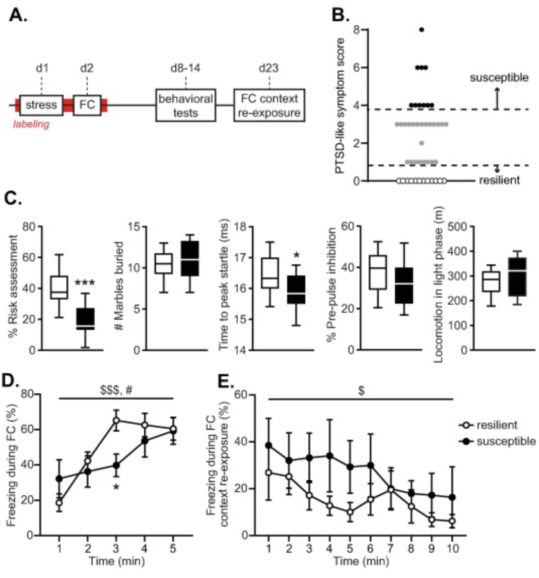
Fig 1

Fig. 1. Experimental schedule and behavioral assessments. Mice were exposed to a stressor and then subjected to contextual fear conditioning (FC). PTSD-like symptomatology was assessed in a set of behavioral tests, mice were re-exposed to the conditioning context and then sacrificed (A). Susceptible mice were defined by PTSD-like symptom scores >=4 (necessitating extreme behavior in multiple tests), whereas resilient mice did not show any aberrant behavior (score = 0) (B). Susceptible mice displayed significantly reduced risk assessment behavior and shorter latencies to peak startle compared to resilient animals at the group level. No group differences were observed in marble burying, pre-pulse inhibition or locomotor activity during the light phase (C). Increases in freezing behavior during contextual FC differed across groups (shock administration at 1, 2, 3, 4 and 5 min) (D), whereas no differences in freezing levels were observed upon later FC context re-exposure (no shocks) (E). Data are presented as medians with interquartile ranges (C) and mean +/- standard error of the mean (D,E). $$$: p < .001, $: p < .05, main effect of time; #: p < .05, group × time interaction; ***: p < .001, *: p < .05 effect of group.

To induce a PTSD-like phenotype in susceptible mice, all mice were exposed to an initial stressor, followed by fear learning (contextual fear conditioning) the next day. After a week, mice were subjected to a subset of behavioral tests over the course of two weeks to assess PTSD-like symptomatology. One week after the final behavioral test, mice were re-exposed to the conditioning context for 10 min to induce fear memory recall and sacrificed by perfusion-fixation 90 min later.

Tamoxifen

Tamoxifen was dissolved in a 10% ethanol/corn oil solution at a concentration of 10 mg/mL by overnight sonication and stored at −20 °C until further use. Solutions were heated to body temperature and intraperitoneally injected at a dosage of 150 mg/kg to induce activity-dependent neuronal labeling. Mice were injected with tamoxifen on the morning of day 1 - seven hours before the stressor - to induce SEFL-dependent active neuronal labeling. Fear learning was conducted 21 h post-stressor. This allowed both the stressor and fear learning to fall within the 36-h labeling window, capturing neuronal activation during both events. We opted for this approach as it is currently unknown whether the interindividual differences in SEFL and its long-term consequences originate from differential responding to the first stressor or from later fear learning. We hypothesized the latter, as the behavioral consequences of this PTSD-model are not observed to a similar degree if mice are only exposed to the initial stressor (Lebow et al., 2012), emphasizing aberrant fear learning to be at the core of the development of symptomatology. However, others have shown that PTSD-like memories can also be induced by stress exposure post-learning (Kaouane et al., 2012, Al Abed et al., 2020) (though applied immediately post-learning rahter than delayed), leaving this issue unresolved. Moreover, injection of 4-hydroxytamoxifen instead of tamoxifen to ensure a more specific (∼12-hour) labeling window, comes at the downsite of inducing instant labeling of activated neurons, capturing the neurons processing the injection stress as well, increasing noise. Therefore, we opted to inject tamixfen instead, and labeling of neuronal activity that was non-SEFL-related was minimized by keeping the mice undisturbed in their home cage during the rest of the labeling period.

PTSD protocol

Seven hours after the tamoxifen injection, mice were individually placed in Context A boxes, in which they received 14 1 s 1.0 mA shocks (i.e., the stressor) over 85 min in variable intervals. Mice were first moved to the dark experimental room in groups of two to three animals in dark carton boxes before being placed in the fear-conditioning boxes, which were connected to a shock generator (Campden Instruments). Context A consisted of a black, triangular shaped Plexiglas box with a steel grid and metal tray. The boxes were sprayed with 1% acetic acid, not illuminated, and 70 dB background noise was presented. Boxes were equipped with infrared beams at both ends, and beam break data was used to analyze gross locomotor activity during stress exposure.

On the second day, 28 h after the tamoxifen injection, mice were individually placed in Context B boxes, in which they received 5 1 s shocks of 0.7 mA over a period of five minutes (i.e., the fear learning), presented over fixed intervals. For this trigger session, mice were moved to the 70 lux illuminated experimental room in see-through cages in groups of two to three animals. The Context B boxes contained curved white walls and a steel grid with a white tray underneath. The boxes were furthermore cleaned with 70% ethanol and during the session the house lights in the boxes were turned on. No background noise was presented. As such, all sensory features (olfactory, auditory, and visual) differed between contexts A and B, sharing the sensory input of the grid floor as only commonality.

Mice were allowed to recover for a week, after which the behavioral consequences of SEFL exposure were assessed by testing for PTSD-related behavior: impaired risk assessment (dark-light transfer test), increased threat-induced anxiety (marble burying), hypervigilance (acoustic startle), impaired sensorimotor gaiting (pre-pulse inhibition), and disturbed circadian rhythm (locomotor activity during the light phase) (Lebow et al., 2012).

Behavioral testing

Dark-light transfer test. On day 8 of the protocol, mice were tested in the dark-light transfer test. The test was executed in a box that was divided into a dark compartment (DC, 29 × 14 cm) and brightly illuminated (ca. 1100 lux) compartment (LC, 29 × 29 cm), connected by a retractable door. The mice were individually placed in the DC, and the door was opened to initiate a 5-min test session. Movement of the mice was recorded and scored automatically with Ethovision XT (Noldus IT). An additional area of 6 × 3 cm surrounding the opening of the LC was programmed into the software tracking measurements. Time spent in the LC as well as time spent in this ‘risk assessment’ zone were measured. Percentage risk assessment was calculated as the amount of time spent in the risk assessment zone as a percentage of total time spent in the LC.

Marble burying. On day 10, mice were individually placed in a 10 lux illuminated black open box (30 × 28 cm), containing a 5 cm deep layer of corn cobs, on top of which 20 marbles were centrally arranged in a 4 × 5 grid formation. Each mouse was placed in the corner of the box to initiate the task. Mice were videotaped for 25 min. Videos were scored by assessing the number of buried marbles after 25 min.

Startle response and pre-pulse inhibition. On day 12, mice were moved to the experimental room in their home cage and individually placed in small, see-through Plexiglas constrainers mounted on a vibration-sensitive platform inside a ventilated cabinet that contained two high-frequency loudspeakers (SR-LAB, San Diego Instruments). Movements of the mice were measured with a sensor inside of the platform. The pre-pulse inhibition test (PPI) started with an acclimatization period of 5 min, in which a background noise of 70 dB was presented, which was maintained throughout the entire 30-min session. Thirty-two startle cues of 120 dB, 40 ms in duration and with a random varying ITI (12–30 s), were presented with another 36 startle cues preceded by a 20 ms pre-pulse of either 75 dB, 80 dB or 85 dB. Sessions were scored by assessing the latency to peak startle amplitude of the 12 middle startle trials, and the pre-pulse inhibition, i.e., the percentage of startle inhibition response to the different pre-pulse stimuli [1 − (mean pre-pulse startle response/mean startle response without pre-pulse) × 100].

Homecage locomotion. Immediately after the pre-pulse inhibition test, mice were individually housed in Phenotyper cages (45 × 45 cm, Noldus) for 72 h while their locomotion was being recorded by an infrared-based automated system (EthoVision XT, Noldus). The first 24 h were considered habituation time and data were discarded. Total locomotion time during the subsequent two light phases (21:00–09:00 h) was assessed.

Behavioral categorization

In order to categorize mice as either susceptible or resilient, one compound measure was generated based on the five behavioral outcome scores. Mouse behavior on each of the tests was sorted, and the 20% of mice that had the lowest values were attributed three points for percentage risk assessment, three points for latency to peak startle amplitude, and two points for percentage PPI. Similarly, the 20% of mice showing the highest values were attributed one point for light phase locomotor activity and marble burying (Henckens et al., 2017). Points for each test were determined by factor analysis in which tests were clustered in three separate groups: (1) latency to peak startle amplitude and percentage risk assessment, (2) percentage PPI, and (3) marble burying and total light activity (Lebow et al., 2012) (Table 1). Ties in the marble burying test were resolved by also assessing the number of marbles buried after 15 min, and assigning points to the mice that buried most marbles then. The points per animal were tallied to generate and overall PTSD-like symptom score. Mice that had a total of four or more points (necessitating extreme behavior in multiple tests) were termed susceptible. Only mice that had zero points (indicating no abnormal behavior within any of the tests) were termed resilient.

Table 1.
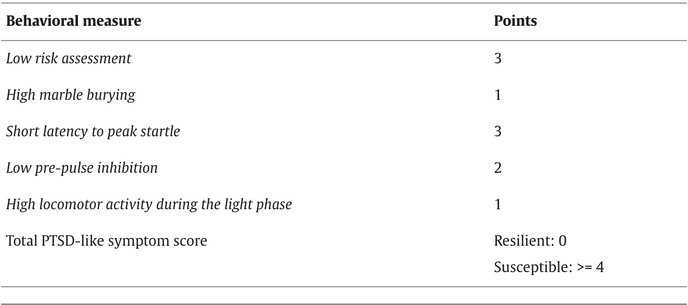
Table 1

Behavioral classification of resilient and susceptible mice. The 20% of mice that displayed the strongest phenotype on each test was assigned points, and points were tallied to generate a total PTSD-like symptom score Re-exposure and sacrifice

On the final day of the experiment, day 23, mice were re-exposed to the Context B box (i.e., the fear conditioning context) for 10 min to induce fear memory recall, following the exact same procedures as during the fear conditioning session. However, no shocks were administered during this context re-exposure session. Mice were sacrificed 90 min post re-exposure under anesthesia (5% isoflurane inhalation followed by intraperitoneal injection of 200 μL pentobarbital) by perfusion with phosphate buffered saline (PBS) followed by 4% paraformaldehyde solution (PFA). The brains were surgically removed and post-fixed for 24 h in 4% PFA, after which they were transferred to 0.1 M PBS with 0.01% sodium azide and stored at 4 °C.

Freezing behavior

Mice were videotaped during fear conditioning (day 2) and the re-exposure to the conditioning context (day 23) to assess fear memory encoding and remote recall. Freezing behavior was manually scored by an observer blinded to the experimental condition using The Observer XT12 software (Noldus). Consistent with previous literature, mice were considered to freeze when they were immobile for more than two consecutive seconds (Patel et al., 2014, Shoji et al., 2014).

Immunofluorescence

Right hemispheres of susceptible (n = 10) and resilient (n = 12) animals were sliced at 30 µm thickness using a freezing sliding microtome (Microm HM440E, GMI Inc., Ramsey, MN, USA) and stored in PBS with 0.01% sodium azide. Floating sections were used for immunohistochemistry of the hippocampus. For each animal, 4–6 sections were collected between anterior-posterior coordinates −1.46 mm and −1.94 mm relative to Bregma for the dorsal hippocampus, and between −2.92 mm and −3.52 mm relative to Bregma for the ventral hippocampus. tdTomato, as a proxy for the immediate early gene Arc, was used to measure neuronal activity during SEFL, while cFos immunofluorescence was assessed to measure recall-related activity. We used cFos, rather than Arc, because Arc labeling is primarily dendritic in some hippocampal subregions (Denny et al., 2014) complicating quantification of activated neurons, and both cFos and Arc expression have earlier been found to strongly overlap in neurons (Nakagami et al., 2013, Mahringer et al., 2019) - and specifically in the hippocampus (Stone et al., 2011, Jiang and VanDongen, 2021) - in response to a challenge.

Immunolabeling of cFos and parvalbumin (PV) or histone deacetylase (HDAC) 2. Sections were washed three times in 1x PBS and blocked in PBS-BT (1x PBS with 0.3% Triton X-100 and 1% bovine serum albumin) for 30 min at room temperature (RT). Incubation of the primary antibodies was performed overnight (guinea pig anti-cFos, 1:750, #226004, Synaptic Systems; rabbit anti-PV, 1:1000, #ab11427, ITK; or rabbit anti-HDAC2, 3 µg/µL, #AB_2533908, Thermo Fisher) in PBS-BT for 18 h at RT. Then, sections were washed three times in 1x PBS, and incubated with the secondary antibodies (Alexa 647-conjugated donkey anti-guinea pig, 1:200, #AP193SA6, Merck Chemicals; Alexa 488-conjugated donkey anti-rabbit, 1:200, #A-21206, Thermo Fisher) in PBS-BT for 3 h at RT. Lastly, slices were washed three times in 1x PBS, mounted on gelatin-coated slides using FluorSaveTM reagent (#345789, Merck Chemicals) and cover slipped. The slices were stored at −20 °C until image acquisition and cell counting.

Immunolabeling of cFos, 5-methylcytosine (5mC) and 5-methylhydroxycytosine (5hmC). Sections were washed three times in 1x PBS and permeabilized in 1x PBS with 0.1% Triton X-100 for 5 min at RT. Then, slices were incubated in 1 M HCl for 2 h, washed three times in 1x PBS and blocked in PBS-NT (1x PBS with 0.3% Triton X-100 and 8% normal goat serum) for 50 min, all at RT. Because this process bleaches endogenous fluorescence - here the tdTomato fluorescent signal - these slices had to be immunolabeled for red fluorescent protein (RFP) in addition to the other markers. After again washing the slices three times in 1x PBS, incubation of the primary antibodies was performed overnight (guinea pig anti-cFos, 1:750, #226004, Synaptic Systems; rat anti-RFP, 1:1000, #5f8, Chromotek; mouse anti-5mC, 1:500, #GWB-BD5190, GenWay Biotech; rabbit anti-5hmC, 1:1000, #AB_10013602, Active Motif) in PBS-NT for 18 h at 4 °C. Then, sections were washed three times in 1x PBS, and incubated with the secondary antibodies (Alexa 647-conjugated donkey anti-guinea pig, 1:200, #AP193SA6, Merck Chemicals; Alexa 555-conjugated donkey anti-rat, 1:200, #ab150154, Abcam; Alexa 488-conjugated goat anti-mouse, 1:200, #A11001, Thermo Fisher; Alexa 405-conjugated anti-rabbit, #ab175651, Abcam) in PBS-NT for 2 h at RT. Lastly, slices were washed three times in 1x PBS, mounted on gelatin-coated slides using FluorSaveTM reagent (345789, Merck Chemicals) and cover slipped. The slices were stored at −20 °C until image acquisition and cell counting.

Image acquisition and cell counting

Images of the tdTomato/cFos/PV and tdTomato/cFos/HDAC2 signals were captured through a light microscope (Axio Imager 2, Zeiss) using a 10x (for tdTomato/cFos/PV) or 40x (for tdTomato/cFos/HDAC2) objective lens and a LED module (Colibri 2, Zeiss). Images of the tdTomato/cFos/5mC/5hmC staining were captured through a confocal microscope (LSM900, Zeiss) using a 40x objective lens. For the tdTomato/cFos/PV signal, as well as the tdTomato/cFos/HDAC2 signal, whole hippocampi were photographed. For the tdTomato/cFos/5mC/5hmC staining, the entire DG was photographed, while for the CA1 and CA3 regions three representative photos each were taken, with locations being consistent across slices and animals (Fig. S1). Separate photos were stitched and cFos+, tdTomato+ and PV+ cells were manually counted per region in Fiji software (Schindelin et al., 2012) by an experimenter blinded to the experimental group. Hippocampal surface areas in each slice were assessed and corrected for to obtain standardized measures of cell density. Normalized cell counts were averaged per hippocampal subregion per animal and subjected to statistical testing. Note that the CA2 and CA1 regions were segmented together. This combined region will henceforth be referred to as ‘CA1′.

Fluorescent signal intensity analysis

Expression levels of HDAC2, 5mC and 5hmC per cell were assessed by measuring signal intensity, and four cell types were identified by masks per hippocampal subregion per slice (Schindelin et al., 2012): 1) all tdTomato+cFos- cells, 2) all cFos+tdTomato- cells, 3) all tdTomato+cFos+ cells, and 4) all tdTomato-cFos- DAPI+ cells. Furthermore, a mask was generated for the background signal, which was obtained by inverting the DAPI+ mask. Within mask 1–4, the mean signal intensity of HDAC2, 5mC and 5hmC was assessed. Here, masks 1–3 define the fear memory engram cells, while mask 4 defines the non-engram cells. In the background mask, the mean of the signal intensity of HDAC2, 5mC and 5hmC was assessed to exclude potential inter-slice differences in background intensity. Analyses revealed that background HDAC2 signal was very consistent across slices and did not differ across the hippocampal axis (F(1,46.241) = 1.113, p = .297), hippocampal subregions (F(2,42.178) =0.698, p = .503) or groups (group main effect; F(1,19.324) = .007, p = .936, all group interactions; p’s >0.508). Background 5mC levels did depend on the hippocampus subregion (F(2,40.945) = 9.363, p <.001), but not axis (F(1,58.531) = .022, p = .884) or group (group main effect; F(1,9.793) = 1.994, p = .189, all group interactions; p’s > .068). A similar pattern was observed for background 5hmC levels that depended on hippocampal subregion (F(2,31.234) = 4.589, p =.018), but not axis (F(1,42.726) = 3.509, p = .068) or group (group main effect: F(1,14.919) = .496, p = .492, all group interactions: p’s > .832). Since no potentially confounding effects of background signals were detected, fluorescent signals were not background-corrected.

Statistical analyses

Data were analyzed using IBM SPSS Statistics 23. Normality was checked using the Shapiro-Wilk test. One resilient animal displayed deviant behavior (deviating more than two standard deviations from the groups’ mean) during the acoustic startle test, and was excluded from further analyses of the latency to peak startle and pre-pulse inhibition. For normally distributed data, statistical testing was performed by independent t-tests or one-way ANOVAs. Freezing behavior over time was analyzed by repeated measures ANOVA (with time as within-subjects factor, and group as between-subjects factor), whereas immunohistochemistry data was analyzed using linear mixed modelling implementing the restricted maximum likelihood estimation. In the latter, the factors axis (dorsal, ventral) and region (DG, CA3, CA1) were included as within-subjects variables, group as between-subjects variable and mouse as random intercept. For the epigenetic data, the factor engram type (non-engram (tdTomato-cFos-), encoding (tdTomato+), recall (cFos+), reactivated (tdTomato+cFos+) engram) was additionally included as within-subjects variable. For non-parametric data, the Mann-Whitney U test or Kruskal-Wallis test were used. Differences were considered statistically significant if p < .05.

Results

Behavioral differences between susceptible and resilient animals

To assess potential differences in hippocampal trauma-related engram activity associated with differential susceptibility to PTSD-like symptoms, a cohort of 44 ArcTRAP mice was exposed to the PTSD induction protocol. Following a week of recovery, mice were assessed for PTSD-like symptomatology to yield a group of susceptible (n = 10) and resilient animals (n = 12), which significantly differed on their overall PTSD-like symptom score (U = 120, p <.001) (Fig. 1(B)). Symptomatology was rather heterogeneous across susceptible animals (Fig. 1(C)), sharing some symptoms (percentage risk assessment (t(19) = 4.280, p <.001) and reaction time to peak startle (t(18) = 2.110, p = .025)), yet differing on others (marble burying (t(20) = 0.739, p = .234), percentage pre-pulse inhibition (t(17) = 1.210, p = .121) and locomotor activity in the light phase (t(14.633) = 0.864, p = .201)). Thus, individual symptom profiles across susceptible mice differed.

Behavior during stress exposure was checked by assessing beam break data as proxy for locomotor activity. Susceptible and resilient mice did not differ in their overall locomotor activity during the stressor (F(1,14) = .041, p = .843), nor in its reduction over time (main effect of time: F(9.490, 132.857) = 25.682, p <.001, group × time interaction: F(9.490, 132.857) = 1.022, p = .427), indicating no gross behavioral differences during initial stress exposure. During the subsequent fear learning session, no overall group differences were observed in freezing rates (F(1,18) = .629, p = .438), yet the increase in freezing behavior over time (F(4,72) = 13.534, p <.001) significantly differed across groups (F(4,72) = 3.172, p = .019) (Fig. 1(D)). Freezing levels tended to start lower in resilient mice, but also seemed to plateau sooner. Post hoc tests revealed only significant differences in the third minute of the fear learning session, when resilient mice displayed higher freezing levels than susceptible mice (t(18) = 2.870, p = .010). Freezing behavior upon re-exposure to the fearful context - to induce fear memory recall - was not different between resilient and susceptible animals (Fig. 1(E)). Neither overall freezing levels (F(1,19) = 1.308, p = .267), nor the observed reduction in freezing over time (F(3.542,67.297) = 3.323, p = .019) differed between groups (group × time interaction: F(3.542,67.297) = .703, p = .576).

Susceptible animals show a smaller activated neuronal ensemble within the vCA1 upon fear memory recall, but not during encoding

In the ArcTRAP mice, the neuronal ensemble active during SEFL, i.e., those neurons expressing the immediate early gene Arc, was permanently labelled by the reporter gene tdTomato (Fig. 2(ABC)). No significant differences in the total number of activated hippocampal neurons during SEFL were observed between susceptible and resilient mice (F(1,33.255) = .715, p = .404), nor was there any interaction effect between group and axis (F(1,40.938) = .880, p = .354), group and hippocampal subregion (F(2,30.033) =0.295, p = .747) or group × axis × subregion interaction (F(2,30.033) =0.255, p = .776), suggesting that hippocampal activity between groups was not different during initial memory formation.
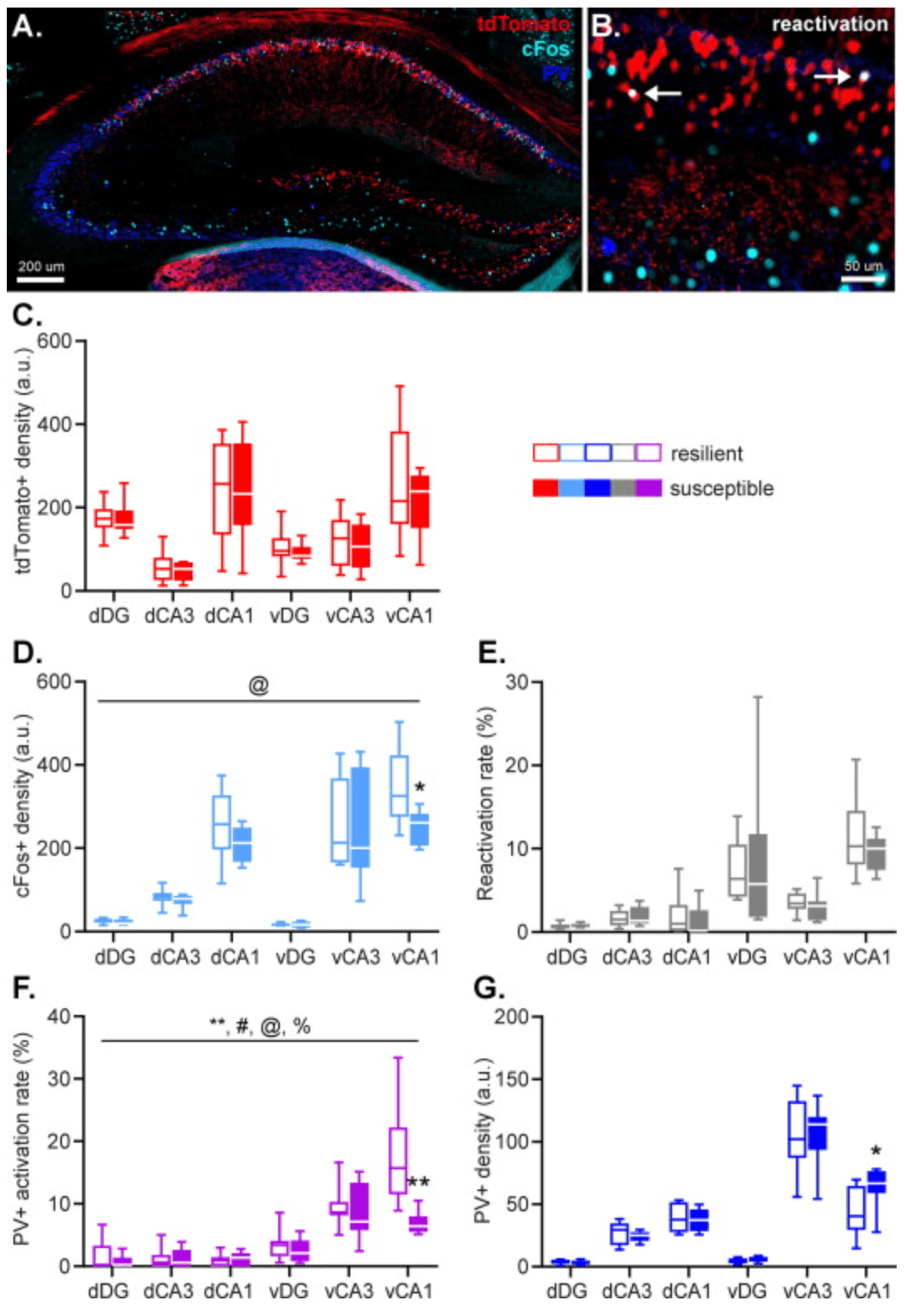
Fig 2

Fig. 2. Hippocampal activity during fear memory encoding (marked by tdTomato expression), remote fear memory recall (marked by cFos expression), as well as parvalbumin (PV) interneuron density were assessed by immunohistochemistry. Arrows indicate tdTomato+cFos+ double-positive cells. (AB). No group differences were observed in the size of the engram recruited during stress-enhanced fear learning (C). However, susceptible animals displayed a smaller population of ventral hippocampal CA1 neurons active during remote fear memory recall (D), without any group differences in neuronal reactivation rate (E). Additionally, susceptible animals showed a decrease in activity of PV+ neurons in the ventral hippocampal CA1 specifically during remote memory recall (F), which was joined by an increase in local PV+ density (G). Data represent medians with interquartile ranges. **: p < .01, *: p < .05, main effect of group, @: p < .05, group × subregion interaction, %: p < .01, group × axis interaction, #: p < .05, group × axis × subregion interaction.

Neuronal activity associated with remote fear memory recall was measured by immunolabelling cFos+ neurons (Fig. 2(ABD)); cells that were active during remote fear memory recall induced by re-exposure to the conditioning context. For the number of hippocampal neurons active upon recall, a significant group × hippocampal subregion interaction (F(2,20.586) = 5.055, p = .016) was found, and a trend towards a main effect of group (F(1,4.034) = 4.100, p = .051). All other group interaction effects failed to reach significance (all p’s >0.577). These effects were caused by lower neuronal activity during remote fear memory recall in the CA1 of susceptible vs. resilient animals (F(1,14.693) = 5.298, p = .036), most notably within the vCA1 (vCA1; p = .013, dCA1; p = .100).

Susceptible and resilient animals show no difference in hippocampal remote fear memory reactivation

To investigate which encoding-related (i.e., tdTomato+) cells eventually remained incorporated in the hippocampal memory engram for the fearful experience, overlap between the tdTomato+ and cFos+ neurons was assessed. These overlapping signals represent neurons that were active both during fear memory encoding and recall, and therefore reflect the stable memory trace. Neuronal reactivation is expressed as the Reactivation Rate (RR), which is calculated by dividing the number of cFos+tdTomato+ overlapping neurons by the number of tdTomato+ neurons (Cowansage et al., 2014, Milczarek et al., 2018).

An average of 4.2% of hippocampal tdTomato+ neurons were reactivated during the trigger context re-exposure, with RRs in the different subregions ranging between 1% (dDG) to 12% (vCA1). Reactivation rates were not statistically different between the groups, and did not show any significant interactions between group, subregion, and/or axis (all p’s = 1.00) (Fig. 2(E)).

Susceptible animals show an increased number of vCA1 PV neurons that is recruited relatively less during remote fear memory recall

Given the influence of PV interneuronal activity on the excitability and firing behavior of surrounding neurons, we investigated PV activity during fear memory recall. For this we calculated PV ‘Activation Rate’ (AR) as the number of PV+cFos+ overlapping neurons divided by the number of PV+ neurons × 100%, reflecting the percentage of the total interneuronal PV population that was active during remote fear memory recall. In line with prior work indicating that Arc-expression in response to behavioral challenges is largely restricted to glutamatergic neurons (Vazdarjanova et al., 2006), the population of tdTomato-labeled (‘TRAPped’) neurons in this mouse line did not overlap with PV expression, which prevented us from also investigating relative activity of PV neurons during SEFL. Moreover, we also calculated the overall density of PV neurons to account for potential structural differences across groups.

PV ARs revealed a significant main effect of group (F(1,35.454) = 8.613, p = .006), together with a group × axis (F(1,35.681) = 9.045, p = .005), group × subregion (F(2,33.512) = 4.728, p = .016), and group × axis × subregion (F(2,33.512) = 5.172, p = .011) interaction effect (Fig. 2(F)). Follow up tests revealed no significant group effects in the dorsal hippocampus (p’s > .543), but a significant main effect of group (F(1,18.823) = 14.541, p = .001) as well as a group × subregion interaction (F(2,15.804) = 7.056, p = .006) in the ventral hippocampus. This interaction effect was driven by significantly reduced PV activation in the vCA1 of susceptible mice (p = .001), but not other hippocampal subregions (both p’s > .340). The overall number of hippocampal PV neurons was not found to be significantly affected by group (F(1,35.978) = 1.533, p = .224) However, exploratory analyses to test whether the activity differences in vCA1 PV neurons were associated with different PV neuron numbers in susceptible mice, revealed a significantly higher PV neuron density in their vCA1 (p = .026), which was not observed in the other hippocampal subregions (all p’s >0.227). These findings suggest that trauma susceptibility is associated with an increase in PV neurons in the vCA1, of which a relatively smaller part is active during remote fear memory recall.

Susceptible mice display altered HDAC2 expression patterns in the ventral hippocampus

The intensity of HDAC2 fluorescence in engram and non-engram cells was measured to quantify HDAC2 expression within these neurons (Fig. 3(AB)) (Ververis and Karagiannis, 2012, Toki et al., 2017). HDAC2 expression was dependent on hippocampal axis (F(1,207.453) = 159.497, p < .001), subregion (F(2,145.718) = 41.926, p < .001) and engram type (F(3,142.060) = 28.686, p < .001), but did not reveal a significant main effect of group (F(1,18.223) = 2.496, p = .131) (Fig. 3(C)). Pair wise comparisons revealed that engram type effects were caused by significantly higher HDAC2 expression in memory encoding (tdTomato+, p < .001), recall (cFos+,p < .001) and reactivated (tdTomato+cFos+; p < .001) neurons, compared to non-engram cells, whereas the engram types amongst each other did not show overall differences in HDAC2 expression (all p’s >0.320) (Fig. 3(CDE)), suggesting histone acetylation is overall reduced in memory engram-related cells compared to non-engram cells.
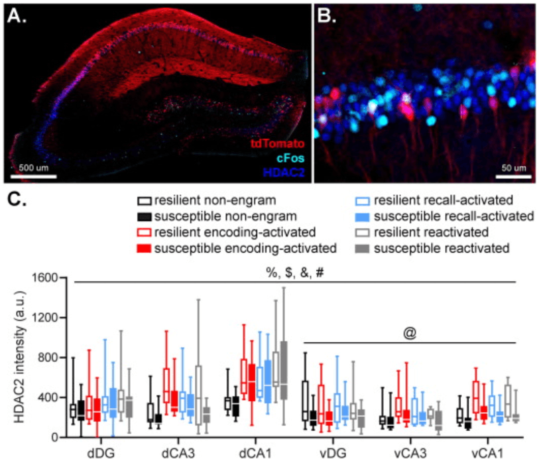
Fig 3

Fig. 3. Hippocampal HDAC2 fluorescence in cells active during fear memory encoding (marked by tdTomato expression) and remote fear memory recall (marked by cFos expression) were assessed by immunohistochemistry (AB). Neurons involved in memory encoding (tdTomato+) and recall (cFos+), as well as reactivated (tdTomato+cFos+; indicated by arrows) neurons, were characterized by overall higher HDAC2 fluorescence than non-engram cells. HDAC2 levels in the ventral hippocampus were modulated by a subregion × group interaction, which seemed to be caused by a tendency towards lower HDAC2 levels in the vCA1 in susceptible animals (CDE). Data represent medians with interquartile ranges. %: p < .001, main effect of axis, $: p < .001, main effect of subregion, &: p < .001, main effect of engram type, #: p < .05, group × axis × subregion interaction, @: p < .05, group × subregion interaction.

Critically, we observed a significant group × hippocampal axis × subregion interaction in HDAC2 levels (F(2,145.718) = 3.467, p = .034). Follow up tests revealed no significant effects of group in the dorsal hippocampus (all p’s >0.227), but a significant group × hippocampal subregion interaction (F(2,80.421) = 3.368, p = .039) in the ventral hippocampus. This interaction seemed to be caused by a tendency towards reduced HDAC2 levels in the vCA1 (p = .057) in susceptible compared to resilient mice, in the absence of differences in the vDG (p = .169) and vCA3 (p = .382).

Susceptible animals show rather generic increases in hippocampal 5mC and 5hmC levels

The intensity of 5mC and 5hmC fluorescence in engram and non-engram cells was measured to determine the DNA methylation status of these neurons (Ramsawhook et al., 2017, Toki et al., 2017) (Fig. 4(AB)). 5mC levels appeared to be modulated by hippocampal subregion (F(2,96.738) = 3.116, p = .049), engram type (F(3,46.479) = 27.426, p < .001) and group (F(1,14.271) = 5.324, p = .037), without a main effect of hippocampal axis (p = .567) (Fig. 4(C)). Moreover, a significant group × engram type interaction was observed (F(3,46.479) = 3.389, p = .026), but no other significant interactions (all p’s >0.062). Post hoc comparisons revealed significant differences in 5mC levels between all types of engram cells, with memory encoding and reactivation cells displaying higher 5mC levels than non-engram cells (p < .001 and p = .004, respectively), whereas memory recall cells displayed significantly lower 5mC levels compared to non-engram cells (p = .005). Follow up tests on the group × engram type interaction revealed significant upregulation of 5mC levels of susceptible mice in memory encoding (p = .019), recall (p = .015) and non-engram cells (p = .029), without significant differences in reactivated cells (p = .107).
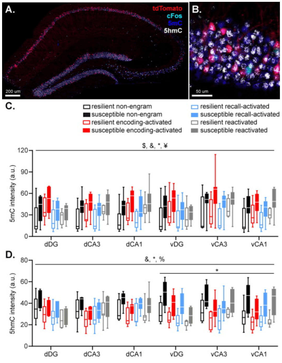
Fig 4

Fig. 4. Hippocampal 5mC and 5hmC fluorescence in cells active during fear memory encoding (marked by tdTomato expression), remote fear memory recall (marked by cFos expression) and both were assessed by immunohistochemistry and compared to non-engram cells (tdTomato and cFos negative cells) (AB). 5mC levels were higher in encoding and recall cells compared to non-engram cells. In contrast, reactivated cells displayed lower 5mC levels than non-engram cells. Importantly, susceptible mice displayed higher 5mC levels in memory encoding, recall, and non-engram cells, compared to resilient mice, without significant differences in reactivated cells (C). 5hmC levels were lower in all types of engram cells compared to non-engram cells, and susceptible mice displayed higher 5hmC levels in the ventral hippocampus (D). Data represent medians with interquartile ranges. $: p < .05, main effect of subregion, &: p < .001, main effect of engram type, *: p < .05, main effect of group, ¥: p < .05, group × engram type interaction, %: p < .001, group × axis interaction.

5hmC levels depended on engram type (F(3,97.708) = 24.770, p < .001) and group (F(1,16.006) = 6.837, p = .019), without a main effect of hippocampal axis (p = .457) or subregion (p = .431) (Fig. 4(D)). Moreover, a significant group × axis interaction was observed (F(1,183.505) = 28.105, p < .001). All other interactions with group were non-significant (all p’s >0.063). Pair wise comparisons revealed significantly lower 5hmC levels in all types of engram cells compared to non-engram cells (all p’s < .001), whereas the different type of engram cells (encoding, recall and reactivation) did not differ from each other (all p’s > .425). Follow up tests for the group × hippocampal axis interaction revealed that susceptible displayed significantly higher 5hmC levels in the ventral hippocampus (F(1,14.694) = 8.419, p = .011), but not dorsal hippocampus (F(1,14.190) = 3.295, p = .091).

While 5mC and 5hmC levels have been linked to decreased and increased gene expression respectively (Razin and Cedar, 1991, Mendonca et al., 2014), the 5hmC/5mC ratio might actually be most informative with regard to a cell’s gene expression profile, with high ratios coding increased gene expression(Mellen et al., 2012). Therefore, 5hmC/5mC ratios were calculated as well (Fig. S2). 5hmC/5mC ratio data revealed a significant effect of engram type (F(3,71.552) = 65.954, p < .001), without any effects of hippocampal axis (p = .194), subregion (p = .540) or group (p = .210). Moreover, a significant group × engram type interaction was found (F(3,71.552) = 6.833, p < .001). Pair wise comparisons of 5hmC/5mC ratios revealed significantly lower ratio in engram vs. non-engram cells (encoding; p < .001, recall; p = .010, reactivation; p < .001), with encoding and reactivation cells displaying lowest ratio’s (both p’s <0.001 compared to recall cells). This suggests that engram neurons are transcriptionally less active than neurons that are not incorporated into the engram, which is in line with previous studies marking increased DNA methylation in engram cells as a key mechanism in stabilizing memory engrams during memory consolidation (Gulmez Karaca et al., 2020). Follow up analyses on the group × engram type interaction however failed to indicate clear differences between susceptible and resilient mice (all p’s > .195).

Discussion

Here, we tested the hypothesis that susceptibility to traumatic stress is characterized by interindividual differences in the trauma-related hippocampal memory engram and its epigenetic regulation. We examined potential alterations in the hippocampal memory engram for a stress-enhanced fear memory in mice that were susceptible and resilient to developing PTSD-like symptoms as a consequence of it. While no differences in the size of the neuronal ensemble activated during fear memory encoding were observed between the groups, susceptible mice displayed a smaller ensemble activated in the vCA1 upon remote fear memory recall, as well as higher PV neuronal density and a relatively lower activity of PV neurons in the vCA1 upon remote memory recall. Epigenetic data revealed rather generic than engram-specific differences across groups, with susceptible animals displaying lower hippocampal HDAC2 expression, as well as higher hippocampal 5mC and 5hmC signal, without clear overall differences in 5hmC/5mC ratio.

Mice were classified as susceptible or resilient based on a compound score comprising multiple behavioral PTSD-like symptoms (i.e., impaired risk assessment, increased threat-induced anxiety, hypervigilance, impaired pre-pulse inhibition and higher activity during the inactive phase, potentially linking to sleep disturbances), rather than single behavioral features. Because of obvious limitations in capturing true PTSD-symptomatology in mice, these behavioral features should merely be considered as proxy’s for the complex behavioral symptomatology as observed in patients. Yet, the behavioral classification in which mice are categorized based on a compound score of symptomatology, resembles the situation in PTSD patients (Zoellner et al., 2014). No large differences were observed in how susceptible and resilient mice behaved during the encoding and recall of the fear memory. Whereas the susceptible mice showed a somewhat different temporal development of freezing during fear conditioning, no differences in freezing levels were observed during remote fear memory recall. Prior work has indicated that stress susceptible mice show exaggerated and extinction-resistant fear memory in a stress-enhanced cued fear learning paradigm (Sillivan et al., 2017). This fits observations of emotional hypermnesia in PTSD patients, as well as their strongly cue-based rather than context-specific recall of the trauma memory. Importantly, PTSD-like memory alterations also include contextual amnesia (Kaouane et al., 2012, Desmedt et al., 2015). We here implemented stress-enhanced contextual fear learning (Rau et al., 2005), making that one could speculate on impaired contextual fear memory recall canceling out excessive fear upon successful cue-induced recall in susceptible mice. Future studies need to confirm this speculation of a maladaptive fear memory in susceptible mice by dissociating both aspects of fear memory by re-exposing mice to partial fear reminders (cues vs context) only. Moreover, it would be valuable to include a regular (not stress-enhanced) fear learning group as control in future experiments, to both verify the effect of prior stress exposure overall and to determine whether these effects differ in resilient vs. susceptible mice.

Despite the absence of differential freezing behavior, we did find a significant reduction in vCA1 activity and a relative decrease in PV cell activation during remote fear memory recall in susceptible animals. Previous work has implicated the vCA1 in contextual fear memory (Maren and Fanselow, 1995, Rudy et al., 2004, Rogers et al., 2006, Zhu et al., 2014, Kim and Cho, 2017) and the subsequent contextual modulation of fear recall and expression (Orsini et al., 2011, Xu et al., 2016). Ventral CA1 neurons have been shown to convey contextual information through monosynaptic projections to the basolateral amygdala (Maren and Fanselow, 1995, Maren et al., 2013, Kim and Cho, 2017, Kim and Cho, 2020). As such, the reduction in the vCA1 ensemble activated during recall might reflect impaired functionality in the recall of contextual information, which may lie at the core of the context-nonspecific recall of trauma memories as observed in PTSD (Liberzon and Abelson, 2016, Asok et al., 2018, Zinn et al., 2020) as well as the reported contextual amnesia (Desmedt, 2021). As we did not find any differences in engram size during fear memory encoding (and reactivation), data suggest that initial memory encoding is not different between groups, but it is rather the (systems) consolidation process during which differences arise. The memory engram is not static, but rather dynamic over time, reorganizing both within and across brain regions (Davis and Reijmers, 2018; Ziv et al., 2013, Wang et al., 2015), ultimately resulting in different storage sites of the memory following its consolidation (Frankland et al., 2004, Tayler et al., 2013, Ziv et al., 2013, Tonegawa et al., 2015, Wang et al., 2015, Davis and Reijmers, 2018). This is especially relevant as we employed a remote recall paradigm, whereas most previous studies focused on more recent memory recall and might explain why we only observed differential vCA1 activity and not reactivity during recall.

Parvalbuminergic network plasticity has been shown critical in the regulation of learning (Donato et al., 2013), with PV interneurons contributing to memory consolidation by stabilizing functional connectivity patterns among CA1 neurons (Ognjanovski et al., 2017) and mediating coherent hippocampal-neocortical communication (Xia et al., 2017). We observed a higher number of PV neurons in the vCA1 of susceptible animals as well as a smaller portion of these being activated during memory recall. Seemingly contradictory to our findings, prior research has reported on a loss of PV neurons following chronic stress (Holm et al., 2011, Czeh et al., 2015), although this has not consistently been reported (Nowak et al., 2010, Holm et al., 2011). Yet, notably, previous studies ignored interindividual differences in terms of anxiety-like behavior, posing the possibility that this observed reduction in PV density may actually reflect an adaptive response to stress. Alternatively, these differences between susceptible and resilient mice may have been present already before PTSD induction, and as such do not reflect a differential effect of trauma itself. The lower recruitment of these neurons in susceptible mice may indicate a compensatory effect, resulting in similar absolute activity levels of the total PV population in both susceptible and resilient animals. Regardless, these alterations in PV interneuron presence and recruitment might relate to disrupted consolidation of the traumatic memory in PTSD (Parsons and Ressler, 2013), proposing it as a target for dedicated future studies.

Hippocampal HDAC2 expression was higher in engram compared to non-engram cells and reduced in susceptible compared to resilient animals. Histone acetylation is most robustly associated with promoting memory formation. It is increased following neuronal activity, and promotes a chromatin structure permissive to gene transcription (Eberharter and Becker, 2002), necessary for synaptic plasticity (Marco, 2022). HDACs, in particular HDAC2, induce the removal of acetyl groups, suppressing gene transcription, and their pharmacological or genetic inhibition was found to facilitate learning and memory (Guan et al., 2009) and improve extinction learning (Vecsey et al., 2007, Morris et al., 2013, Gräff et al., 2014). Our finding of increased HDAC2 levels in engram vs. non-engram cells seems to be at odds with these reports. Yet, one could speculate that plasticity should be suppressed once a memory is formed, with memory-related gene silencing serving to stabilize the memory engram (Alberini and Kandel, 2014). This interpretation is supported by our findings in terms of DNA methylation patterns, with engram cells having overall higher levels of 5mC (generally suppressing gene transcription) and lower levels of 5hmC (typically increasing gene transcription), decreasing the 5hmC/5mC ratio, suggesting an overall decrease in transcriptional activity within engram neurons. Prior work implicating DNA methylation in stabilizing engrams during consolidation and aiding successful memory recall support this notion (Gulmez Karaca et al., 2020). As such, reduced HDAC2 levels as observed in susceptible mice may indicate a less stable fear memory engram. Whereas this interpretation might fit with the readily re-activated trauma memory in PTSD, it is contrasting behavioral observations of a trauma memory in patients that is very rigid, and less sensitive to extinction. However, our findings are in line with prior reports on HDAC2 downregulation following acute stress being related to increased stress susceptibility (Karnib et al., 2019) and a stronger fear memory (Takei et al., 2011). In terms of methylation, we found susceptible animals to be characterized by rather generic increases in hippocampal 5mC and 5hmC levels, both in engram and non-engram cells. As these markers are inversely related to gene expression and their ratio was not consistently affected, we conclude that both groups, despite the slight differences in hippocampal methylation profile, do likely not differ in terms of overall gene expression as a consequence of this. Prior research has reported on changes into hippocampal global methylation levels as a consequence of stress exposure, with both increases (Sales and Joca, 2018, Hammels et al., 2015) and decreases (Rodrigues et al., 2015) being reported. We add to this existing literature by relating methylation patterns to interindividual differences in stress susceptibility, which match reports on increased global methylation in PTSD patients (Smith et al., 2011). Yet, future studies should investigate these differences in more detail, by assessing specific methylation sites, as well as potential mediators of these differences (e.g., DNA methyltransferases). Moreover, as the observed epigenetic differences cannot readily explain the reduced number of cFos expressing cells upon memory recall in susceptible mice, it would be worthwhile investigating other epigenetic regulators. One of these might be HDAC5, which was previously found to be upregulated post-trauma in the bed nucleus stria terminalis of susceptible animals in this same mouse model (Lebow et al., 2012).

Some limitations should be noted. Firstly, assessment of the memory engram related to memory encoding was largely restricted to glutamatergic neurons in the ArcTRAP mice (Vazdarjanova et al., 2006), leaving the role of GABAergic neurons in the engram and traumatic stress susceptibility to be elucidated. We preferred ArcTRAP mice over the available FosTRAP mice based on superior labeling sensitivity in the hippocampal CA3 and CA1, which are typically devoid of labeled cells in the FosTRAP mouse lines (Guenthner et al., 2013, Dirven et al., 2022). Secondly, the ArcTRAP line has substantial background labeling (i.e., fluorescent tagging of neurons in the absence of tamoxifen) in the hippocampal DG (Guenthner et al., 2013), which may explain why we did not recapitulate prior findings of peri-trauma DG activation being predictive of fear memory generalization and stress susceptibility in general (Lesuis et al., 2021, Dirven et al., 2022). Furthermore, tdTomato-labeling captured both neuronal activity during the initial stressor and subsequent fear learning experience. Future studies should separate these two episodes by making use of 4-hydroxytamoxifen injection, restricting the labeling period. Moreover, while we assume that the tdTomato-tagged and cFos-labelled neurons represent the fear memory, it will require experimental manipulation of these populations to show that their activity is necessary and/or sufficient for memory expression. Finally, while immunofluorescence of epigenetic markers is more often used to draw preliminary conclusions about changes in transcriptional processes (Chouliaras et al., 2013, Demyanenko and Uzdensky, 2019), it is not possible to draw a one-to-one relationship between the observed differences in HDAC2, 5mC and 5hmC levels and actual alterations in histone acetylation and gene expression. Different studies have shown transcriptional alterations in response to stress (Floriou-Servou et al., 2018), and in PTSD specifically (Girgenti et al., 2021, Zhang et al., 2021), but it would require future studies to causally link such changes to the alterations in histone acetylation, DNA methylation and hydroxymethylation that have been observed.

Concluding, we have shown PTSD-like symptomatology in mice to be related to alterations in remote fear recall-induced activation and PV interneuronal activity - as well as overall PV density - in the ventral CA1. These findings propose an important role for aberrant remote fear memory recall, resulting from an altered (systems) consolidation process, in mediating traumatic stress susceptibility. Future assessments should investigate whether this can also translate into an aberrant behavioral manifestation of the fear memory. Epigenetically, we found marked differences in HDAC2 expression and DNA methylation and hydroxymethylation between susceptible vs. resilient mice, suggestive of net higher hippocampal transcriptional activity. These changes were however not restricted to neurons involved in the memory engram, indicating epigenetic changes throughout the entire hippocampus as an important target for further research into the pathophysiology of PTSD. These overall alterations could potentially contribute to deviations in memory consolidation by destabilizing hippocampal memory representations, although future research is needed to determine such causal relationship.

Open Article as PDF

Abstract

While the majority of the population is ever exposed to a traumatic event during their lifetime, only a fraction develops posttraumatic stress disorder (PTSD). Disrupted trauma memory processing has been proposed as a core factor underlying PTSD symptomatology. We used transgenic Targeted-Recombination-in-Active-Populations (TRAP) mice to investigate potential alterations in trauma-related hippocampal memory engrams associated with the development of PTSD-like symptomatology. Mice were exposed to a stress-enhanced fear learning paradigm, in which prior exposure to a stressor affects the learning of a subsequent fearful event (contextual fear conditioning using foot shocks), during which neuronal activity was labeled. One week later, mice were behaviorally phenotyped to identify mice resilient and susceptible to developing PTSD-like symptomatology. Three weeks post-learning, mice were re-exposed to the conditioning context to induce remote fear memory recall, and associated hippocampal neuronal activity was assessed. While no differences in the size of the hippocampal neuronal ensemble activated during fear learning were observed between groups, susceptible mice displayed a smaller ensemble activated upon remote fear memory recall in the ventral CA1, higher regional hippocampal parvalbumin neuronal density and a relatively lower activity of parvalbumin interneurons upon recall. Investigation of potential epigenetic regulators of the engram revealed rather generic (rather than engram-specific) differences between groups, with susceptible mice displaying lower hippocampal histone deacetylase 2 expression, and higher methylation and hydroxymethylation levels. These finding implicate variation in epigenetic regulation within the hippocampus, as well as reduced regional hippocampal activity during remote fear memory recall in interindividual differences in susceptibility to traumatic stress.

Summary

Post-traumatic stress disorder (PTSD) can develop after a traumatic event. A key feature is re-experiencing the trauma through flashbacks and nightmares. Treatments focusing on trauma memory are effective, suggesting that problems with how these memories are processed play a role in PTSD. While many people experience trauma, only a small number develop PTSD. It is thought that resilient individuals process trauma memories adaptively, while susceptible individuals do not. Memory formation involves strengthening connections between brain cells, creating a physical "memory engram." New tools allow scientists to study these engrams. This study used a method called TRAP to see if PTSD-like symptoms are linked to an abnormal memory engram in the hippocampus, a brain area important for memory.

The hippocampus is known for its role in contextual memory and is affected by emotional events. Brain scans of PTSD patients show smaller hippocampal volume and reduced function. Animal studies of PTSD also point to abnormal hippocampal function. Reduced activity in the hippocampus when exposed to trauma-related cues has been linked to PTSD severity and memory problems. However, it is not clear how these general hippocampal issues relate to changes in the trauma memory engram itself. This study explored if changes in the hippocampal fear memory engram are linked to vulnerability to long-term trauma effects and PTSD-like symptoms in mice. Researchers looked at different parts of the hippocampus: the ventral and dorsal regions, and subregions like the dentate gyrus (DG), Cornu Ammonis areas 1 (CA1), and 3 (CA3).

The study also looked at parvalbumin (PV) interneurons, which are brain cells that help coordinate activity among other neurons and are known to be affected by stress. Their activity is important for stabilizing connections in the hippocampus during learning. Additionally, epigenetic regulation was investigated. This refers to changes in gene activity that don't involve altering the DNA sequence itself, but can be influenced by environmental stress and impact memory formation and long-term behaviors. Histone acetylation, a type of epigenetic change, is strongly linked to memory. For example, the protein HDAC2, found in the hippocampus, is negatively related to memory and brain plasticity. While some studies suggest stress reduces HDAC2 levels, others point to a protective effect. DNA methylation and hydroxymethylation, other epigenetic changes, are also known to be affected by stress.

A mouse model was used to test if changes in trauma-related hippocampal engrams are linked to PTSD-like symptoms, and if these key engram regulators (PV interneurons and epigenetic changes) are involved. The mouse model involved exposing mice to severe stress, followed by fear conditioning. Mice were then tested for PTSD-like symptoms to identify susceptible and resilient groups. Engram neurons active during fear memory formation were identified using the TRAP model, and those involved in recalling the memory were identified later. Researchers also examined the presence and activity of PV interneurons, and levels of HDAC2, 5mC, and 5hmC in both engram and non-engram neurons.

Experimental Procedures

Animals

This study used ArcTRAP mice, a specific genetically modified strain. These mice were bred to allow active neurons to be labeled with a fluorescent marker (tdTomato) for 36 hours after a special injection. ArcTRAP mice were chosen over another type (FosTRAP) because they showed better labeling in certain hippocampal areas. Only male mice were used because the PTSD model had only been proven valid in males. Mice were housed in groups, with food and water always available. Behavioral tests were done during the animals' active period. All experimental procedures followed strict ethical guidelines and were approved by relevant animal welfare committees.

General Procedure

Forty-four ArcTRAP mice were injected with a substance to label active neurons. They were then put through a PTSD mouse model that involves stress-enhanced fear learning (SEFL). This model is based on the idea that prior stress makes people more likely to develop PTSD. SEFL typically strengthens fear memories, but in a specific group of mice, it also causes persistent anxiety and arousal, similar to PTSD symptoms. These susceptible mice also show changes in stress hormones, mirroring observations in some PTSD patients. Importantly, these behavioral and hormonal changes are not seen if mice only experience the initial stressor, suggesting that abnormal fear learning is central to developing symptoms.

The procedure involved an initial stressor, followed by fear learning the next day. After a week of recovery, mice underwent behavioral tests for two weeks to assess PTSD-like symptoms. One week after these tests, mice were re-exposed to the fear conditioning context for 10 minutes to trigger memory recall, and then euthanized for brain tissue analysis.

Tamoxifen

Tamoxifen was injected to label active neurons. It was given seven hours before the stressor to ensure that both the stressor and the subsequent fear learning (which happened 21 hours after the stressor) fell within the 36-hour labeling window. This approach was chosen because it's unclear if differences in SEFL and its long-term effects come from how mice respond to the initial stress or to later fear learning. The researchers hypothesized it was the latter. Non-SEFL related neuronal activity was minimized by keeping the mice undisturbed during the rest of the labeling period.

PTSD Protocol

Seven hours after tamoxifen injection, mice were placed individually in a "Context A" box where they received 14 mild electrical shocks over 85 minutes at varying intervals. This was the stressor. Context A had specific visual, olfactory, and auditory features. Locomotor activity was measured during this time.

The next day, 28 hours after tamoxifen, mice were placed individually in a "Context B" box for fear conditioning. They received 5 mild electrical shocks over five minutes at fixed intervals. Context B had different sensory features from Context A, except for a grid floor.

After a week of recovery, mice underwent a series of behavioral tests over two weeks to assess PTSD-like symptoms. These tests measured impaired risk assessment, increased anxiety, hypervigilance, impaired sensorimotor gating, and disturbed circadian rhythm.

Behavioral Testing

Dark-light transfer test: Mice were placed in a dark box connected to a brightly lit one. Time spent in the light and a "risk assessment" zone was measured for 5 minutes.

Marble burying: Mice were placed in a box with corn cobs and 20 marbles. The number of buried marbles after 25 minutes was counted.

Startle response and pre-pulse inhibition: Mice were placed in a special chamber. They were exposed to loud startle sounds, some of which were preceded by quieter "pre-pulse" sounds. The latency to peak startle and the amount of startle inhibition (PPI) by the pre-pulse were measured.

Homecage locomotion: Immediately after the startle test, mice were housed individually in special cages for 72 hours, and their movement was recorded. Total locomotion time during the two subsequent light phases was analyzed.

Behavioral Categorization

Mice were classified as susceptible or resilient based on a combined score from the five behavioral tests. The 20% of mice with the most extreme behavior on certain tests received points (e.g., lowest risk assessment, shortest startle latency). The points were summed, and mice with four or more points were labeled "susceptible" (meaning extreme behavior in multiple tests). "Resilient" mice had zero points (no abnormal behavior in any test). In cases of ties in marble burying, the number of marbles buried after 15 minutes was used to differentiate.

Re-exposure and Sacrifice

On day 23, mice were re-exposed to Context B (the fear conditioning context) for 10 minutes to trigger fear memory recall, but no shocks were given. They were euthanized 90 minutes later. Brains were removed and preserved for further analysis.

Freezing Behavior

Freezing behavior was manually scored from video recordings during fear conditioning and context re-exposure. Freezing was defined as immobility lasting more than two consecutive seconds.

Immunofluorescence

Brain tissue from susceptible and resilient animals was sliced and stained to visualize specific proteins. tdTomato, representing the gene Arc, marked neurons active during SEFL (stress-enhanced fear learning). cFos marked neurons active during fear memory recall. cFos was chosen over Arc for recall activity because Arc labeling can be difficult to quantify in some hippocampal areas.

Immunolabeling of cFos and parvalbumin (PV) or histone deacetylase (HDAC) 2: Sections were treated with specific antibodies to label cFos, PV, or HDAC2 proteins. This process involved washing, blocking, overnight incubation with primary antibodies, and then incubation with secondary fluorescent antibodies. Finally, the slices were mounted and stored for imaging.

Immunolabeling of cFos, 5-methylcytosine (5mC) and 5-methylhydroxycytosine (5hmC): This procedure involved similar steps, but required additional treatment (hydrochloric acid) that bleached the tdTomato signal. Therefore, these slices were also stained for red fluorescent protein (RFP) to re-label the tdTomato-positive neurons. Antibodies were used to target cFos, RFP, 5mC, and 5hmC.

Image Acquisition and Cell Counting

Images of the stained brain sections were captured using microscopes. For some stains, whole hippocampi were photographed, while for others, specific regions like the dentate gyrus (DG), CA1, and CA3 were imaged. Separate photos were combined, and cFos+, tdTomato+, and PV+ cells were manually counted in Fiji software by an experimenter who did not know which group the animal belonged to. Cell counts were adjusted for the size of the hippocampal areas.

Fluorescent Signal Intensity Analysis

The brightness of HDAC2, 5mC, and 5hmC signals within different cell types was measured. Four types of cells were identified: those active only during encoding (tdTomato+ cFos-), only during recall (cFos+ tdTomato-), during both (tdTomato+ cFos+), and those not involved in either (tdTomato- cFos-). These represent different parts of the fear memory engram and non-engram cells. Background signal intensity was also measured to ensure that differences were due to actual protein expression, not imaging variations.

Statistical Analyses

Data were analyzed using specialized statistical software. Tests were performed to check for normal distribution of data. Depending on the data distribution, t-tests, ANOVAs, or linear mixed models were used. Freezing behavior over time was analyzed with repeated measures ANOVA. Immunohistochemistry data used linear mixed models to account for different factors like brain region and group. Non-parametric tests were used for data that were not normally distributed. A p-value less than 0.05 was considered statistically significant.

Results

Behavioral Differences Between Susceptible and Resilient Animals

Mice were categorized as susceptible or resilient based on their PTSD-like symptom scores, with susceptible mice showing significantly higher scores. However, the specific symptoms varied among susceptible mice. For instance, both susceptible and resilient mice showed differences in risk assessment and reaction time to peak startle, but not in marble burying, pre-pulse inhibition, or activity during the light phase.

During the initial stress exposure, both groups showed similar locomotor activity and its reduction over time, indicating no major behavioral differences during the stressor itself. During subsequent fear learning, there were no overall differences in freezing, but the increase in freezing over time differed between groups. Resilient mice showed higher freezing levels than susceptible mice during the third minute of fear learning. When re-exposed to the fear context for memory recall, there were no differences in freezing behavior between resilient and susceptible animals.

Susceptible Animals Show a Smaller Activated Neuronal Ensemble Within the vCA1 Upon Fear Memory Recall, But Not During Encoding

In ArcTRAP mice, neurons active during stress-enhanced fear learning were permanently marked with tdTomato. No significant differences were found between susceptible and resilient mice in the total number of hippocampal neurons activated during initial memory formation. This suggests that initial hippocampal activity was similar between groups.

However, neuronal activity during remote fear memory recall, marked by cFos expression, showed a significant interaction between group and hippocampal subregion. There was also a trend for a main effect of group, indicating lower neuronal activity during recall in the CA1 region of susceptible mice, particularly in the ventral CA1 (vCA1).

Susceptible and Resilient Animals Show No Difference in Hippocampal Remote Fear Memory Reactivation

To see which neurons involved in encoding remained part of the memory engram, the overlap between tdTomato+ and cFos+ neurons was assessed. This overlap represents neurons active during both memory encoding and recall, reflecting a stable memory trace. The average reactivation rate of tdTomato+ neurons during recall was about 4.2% across the hippocampus, with variations in different subregions. There were no statistically significant differences in reactivation rates between susceptible and resilient groups, nor any interactions with brain regions.

Susceptible Animals Show an Increased Number of vCA1 PV Neurons That Is Recruited Relatively Less During Remote Fear Memory Recall

The study investigated the activity of PV interneurons during fear memory recall by calculating their "Activation Rate" (AR), which is the percentage of PV neurons that were active. PV interneurons did not overlap with tdTomato-labeled neurons, so their activity during SEFL could not be studied. The overall density of PV neurons was also examined.

PV ARs showed a significant group effect and interactions involving group, brain axis (dorsal/ventral), and subregion. Specifically, in the ventral hippocampus, susceptible mice showed significantly reduced PV activation in the vCA1, but not in other subregions. While the overall number of hippocampal PV neurons was not significantly different between groups, exploratory analysis revealed a significantly higher PV neuron density in the vCA1 of susceptible mice. These findings suggest that susceptibility to trauma is linked to more PV neurons in the vCA1, with a smaller proportion of them active during remote fear memory recall.

Susceptible Animals Display Altered HDAC2 Expression Patterns in the Ventral Hippocampus

HDAC2 protein levels were measured in engram and non-engram cells. HDAC2 expression varied across hippocampal axis, subregion, and engram type. HDAC2 levels were significantly higher in memory encoding, recall, and reactivated neurons compared to non-engram cells. This suggests that histone acetylation is generally reduced in memory-related cells compared to non-engram cells.

A significant interaction was found between group, hippocampal axis, and subregion for HDAC2 levels. While no significant group effects were seen in the dorsal hippocampus, there was a group × hippocampal subregion interaction in the ventral hippocampus. This interaction suggested a tendency towards reduced HDAC2 levels in the vCA1 of susceptible mice compared to resilient mice.

Susceptible Animals Show Rather Generic Increases in Hippocampal 5mC and 5hmC Levels

Levels of 5mC and 5hmC, markers of DNA methylation status, were measured in engram and non-engram cells. 5mC levels were affected by hippocampal subregion, engram type, and group. Encoding and reactivation cells had higher 5mC levels than non-engram cells, while recall cells had lower 5mC levels. Susceptible mice showed generally higher 5mC levels in memory encoding, recall, and non-engram cells.

5hmC levels were dependent on engram type and group. All types of engram cells had lower 5hmC levels compared to non-engram cells. Susceptible mice displayed significantly higher 5hmC levels in the ventral hippocampus, but not the dorsal hippocampus.

The ratio of 5hmC/5mC, which can indicate gene expression, was also calculated. This ratio was significantly lower in engram cells compared to non-engram cells, suggesting that engram neurons are less transcriptionally active. However, no clear differences in this ratio were found between susceptible and resilient mice.

Discussion

This study investigated whether susceptibility to traumatic stress is linked to differences in the hippocampal memory engram and its epigenetic regulation. Researchers found that mice prone to developing PTSD-like symptoms (susceptible mice) showed a smaller group of neurons activated in the vCA1 during remote fear memory recall. These susceptible mice also had a higher density of PV neurons in the vCA1, with fewer of them active during recall. Epigenetic analysis showed that susceptible mice had lower hippocampal HDAC2 expression and higher 5mC and 5hmC levels, but these changes were not specific to engram neurons.

Mice were categorized as susceptible or resilient using a combined score of multiple PTSD-like behaviors, which is a more realistic approach than relying on a single behavior. While there were no major differences in how susceptible and resilient mice behaved during the encoding and recall of fear memories, susceptible mice showed a slightly different pattern of freezing during fear conditioning. Previous research suggests that stress-susceptible mice might have exaggerated and persistent fear memories, similar to what is seen in PTSD patients. PTSD can also involve forgetting specific contexts related to the trauma. This study used stress-enhanced contextual fear learning, so it's possible that impaired contextual memory recall in susceptible mice might counterbalance excessive fear. Future studies should separate cue-based and context-based fear memories and include a control group with regular (non-stress-enhanced) fear learning.

Despite similar freezing behavior during recall, susceptible mice showed reduced vCA1 activity and less PV cell activation during remote fear memory recall. The vCA1 is important for contextual fear memory and how context influences fear recall. The reduced vCA1 activity in susceptible mice during recall might mean they have trouble recalling contextual information, which could explain the context-nonspecific recall of trauma memories and contextual amnesia seen in PTSD. Since no differences were found in engram size during initial memory encoding, these findings suggest that differences emerge during the process of memory consolidation over time. Memory engrams are dynamic and reorganize over time, which is relevant because this study looked at remote memory recall.

PV interneurons are crucial for learning and memory consolidation by stabilizing connections in the CA1. Susceptible animals had more PV neurons in the vCA1, but a smaller proportion of them were active during recall. This seems to contradict some studies that reported a loss of PV neurons after chronic stress, but those studies did not account for individual differences in anxiety. It's also possible these differences existed before trauma exposure. The lower recruitment of PV neurons in susceptible mice might be a compensatory mechanism. Regardless, these changes in PV interneurons could be linked to problems with consolidating traumatic memories in PTSD.

HDAC2 expression in the hippocampus was higher in engram cells than in non-engram cells, and lower in susceptible compared to resilient animals. Histone acetylation is generally associated with promoting memory formation and gene activity, while HDACs suppress it. The finding of increased HDAC2 in engram cells might suggest that once a memory is formed, plasticity is suppressed to stabilize it. This is supported by DNA methylation patterns, where engram cells had higher 5mC and lower 5hmC, suggesting reduced gene activity to stabilize memory. Therefore, the reduced HDAC2 in susceptible mice might indicate a less stable fear memory engram. This interpretation aligns with the easily reactivated trauma memories in PTSD, but contrasts with the rigid, extinction-resistant nature of trauma memories. However, it is consistent with previous reports linking HDAC2 downregulation to increased stress susceptibility and stronger fear memory. Regarding methylation, susceptible mice showed widespread increases in hippocampal 5mC and 5hmC. Since these markers have opposing effects on gene expression and their ratio was not consistently affected, it's unclear if there are overall differences in gene expression between groups. However, these findings are in line with reports of increased global methylation in PTSD patients. Future studies should examine specific methylation sites and the factors influencing these changes. Other epigenetic regulators, like HDAC5, might also be important.

Several limitations should be noted. The study primarily focused on glutamatergic neurons, so the role of other types of neurons in the engram needs further investigation. The ArcTRAP mice also had significant background labeling in the DG, which might affect results related to that region. The tdTomato labeling captured activity during both the initial stressor and fear learning, and future studies should separate these two events. While the tagged neurons are assumed to represent fear memory, direct experimental manipulation is needed to confirm their necessity and sufficiency for memory expression. Finally, while immunofluorescence can suggest changes in gene activity, it cannot definitively link them to actual alterations in gene expression. Future studies are needed to establish causal links between observed epigenetic changes and transcriptional alterations in PTSD.

In conclusion, this study found that PTSD-like symptoms in mice are linked to changes in remote fear memory recall activation and PV interneuron activity, as well as overall PV density, in the ventral CA1. These findings suggest that altered remote fear memory recall, possibly due to issues in memory consolidation, contributes to susceptibility to traumatic stress. Future research should explore how these findings relate to the behavioral expression of fear memory. Epigenetically, susceptible mice showed differences in HDAC2 expression and DNA methylation/hydroxymethylation in the hippocampus, suggesting higher overall hippocampal transcriptional activity. These changes were not specific to engram neurons, indicating that broad epigenetic changes throughout the hippocampus are important for understanding PTSD. These overall alterations could contribute to problems with memory consolidation by destabilizing hippocampal memory representations, but this causal relationship needs further investigation.

Open Article as PDF

Abstract

While the majority of the population is ever exposed to a traumatic event during their lifetime, only a fraction develops posttraumatic stress disorder (PTSD). Disrupted trauma memory processing has been proposed as a core factor underlying PTSD symptomatology. We used transgenic Targeted-Recombination-in-Active-Populations (TRAP) mice to investigate potential alterations in trauma-related hippocampal memory engrams associated with the development of PTSD-like symptomatology. Mice were exposed to a stress-enhanced fear learning paradigm, in which prior exposure to a stressor affects the learning of a subsequent fearful event (contextual fear conditioning using foot shocks), during which neuronal activity was labeled. One week later, mice were behaviorally phenotyped to identify mice resilient and susceptible to developing PTSD-like symptomatology. Three weeks post-learning, mice were re-exposed to the conditioning context to induce remote fear memory recall, and associated hippocampal neuronal activity was assessed. While no differences in the size of the hippocampal neuronal ensemble activated during fear learning were observed between groups, susceptible mice displayed a smaller ensemble activated upon remote fear memory recall in the ventral CA1, higher regional hippocampal parvalbumin neuronal density and a relatively lower activity of parvalbumin interneurons upon recall. Investigation of potential epigenetic regulators of the engram revealed rather generic (rather than engram-specific) differences between groups, with susceptible mice displaying lower hippocampal histone deacetylase 2 expression, and higher methylation and hydroxymethylation levels. These finding implicate variation in epigenetic regulation within the hippocampus, as well as reduced regional hippocampal activity during remote fear memory recall in interindividual differences in susceptibility to traumatic stress.

Introduction

Posttraumatic stress disorder (PTSD) is a severe condition that can develop after experiencing a traumatic event. A key feature of PTSD is re-experiencing the trauma through flashbacks, spontaneous memories, and recurrent nightmares, affecting more than 90% of individuals with the disorder. Behavioral treatments that focus on the trauma memory are highly effective for PTSD, suggesting that disrupted processing of trauma memories plays a role. While most people experience a traumatic event in their lifetime, only a small percentage develop PTSD. This leads to the hypothesis that resilience involves adaptive processing of trauma memories, which becomes maladaptive in individuals prone to the disorder.

During trauma processing, complex trauma-related information activates groups of brain cells (neural ensembles). These cells communicate through connections called synapses, which are strengthened and stabilized through changes in brain cell and circuit function (synaptic plasticity). These neural ensembles, where memories are physically stored, are known as memory engrams. New genetic tools allow for the capture and study of these engrams in unprecedented ways. Researchers are using a technique called Targeted-Recombination-in-Active-Populations (TRAP) to investigate whether PTSD-like symptoms are linked to an abnormal hippocampal memory engram related to trauma.

For decades, the hippocampus has been recognized as important for memory engrams, especially for processing contextual memories. Its function is also influenced by the amygdala during emotionally significant events. Brain imaging studies have shown that individuals with PTSD often have smaller hippocampi and impaired hippocampal function. Animal studies modeling PTSD have also revealed changes in the hippocampus, such as increased cell death, reduced levels of a growth factor called brain-derived neurotrophic factor, and increased expression of glucocorticoid receptors, all suggesting abnormal hippocampal function in PTSD. Additionally, reduced hippocampal activity when exposed to trauma-related cues has been linked to PTSD severity and memory distortions in combat veterans with PTSD. However, it is not yet clear how these general hippocampal abnormalities relate to possible changes in the trauma memory engram itself. This research explored whether deviations in the hippocampal fear memory engram predict vulnerability to long-term trauma effects, specifically PTSD-like symptoms in mice. The study examined different parts of the hippocampus, including the ventral and dorsal regions, as well as subregions like the dentate gyrus (DG) and Cornu Ammonis areas 1 (CA1) and 3 (CA3).

Potential factors that influence the engram were also investigated. One factor examined was parvalbumin (PV) interneurons, which connect with many hippocampal pyramidal neurons and are positioned to coordinate brain cell activity. Their activity is necessary for stabilizing hippocampal connections during new learning, and PV neurons are known to be affected by prolonged stress. Another area of investigation was epigenetic regulation, which involves changes in gene expression that do not alter the underlying DNA sequence. Epigenetic changes create a "transcriptional memory" of environmental stress, regulate memory formation, and shape long-term behavioral adaptations. Histone acetylation is strongly linked to memory formation, and the expression of hippocampal histone deacetylase (HDAC) 2 is negatively associated with memory performance and hippocampal plasticity. While some studies show that chronic stress reduces hippocampal HDAC2 levels, leading to depression-like symptoms in mice, others suggest a protective effect from HDAC2 reductions. Stress exposure also changes DNA methylation, with both increases and decreases in hippocampal DNA methylation being observed. Levels of 5-hydroxymethylcytosine (5hmC), a stable epigenetic modification that influences gene transcription, have also been shown to be affected by stress.

Researchers used a mouse model to test the hypothesis that changes in trauma-related hippocampal engrams are linked to the development of PTSD-like symptoms. The study also explored the aforementioned engram regulators that might be central to these changes. The PTSD mouse model used is based on stress-enhanced fear learning (SEFL), where prior stress affects fear learning and memory. Mice were first exposed to a severe, uncontrollable, unpredictable stressor (foot shocks), followed by a milder contextual fear conditioning the next day. In this model, mice were tested for PTSD-like symptoms to distinguish those that were susceptible from those that were resilient, allowing for the observation of different SEFL memory formation and recall in these subgroups. Engram neurons active during the initial learning of SEFL were identified using a TRAP transgenic mouse model. Neurons supporting long-term fear memory recall were identified by re-exposing mice to the conditioning context three weeks later and then examining the immediate early gene cFos using a technique called immunohistochemistry. Additionally, the presence and activity of PV interneurons, as well as HDAC2, 5mC, and 5hmC expression levels in both engram and non-engram neurons (neurons not active during either memory encoding or recall) were assessed using immunohistochemistry.

Experimental Procedures

Animals

Two original mouse strains, ArcCreERT2 and conditional tdTomato, were acquired and bred to produce ArcCreERT2xtdTomato offspring, referred to as ArcTRAP mice. This genetic setup allows for a fluorescent protein called tdTomato to label active neurons (those expressing the Arc gene) within 36 hours of a tamoxifen injection. ArcTRAP mice were chosen over FosTRAP mice because they showed better labeling sensitivity in the hippocampal CA3 and CA1 regions, which typically have few labeled cells in FosTRAP lines. Since the PTSD model had only been validated in male mice, experiments were limited to males. Mice were housed in groups (3–4 per cage) in individually ventilated cages with a reverse 12-hour light/dark cycle (09:00–21:00 h) at a central animal facility, following institutional guidelines. Food and water were freely available. Behavioral testing was generally conducted during the animals' active period (dark phase) between 13:00–18:00 h. All experimental procedures followed international guidelines and were approved by the Central Committee for Animal Experiments in The Netherlands.

General Procedure

Forty-four ArcTRAP mice received tamoxifen injections to label active neurons and were then exposed to a PTSD mouse model, as previously described. This model relies on stress-enhanced fear learning (SEFL), which is based on the clinical observation that prior stress can lead to PTSD. SEFL has been shown to strongly influence future aversive learning, often adaptively, creating stronger fear memories that are harder to extinguish. However, it also induces persistent anxiety and arousal-related behaviors (such as impaired risk assessment, increased marble burying, quicker startle responses, impaired pre-pulse inhibition, and increased movement during the inactive phase) in a specific group of susceptible mice. The behavioral profile of susceptible animals resembles that of PTSD patients, including certain abnormalities in the hypothalamic–pituitary–adrenal (HPA)-axis. Importantly, susceptible mice do not show different behavior before SEFL, suggesting a unique, potentially maladaptive, response to the SEFL procedure. These behavioral and neuroendocrine effects are not seen when mice are only exposed to the initial stressor, highlighting that differences in subsequent fear learning are central to the development of symptoms.

To create a PTSD-like phenotype in susceptible mice, all mice were first exposed to a stressor, followed by fear learning (contextual fear conditioning) the next day. After a week, mice underwent a series of behavioral tests over two weeks to assess PTSD-like symptoms. One week after the final behavioral test, mice were re-exposed to the conditioning context for 10 minutes to trigger fear memory recall, and then euthanized 90 minutes later.

Tamoxifen

Tamoxifen was prepared in a 10% ethanol/corn oil solution at a concentration of 10 mg/mL, sonicated overnight, and stored at -20 °C. Solutions were warmed to body temperature and injected into the abdomen at a dose of 150 mg/kg to induce activity-dependent neuronal labeling. Mice received tamoxifen injections on the morning of day 1, seven hours before the stressor, to ensure that the labeling captured neurons active during SEFL. Fear learning was conducted 21 hours after the stressor. This timing allowed both the stressor and fear learning to occur within the 36-hour labeling window, capturing neuronal activation during both events. This approach was chosen because it is unknown whether individual differences in SEFL and its long-term effects stem from responses to the initial stressor or later fear learning. While it was hypothesized that aberrant fear learning is key to symptom development, as evidenced by studies where only the initial stressor did not cause similar behavioral consequences, other research has shown that PTSD-like memories can also be induced by stress after learning. Additionally, using 4-hydroxytamoxifen for a more specific (approximately 12-hour) labeling window would cause immediate labeling of activated neurons, including those processing the injection stress itself, increasing background noise. Therefore, tamoxifen was used, and non-SEFL related neuronal activity labeling was minimized by keeping mice undisturbed in their home cages during the remainder of the labeling period.

PTSD Protocol

Seven hours after the tamoxifen injection, mice were individually placed in Context A boxes. They received 14 foot shocks (1 second, 1.0 mA) over 85 minutes at variable intervals, serving as the stressor. Mice were moved to a dark experimental room in small groups in dark boxes before being placed in the fear-conditioning boxes, which were connected to a shock generator. Context A was a black, triangular Plexiglas box with a steel grid floor and metal tray. The boxes were sprayed with 1% acetic acid, kept dark, and had 70 dB background noise. Infrared beams in the boxes recorded general movement during stress exposure.

On the second day, 28 hours after the tamoxifen injection, mice were individually placed in Context B boxes. They received 5 foot shocks (1 second, 0.7 mA) over five minutes at fixed intervals, which constituted the fear learning. For this session, mice were moved to a 70 lux illuminated experimental room in clear cages in small groups. Context B boxes had curved white walls and a steel grid floor with a white tray underneath. The boxes were cleaned with 70% ethanol, and internal lights were on during the session. No background noise was present. Thus, all sensory features (smell, sound, sight) differed between Context A and Context B, with only the grid floor being a shared sensory input.

Mice were allowed to recover for one week. After recovery, the behavioral effects of SEFL exposure were assessed over two weeks by testing for PTSD-related behaviors: impaired risk assessment (dark-light transfer test), increased threat-induced anxiety (marble burying), hypervigilance (acoustic startle), impaired sensorimotor gating (pre-pulse inhibition), and disrupted circadian rhythm (locomotor activity during the light phase).

Behavioral Testing

Dark-light transfer test. On day 8, mice were tested in a box divided into a dark compartment (DC, 29 × 14 cm) and a brightly lit compartment (LC, 29 × 29 cm), connected by a retractable door. Mice were placed in the DC, the door opened, and a 5-minute test session began. Movement was recorded and analyzed automatically. An additional 6 × 3 cm area around the LC opening was defined as a "risk assessment" zone. Time spent in the LC and the risk assessment zone was measured. Percentage risk assessment was calculated as the time in the risk assessment zone as a percentage of total time in the LC.

Marble burying. On day 10, mice were individually placed in a 10 lux illuminated black open box (30 × 28 cm) containing a 5 cm deep layer of corn cobs with 20 marbles centrally arranged in a 4 × 5 grid. Each mouse was placed in a corner to start the task. Mice were videotaped for 25 minutes, and the number of buried marbles was scored at the end.

Startle response and pre-pulse inhibition. On day 12, mice were moved to the experimental room in their home cages and individually placed in small, clear Plexiglas restrainers on a vibration-sensitive platform inside a ventilated cabinet with two high-frequency loudspeakers (SR-LAB, San Diego Instruments). Mouse movements were measured by a sensor. The pre-pulse inhibition (PPI) test began with a 5-minute acclimatization period with 70 dB background noise, maintained throughout the 30-minute session. Thirty-two startle cues (120 dB, 40 ms, random 12–30 s interval) were presented, along with 36 other startle cues preceded by a 20 ms pre-pulse of 75 dB, 80 dB, or 85 dB. Sessions were scored by assessing the latency to peak startle amplitude of the 12 middle startle trials and the pre-pulse inhibition, which is the percentage of startle inhibition in response to pre-pulse stimuli.

Homecage locomotion. Immediately after the pre-pulse inhibition test, mice were individually housed in Phenotyper cages (45 × 45 cm) for 72 hours, with locomotion recorded by an infrared automated system. The first 24 hours were for habituation, and data were discarded. Total locomotion time during the subsequent two light phases (21:00–09:00 h) was assessed.

Behavioral Categorization

To classify mice as susceptible or resilient, a single combined measure was created from the five behavioral test scores. For each test, mouse behavior was ranked. The 20% of mice with the lowest values received three points for percentage risk assessment, three points for latency to peak startle amplitude, and two points for percentage PPI. Similarly, the 20% of mice with the highest values received one point for light phase locomotor activity and marble burying. Points for each test were determined by factor analysis, grouping tests into three clusters: (1) latency to peak startle amplitude and percentage risk assessment, (2) percentage PPI, and (3) marble burying and total light activity. Ties in the marble burying test were resolved by also assessing the number of marbles buried after 15 minutes, with points assigned to mice that buried the most marbles at that time. The points per animal were added to create an overall PTSD-like symptom score. Mice with a total of four or more points (requiring extreme behavior in multiple tests) were labeled susceptible. Only mice with zero points (indicating no abnormal behavior in any test) were labeled resilient.

Re-exposure and Sacrifice

On day 23, mice were re-exposed to Context B (the fear conditioning context) for 10 minutes to induce fear memory recall, following the same procedures as during the initial fear conditioning session, but without shocks. Mice were euthanized 90 minutes post re-exposure under anesthesia (5% isoflurane followed by intraperitoneal pentobarbital injection) by perfusion with phosphate-buffered saline (PBS) followed by 4% paraformaldehyde (PFA) solution. Brains were surgically removed, post-fixed for 24 hours in 4% PFA, and then stored in 0.1 M PBS with 0.01% sodium azide at 4 °C.

Freezing Behavior

Mice were videotaped during fear conditioning (day 2) and re-exposure to the conditioning context (day 23) to measure fear memory encoding and long-term recall. Freezing behavior was manually scored by an observer who was unaware of the experimental conditions. Consistent with prior research, mice were considered to be freezing if they remained immobile for more than two consecutive seconds.

Immunofluorescence

The right hemispheres of susceptible (n = 10) and resilient (n = 12) animals were sliced at 30 µm thickness using a freezing sliding microtome and stored in PBS with 0.01% sodium azide. Floating sections were used for immunohistochemistry of the hippocampus. For each animal, 4–6 sections were collected from specific anterior-posterior coordinates for the dorsal and ventral hippocampus. tdTomato, representing the immediate early gene Arc, was used to measure neuronal activity during SEFL, while cFos immunofluorescence was assessed for recall-related activity. cFos was chosen over Arc because Arc labeling is primarily in dendrites in some hippocampal subregions, making cell quantification difficult. Both cFos and Arc expression have been shown to largely overlap in neurons, especially in the hippocampus, in response to challenges.

Immunolabeling of cFos and parvalbumin (PV) or histone deacetylase (HDAC) 2. Sections were washed, blocked, and incubated overnight with primary antibodies (guinea pig anti-cFos, rabbit anti-PV, or rabbit anti-HDAC2) in PBS-BT at room temperature (RT). After washing, sections were incubated with secondary antibodies (Alexa 647-conjugated donkey anti-guinea pig, Alexa 488-conjugated donkey anti-rabbit) in PBS-BT for 3 hours at RT. Finally, slices were washed, mounted on gelatin-coated slides using FluorSaveTM, and cover-slipped. Slices were stored at −20 °C until image acquisition and cell counting.

Immunolabeling of cFos, 5-methylcytosine (5mC), and 5-methylhydroxycytosine (5hmC). Sections were washed, permeabilized, incubated in 1 M HCl, washed again, and blocked in PBS-NT, all at RT. Since this process bleaches endogenous fluorescence (tdTomato), these slices also required immunolabeling for red fluorescent protein (RFP). After further washing, primary antibodies (guinea pig anti-cFos, rat anti-RFP, mouse anti-5mC, rabbit anti-5hmC) were incubated overnight in PBS-NT at 4 °C. Sections were then washed and incubated with secondary antibodies (Alexa 647-conjugated donkey anti-guinea pig, Alexa 555-conjugated donkey anti-rat, Alexa 488-conjugated goat anti-mouse, Alexa 405-conjugated anti-rabbit) in PBS-NT for 2 hours at RT. Lastly, slices were washed, mounted, cover-slipped, and stored at −20 °C until imaging and cell counting.

Image Acquisition and Cell Counting

Images of the tdTomato/cFos/PV and tdTomato/cFos/HDAC2 signals were captured using a light microscope (Axio Imager 2, Zeiss) with 10x or 40x objective lenses and an LED module (Colibri 2, Zeiss). Images of the tdTomato/cFos/5mC/5hmC staining were captured using a confocal microscope (LSM900, Zeiss) with a 40x objective lens. For the tdTomato/cFos/PV and tdTomato/cFos/HDAC2 signals, whole hippocampi were photographed. For the tdTomato/cFos/5mC/5hmC staining, the entire dentate gyrus (DG) was photographed, and three representative photos were taken for the CA1 and CA3 regions, with consistent locations across slices and animals. Separate photos were stitched, and cFos+, tdTomato+, and PV+ cells were manually counted per region using Fiji software by an experimenter blinded to the experimental group. Hippocampal surface areas in each slice were measured and used for correction to obtain standardized cell density measures. Normalized cell counts were averaged per hippocampal subregion per animal for statistical analysis. Note that the CA2 and CA1 regions were analyzed together and are referred to as 'CA1'.

Fluorescent Signal Intensity Analysis

Expression levels of HDAC2, 5mC, and 5hmC per cell were determined by measuring signal intensity. Four cell types were identified by masks per hippocampal subregion per slice: 1) all tdTomato+cFos- cells, 2) all cFos+tdTomato- cells, 3) all tdTomato+cFos+ cells, and 4) all tdTomato-cFos- DAPI+ cells. A mask for background signal was generated by inverting the DAPI+ mask. The mean signal intensity of HDAC2, 5mC, and 5hmC was measured within masks 1-4. Masks 1-3 represent fear memory engram cells, while mask 4 represents non-engram cells. Background signal intensity was measured in the background mask to account for potential inter-slice differences. Analysis showed that background HDAC2 signal was consistent across slices and did not differ across hippocampal regions or groups. Background 5mC levels depended on the hippocampal subregion but not axis or group. A similar pattern was observed for background 5hmC levels. Since no confounding effects of background signals were found, fluorescent signals were not background-corrected.

Statistical Analyses

Data were analyzed using IBM SPSS Statistics 23, with normality checked using the Shapiro-Wilk test. One resilient animal showed abnormal behavior (more than two standard deviations from the group mean) during the acoustic startle test and was excluded from further analysis of latency to peak startle and pre-pulse inhibition. For normally distributed data, independent t-tests or one-way ANOVAs were used. Freezing behavior over time was analyzed by repeated measures ANOVA (with time as a within-subjects factor and group as a between-subjects factor). Immunohistochemistry data were analyzed using linear mixed modeling with restricted maximum likelihood estimation, including factors for axis (dorsal, ventral), region (DG, CA3, CA1) as within-subjects variables, group as a between-subjects variable, and mouse as a random intercept. For epigenetic data, "engram type" (non-engram (tdTomato-cFos-), encoding (tdTomato+), recall (cFos+), reactivated (tdTomato+cFos+) engram) was also included as a within-subjects variable. Non-parametric data used the Mann-Whitney U test or Kruskal-Wallis test. Differences were considered statistically significant if p < .05.

Results

Behavioral Differences Between Susceptible and Resilient Animals

To examine potential differences in hippocampal trauma-related engram activity linked to varying susceptibility to PTSD-like symptoms, a group of 44 ArcTRAP mice underwent the PTSD induction protocol. After a week of recovery, mice were assessed for PTSD-like symptoms, resulting in a group of susceptible (n = 10) and resilient animals (n = 12). These groups significantly differed in their overall PTSD-like symptom scores. The symptoms displayed by susceptible animals were quite varied, with shared symptoms including impaired risk assessment and shorter reaction times to peak startle, but differences in other behaviors such as marble burying, pre-pulse inhibition, and light phase locomotor activity. Thus, individual symptom profiles varied among susceptible mice.

Behavior during stress exposure was monitored by assessing beam break data, which served as an indicator of locomotor activity. Susceptible and resilient mice did not differ in their overall locomotor activity during the stressor, nor in the reduction of activity over time, suggesting no major behavioral differences during the initial stress exposure. During the subsequent fear learning session, there were no overall group differences in freezing rates, but the increase in freezing behavior over time differed significantly between the groups. Post hoc tests revealed significant differences only in the third minute of the fear learning session, where resilient mice showed higher freezing levels than susceptible mice. Freezing behavior during re-exposure to the fearful context, which was intended to induce fear memory recall, did not differ between resilient and susceptible animals. Neither overall freezing levels nor the observed reduction in freezing over time showed differences between the groups.

Susceptible Animals Show Smaller Activated Neuronal Ensemble in vCA1 Upon Fear Memory Recall, But Not During Encoding

In the ArcTRAP mice, the group of neurons active during SEFL (those expressing the Arc gene) was permanently labeled by the reporter gene tdTomato. No significant differences were observed between susceptible and resilient mice in the total number of activated hippocampal neurons during SEFL, nor were there any interaction effects between group and hippocampal regions. This suggests that hippocampal activity during initial memory formation was similar between the groups.

Neuronal activity related to long-term fear memory recall was measured by immunolabeling cFos+ neurons, which were active during re-exposure to the conditioning context. For the number of hippocampal neurons active upon recall, a significant interaction was found between group and hippocampal subregion, with a trend towards a main effect of group. Other group interaction effects were not significant. These effects were driven by lower neuronal activity during long-term fear memory recall in the CA1 region of susceptible compared to resilient animals, most noticeably within the ventral CA1 (vCA1).

Susceptible and Resilient Animals Show No Difference in Hippocampal Long-Term Fear Memory Reactivation

To investigate which encoding-related (tdTomato+) cells remained part of the hippocampal memory engram for the fearful experience, the overlap between tdTomato+ and cFos+ neurons was assessed. These overlapping signals represent neurons active during both fear memory encoding and recall, thus reflecting the stable memory trace. Neuronal reactivation is expressed as the Reactivation Rate (RR), calculated by dividing the number of cFos+tdTomato+ overlapping neurons by the number of tdTomato+ neurons.

An average of 4.2% of hippocampal tdTomato+ neurons were reactivated during re-exposure to the trigger context, with RRs in different subregions ranging from 1% (dDG) to 12% (vCA1). Reactivation rates were not statistically different between the groups, and no significant interactions were found between group, subregion, and/or axis.

Susceptible Animals Show Increased Number of vCA1 PV Neurons, Which Are Recruited Relatively Less During Long-Term Fear Memory Recall

Given the influence of PV interneuronal activity on the excitability and firing patterns of surrounding neurons, PV activity during fear memory recall was investigated. PV 'Activation Rate' (AR) was calculated as the percentage of PV+ neurons that were also cFos+ (active during recall). The population of tdTomato-labeled neurons in this mouse line did not overlap with PV expression, preventing the investigation of PV neuron activity during SEFL. Additionally, the overall density of PV neurons was calculated to account for potential structural differences between groups.

PV ARs showed a significant main effect of group, along with significant interaction effects involving group, axis, and subregion. Follow-up tests revealed no significant group effects in the dorsal hippocampus but a significant main effect of group and a group × subregion interaction in the ventral hippocampus. This interaction was primarily due to significantly reduced PV activation in the vCA1 of susceptible mice, but not in other hippocampal subregions. The overall number of hippocampal PV neurons was not significantly affected by group. However, exploratory analyses revealed a significantly higher PV neuron density in the vCA1 of susceptible mice, which was not observed in other hippocampal subregions. These findings suggest that vulnerability to trauma is linked to an increase in PV neurons in the vCA1, with a relatively smaller proportion of these neurons being active during long-term fear memory recall.

Susceptible Mice Display Altered HDAC2 Expression Patterns in the Ventral Hippocampus

The intensity of HDAC2 fluorescence in engram and non-engram cells was measured to quantify HDAC2 expression within these neurons. HDAC2 expression varied based on hippocampal axis, subregion, and engram type, but there was no significant main effect of group. Comparisons revealed that engram types (memory encoding, recall, and reactivated neurons) showed significantly higher HDAC2 expression compared to non-engram cells, while engram types themselves did not show overall differences in HDAC2 expression. This suggests that histone acetylation is generally reduced in memory engram-related cells compared to non-engram cells.

A significant interaction involving group, hippocampal axis, and subregion was observed in HDAC2 levels. Follow-up tests revealed no significant group effects in the dorsal hippocampus, but a significant group × hippocampal subregion interaction in the ventral hippocampus. This interaction appeared to be driven by a tendency towards reduced HDAC2 levels in the vCA1 of susceptible compared to resilient mice, with no significant differences in the vDG and vCA3.

Susceptible Animals Show Rather Generic Increases in Hippocampal 5mC and 5hmC Levels

The intensity of 5mC and 5hmC fluorescence in engram and non-engram cells was measured to determine the DNA methylation status of these neurons. 5mC levels appeared to be influenced by hippocampal subregion, engram type, and group, with no main effect of hippocampal axis. Additionally, a significant interaction was observed between group and engram type, but no other significant interactions. Post hoc comparisons showed significant differences in 5mC levels between all types of engram cells: memory encoding and reactivation cells had higher 5mC levels than non-engram cells, while memory recall cells had significantly lower 5mC levels than non-engram cells. Follow-up tests on the group × engram type interaction revealed significant upregulation of 5mC levels in susceptible mice in memory encoding, recall, and non-engram cells, with no significant differences in reactivated cells.

5hmC levels depended on engram type and group, without a main effect of hippocampal axis or subregion. A significant interaction between group and axis was also observed. All other interactions with group were not significant. Pairwise comparisons revealed significantly lower 5hmC levels in all types of engram cells compared to non-engram cells, while the different engram cell types did not differ from each other. Follow-up tests for the group × hippocampal axis interaction revealed that susceptible animals displayed significantly higher 5hmC levels in the ventral hippocampus, but not the dorsal hippocampus.

While 5mC and 5hmC levels are linked to decreased and increased gene expression, respectively, the 5hmC/5mC ratio might be more informative about a cell’s gene expression profile, with high ratios indicating increased gene expression. Therefore, 5hmC/5mC ratios were also calculated. The 5hmC/5mC ratio data revealed a significant effect of engram type, without any effects of hippocampal axis, subregion, or group. Furthermore, a significant group × engram type interaction was found. Pairwise comparisons of 5hmC/5mC ratios showed significantly lower ratios in engram compared to non-engram cells, with encoding and reactivation cells displaying the lowest ratios. This suggests that engram neurons are transcriptionally less active than neurons not incorporated into the engram, aligning with previous studies that identify increased DNA methylation in engram cells as a key mechanism for stabilizing memory engrams during consolidation. However, follow-up analyses on the group × engram type interaction did not indicate clear differences between susceptible and resilient mice.

Discussion

This study investigated whether susceptibility to traumatic stress in mice is characterized by differences in the trauma-related hippocampal memory engram and its epigenetic regulation. It examined potential changes in the hippocampal memory engram for a stress-enhanced fear memory in mice that were either susceptible or resilient to developing PTSD-like symptoms. While no differences were observed in the size of the neuronal ensemble activated during fear memory encoding, susceptible mice showed a smaller ensemble activated in the ventral CA1 (vCA1) during long-term fear memory recall. They also displayed a higher density of PV neurons and relatively lower PV neuron activity in the vCA1 during long-term memory recall. Epigenetic data revealed more general, rather than engram-specific, differences between groups, with susceptible animals showing lower hippocampal HDAC2 expression and higher hippocampal 5mC and 5hmC signals, but without clear overall differences in the 5hmC/5mC ratio.

Mice were categorized as susceptible or resilient based on a combined score from multiple behavioral PTSD-like symptoms, which is similar to how PTSD patients are categorized. Given the limitations of modeling true PTSD in mice, these behavioral features are considered proxies for the complex symptoms observed in humans. No major differences were observed in how susceptible and resilient mice behaved during the encoding and recall of fear memory. While susceptible mice showed a slightly different pattern of freezing during fear conditioning, no differences in freezing levels were found during long-term fear memory recall. Previous research suggests that stress-susceptible mice exhibit exaggerated and extinction-resistant fear memories in certain paradigms, which aligns with observations of emotional hypermnesia and cue-based rather than context-specific trauma recall in PTSD patients. PTSD-like memory alterations also include contextual amnesia. The current study used stress-enhanced contextual fear learning, suggesting that impaired contextual fear memory recall might offset excessive cue-induced fear in susceptible mice. Future studies should separate these aspects of fear memory by re-exposing mice to partial fear reminders (cues vs. context). Additionally, including a control group with regular (non-stress-enhanced) fear learning would help clarify the overall effect of prior stress exposure and whether these effects differ in resilient versus susceptible mice.

Despite similar freezing behavior, susceptible animals exhibited a significant reduction in vCA1 activity and a relative decrease in PV cell activation during long-term fear memory recall. The vCA1 is implicated in contextual fear memory and the contextual modulation of fear recall and expression. Neurons in the vCA1 are known to convey contextual information to the basolateral amygdala. Therefore, the reduced vCA1 ensemble activity during recall might indicate impaired contextual information recall, which could underlie the context-nonspecific recall of trauma memories seen in PTSD and reported contextual amnesia. Since no differences were found in engram size during fear memory encoding, it suggests that initial memory encoding is similar between groups, but differences emerge during the (systems) consolidation process. Memory engrams are dynamic, reorganizing over time within and across brain regions, leading to different memory storage sites after consolidation. This is particularly relevant as a long-term recall paradigm was used, which might explain why differential vCA1 activity was observed during recall but not necessarily reactivity.

Parvalbuminergic network plasticity is crucial for learning, with PV interneurons contributing to memory consolidation by stabilizing connections among CA1 neurons and facilitating hippocampal-neocortical communication. This study found a higher number of PV neurons in the vCA1 of susceptible animals, with a smaller proportion activated during memory recall. While prior research has reported PV neuron loss after chronic stress, these studies often did not account for individual differences in anxiety-like behavior, suggesting that observed PV reduction might be an adaptive response. Alternatively, these differences in PV neurons might pre-exist PTSD induction. The lower recruitment of these neurons in susceptible mice could be a compensatory mechanism, leading to similar absolute activity levels of the total PV population in both susceptible and resilient animals. Regardless, these changes in PV interneuron presence and recruitment might relate to disrupted consolidation of traumatic memory in PTSD, making them a potential target for future research.

Hippocampal HDAC2 expression was higher in engram cells compared to non-engram cells and reduced in susceptible mice compared to resilient animals. Histone acetylation is strongly associated with promoting memory formation, increasing after neuronal activity, and fostering a chromatin structure that supports gene transcription necessary for synaptic plasticity. HDACs, especially HDAC2, remove acetyl groups, suppressing gene transcription. Pharmacological or genetic inhibition of HDACs has been shown to facilitate learning and memory and improve extinction learning. The finding of increased HDAC2 levels in engram vs. non-engram cells appears to contradict these reports. However, it could be hypothesized that plasticity should be suppressed once a memory is formed, with memory-related gene silencing serving to stabilize the memory engram. This interpretation is supported by DNA methylation patterns, where engram cells had higher 5mC levels (generally suppressing gene transcription) and lower 5hmC levels (typically increasing gene transcription), resulting in a decreased 5hmC/5mC ratio, suggesting overall decreased transcriptional activity within engram neurons. Previous work supporting DNA methylation's role in stabilizing engrams during consolidation and aiding successful memory recall also aligns with this idea. Thus, reduced HDAC2 levels in susceptible mice might indicate a less stable fear memory engram. While this could fit with the easily reactivated trauma memory in PTSD, it contrasts behavioral observations of a rigid trauma memory in patients that is less sensitive to extinction. However, these findings are consistent with prior reports linking HDAC2 downregulation after acute stress to increased stress susceptibility and a stronger fear memory. Regarding methylation, susceptible animals showed general increases in hippocampal 5mC and 5hmC levels in both engram and non-engram cells. As these markers are inversely related to gene expression and their ratio was not consistently affected, it is concluded that both groups, despite minor differences in hippocampal methylation profiles, likely do not differ in overall gene expression as a result of this. Prior research has reported changes in hippocampal global methylation levels due to stress exposure, with both increases and decreases observed. This study adds to the literature by connecting methylation patterns to individual differences in stress susceptibility, which matches reports of increased global methylation in PTSD patients. However, future studies should investigate these differences in more detail, by assessing specific methylation sites and potential mediators of these differences (e.g., DNA methyltransferases). Moreover, since the observed epigenetic differences do not readily explain the reduced number of cFos-expressing cells during memory recall in susceptible mice, investigating other epigenetic regulators, such as HDAC5, which was previously found to be upregulated in susceptible animals in this same mouse model, would be worthwhile.

Several limitations should be considered. First, memory engram assessment in ArcTRAP mice was largely restricted to glutamatergic neurons, leaving the role of GABAergic neurons in the engram and traumatic stress susceptibility to be explored. ArcTRAP mice were preferred over FosTRAP mice due to superior labeling sensitivity in hippocampal CA3 and CA1. Second, the ArcTRAP line has significant background labeling in the hippocampal DG, which might explain why previous findings of peri-trauma DG activation predicting fear memory generalization and stress susceptibility were not replicated. Furthermore, tdTomato-labeling captured neuronal activity during both the initial stressor and subsequent fear learning. Future studies should separate these two events by using 4-hydroxytamoxifen injections to restrict the labeling period. While it is assumed that tdTomato-tagged and cFos-labeled neurons represent fear memory, experimental manipulation of these populations would be needed to prove their activity is necessary and/or sufficient for memory expression. Finally, while immunofluorescence of epigenetic markers is often used to draw preliminary conclusions about changes in transcriptional processes, it is not possible to establish a direct, one-to-one relationship between observed differences in HDAC2, 5mC, and 5hmC levels and actual alterations in histone acetylation and gene expression. Studies have shown transcriptional changes in response to stress and specifically in PTSD, but future research is needed to causally link such changes to the observed alterations in histone acetylation, DNA methylation, and hydroxymethylation.

In conclusion, PTSD-like symptoms in mice were found to be related to alterations in long-term fear recall-induced activation and PV interneuronal activity, as well as overall PV density, in the ventral CA1. These findings suggest that aberrant long-term fear memory recall, resulting from an altered (systems) consolidation process, plays an important role in mediating susceptibility to traumatic stress. Future assessments should investigate whether this also translates into an aberrant behavioral manifestation of fear memory. Epigenetically, marked differences in HDAC2 expression and DNA methylation and hydroxymethylation were found between susceptible and resilient mice, suggesting generally higher hippocampal transcriptional activity. However, these changes were not limited to neurons involved in the memory engram, indicating that epigenetic changes throughout the entire hippocampus are an important area for further research into the pathophysiology of PTSD. These overall alterations could potentially contribute to deviations in memory consolidation by destabilizing hippocampal memory representations, though future research is required to determine such a causal relationship.

Open Article as PDF

Abstract

While the majority of the population is ever exposed to a traumatic event during their lifetime, only a fraction develops posttraumatic stress disorder (PTSD). Disrupted trauma memory processing has been proposed as a core factor underlying PTSD symptomatology. We used transgenic Targeted-Recombination-in-Active-Populations (TRAP) mice to investigate potential alterations in trauma-related hippocampal memory engrams associated with the development of PTSD-like symptomatology. Mice were exposed to a stress-enhanced fear learning paradigm, in which prior exposure to a stressor affects the learning of a subsequent fearful event (contextual fear conditioning using foot shocks), during which neuronal activity was labeled. One week later, mice were behaviorally phenotyped to identify mice resilient and susceptible to developing PTSD-like symptomatology. Three weeks post-learning, mice were re-exposed to the conditioning context to induce remote fear memory recall, and associated hippocampal neuronal activity was assessed. While no differences in the size of the hippocampal neuronal ensemble activated during fear learning were observed between groups, susceptible mice displayed a smaller ensemble activated upon remote fear memory recall in the ventral CA1, higher regional hippocampal parvalbumin neuronal density and a relatively lower activity of parvalbumin interneurons upon recall. Investigation of potential epigenetic regulators of the engram revealed rather generic (rather than engram-specific) differences between groups, with susceptible mice displaying lower hippocampal histone deacetylase 2 expression, and higher methylation and hydroxymethylation levels. These finding implicate variation in epigenetic regulation within the hippocampus, as well as reduced regional hippocampal activity during remote fear memory recall in interindividual differences in susceptibility to traumatic stress.

Introduction

Post-traumatic stress disorder (PTSD) is a severe condition that can develop after someone experiences a traumatic event. A key symptom of PTSD is reliving the trauma through flashbacks, unplanned memories, and bad dreams, which affects over 90% of people with the disorder. Treatments that focus on the traumatic memory are among the most effective for PTSD. This suggests that how the brain processes traumatic memories is disrupted in people with PTSD.

While most people experience a traumatic event in their lifetime, only a small number develop PTSD. Researchers believe that people who are resilient might process traumatic memories in a healthy way. In contrast, those who are vulnerable might process these memories in an unhealthy way. When a traumatic memory is processed, complex information related to the trauma activates groups of brain cells. These cells communicate through connections called synapses, which become stronger and more stable over time. These groups of cells, where memories are physically stored, are known as memory engrams. New genetic tools now allow scientists to study these engrams in detail. This study uses a method called Targeted-Recombination-in-Active-Populations (TRAP) to see if PTSD-like symptoms are linked to an unusual memory engram in the hippocampus, a brain area important for memory.

For decades, the hippocampus has been recognized as a crucial site for memory engrams due to its role in processing contextual memories. Its function is also influenced by the amygdala, a brain region involved in emotion, during emotionally strong events. Brain imaging studies have shown that people with PTSD often have a smaller hippocampus and problems with its function. Animal studies of PTSD have also found increased cell death in the hippocampus, lower levels of a growth factor called brain-derived neurotrophic factor, and more glucocorticoid receptors, all suggesting abnormal hippocampal function in PTSD. Additionally, reduced activity in the hippocampus when exposed to trauma-related cues has been linked to more severe PTSD symptoms and distorted trauma memories in combat veterans with PTSD. However, it is not yet clear how these general hippocampal issues relate to possible differences in the trauma memory engram itself. This study explored whether differences in the hippocampal fear memory engram predict how vulnerable mice are to long-term PTSD-like symptoms. It also looked at different parts of the hippocampus (ventral vs. dorsal) and specific subregions (dentate gyrus (DG), Cornu Ammonis areas 1 (CA1), and 3 (CA3)).

As possible factors influencing the engram, researchers studied parvalbumin (PV) interneurons. These neurons connect to many pyramidal neurons in the hippocampus and are well-positioned to help organize the activity of cell groups. Their activity is needed to stabilize connections in the hippocampus when learning new things, and PV neurons have been shown to be affected by long-term stress. Additionally, researchers looked at epigenetic regulation, which involves changes to genes that do not alter the DNA sequence itself but can affect how genes are expressed. This regulation helps create a "transcriptional memory" of environmental stress, controls memory formation, and shapes long-term behaviors. Histone acetylation is strongly linked to memory formation, and the expression of histone deacetylase (HDAC) 2 in the hippocampus is negatively related to memory performance and brain plasticity. Some studies show that chronic stress reduces hippocampal HDAC2 levels, leading to depression-like symptoms in mice. However, other studies suggest that reducing HDAC2 can protect against stress. Similarly, stress changes DNA methylation, with both increases and decreases observed in the hippocampus. Levels of 5-hydroxymethylcytosine (5hmC), another stable epigenetic change that affects gene activity, have also been shown to be altered by stress.

In this study, a mouse model was used to investigate if changes in hippocampal engrams related to trauma are linked to the development of PTSD-like symptoms. Researchers also examined key engram regulators that might be at the center of these changes. The PTSD mouse model utilized a process called stress-enhanced fear learning (SEFL), where previous stress affects how fear is learned and remembered. Mice were first exposed to a severe, uncontrollable, and unpredictable stressor (foot shocks), followed by contextual fear conditioning (mild foot shock) the next day. Importantly, in this model, mice were tested for PTSD-like behaviors to distinguish between those that were susceptible and those that were resilient. This allowed for the observation of distinct SEFL memory formation and recall patterns in these subgroups. Engram neurons that were active when the SEFL memory was formed were identified using a special transgenic mouse model called TRAP. Neurons involved in recalling the fear memory later were identified by re-exposing mice to the conditioning context three weeks later and then using a staining method to detect the cFos gene, which is activated in active neurons. Researchers also assessed the presence and activity of PV interneurons, as well as the levels of HDAC2, 5mC, and 5hmC in both engram and non-engram neurons (those not active during memory formation or recall) using a similar staining method.

Experimental Procedures

Animals

Two types of mice, ArcCreERT2 and conditional tdTomato, were acquired from The Jackson Laboratory and bred to create ArcTRAP mice. This genetic setup allows active neurons that express the Arc gene to be marked with a fluorescent protein called tdTomato for 36 hours after an injection of tamoxifen. ArcTRAP mice were chosen over FosTRAP mice because they show better labeling in the CA3 and CA1 regions of the hippocampus. Since the PTSD model has only been confirmed in male mice, only male mice were used in this study. Mice were housed in groups of 3–4 in special cages with a reversed 12-hour light/dark cycle at the Radboud University Nijmegen facility, following institutional guidelines. Food and water were always available. Behavioral tests were generally conducted during the animals’ active phase (dark) between 1:00 PM and 6:00 PM. All experimental procedures followed international guidelines and were approved by the Central Committee for Animal Experiments in The Netherlands.

General procedure

Forty-four ArcTRAP mice were injected with tamoxifen to label active neurons and then exposed to a PTSD mouse model. This model is based on stress-enhanced fear learning (SEFL), reflecting the clinical observation that prior stress can lead to PTSD. SEFL typically makes fear memories stronger and harder to forget. However, it can also cause lasting anxiety and arousal-related symptoms, such as poor risk assessment, increased marble burying, quicker startle responses, and more activity during the inactive phase, in a specific group of susceptible mice. The behavior of these susceptible mice mirrors what is seen in PTSD patients, including abnormal HPA-axis responses (a system involved in stress). Importantly, susceptible mice do not show different behavior before SEFL exposure, suggesting their unique response to the procedure. These behavioral and neuroendocrine effects are not seen if mice are only exposed to the initial stressor, highlighting that differences in later fear learning are central to symptom development.

To create PTSD-like symptoms in susceptible mice, all animals were first exposed to a stressor, followed by fear learning (contextual fear conditioning) the next day. After a week of recovery, mice underwent a series of behavioral tests over two weeks to evaluate PTSD-like symptoms. One week after the final behavioral test, mice were re-exposed to the conditioning context for 10 minutes to trigger fear memory recall, and then humanely euthanized 90 minutes later for tissue collection.

Tamoxifen

Tamoxifen was dissolved in a 10% ethanol/corn oil solution at 10 mg/mL, stored at -20 °C, and heated to body temperature before injection. Mice received an intraperitoneal injection of 150 mg/kg to activate neuron labeling. The tamoxifen was injected on the morning of day 1, seven hours before the stressor, to label neurons active during SEFL. Fear learning occurred 21 hours after the stressor, ensuring both events fell within the 36-hour labeling window. This approach was chosen because it is unknown whether individual differences in SEFL and its long-term effects stem from responses to the initial stressor or from subsequent fear learning. Researchers hypothesized the latter, as the full behavioral effects of this PTSD model are not observed if mice are only exposed to the initial stressor, suggesting that abnormal fear learning is key to developing symptoms. While other studies have shown that PTSD-like memories can be induced by stress after learning (though immediately, not delayed), this issue remains unresolved. Using 4-hydroxytamoxifen for a shorter, more specific labeling window would immediately label neurons activated by the injection stress itself, increasing experimental noise. Therefore, tamoxifen was chosen, and mice were kept undisturbed in their home cages during the rest of the labeling period to minimize labeling of non-SEFL-related neuronal activity.

PTSD protocol

Seven hours after the tamoxifen injection, mice were individually placed in "Context A" boxes and subjected to 14 one-second, 1.0 mA foot shocks (the stressor) over 85 minutes at varied intervals. Before placement, mice were moved to the dark experimental room in small groups. Context A boxes were black, triangular Plexiglas with a steel grid and metal tray. They were sprayed with 1% acetic acid, unlit, and had 70 dB background noise. Infrared beams monitored general movement during stress exposure.

On the second day, 28 hours after tamoxifen injection, mice were individually placed in "Context B" boxes. They received five one-second, 0.7 mA shocks (the fear learning) over five minutes, presented at fixed intervals. For this session, mice were moved to a lit experimental room in clear cages. Context B boxes had curved white walls, a steel grid with a white tray, were cleaned with 70% ethanol, and were brightly lit. No background noise was presented. Thus, Context A and B differed in all sensory features (smell, sound, sight), with only the grid floor being common.

After a one-week recovery period, the behavioral effects of SEFL exposure were evaluated. Mice were tested for PTSD-related behaviors: impaired risk assessment (dark-light transfer test), increased threat-induced anxiety (marble burying), hypervigilance (acoustic startle), impaired sensorimotor gating (pre-pulse inhibition), and disturbed circadian rhythm (locomotor activity during the light phase).

Behavioral testing

The Dark-light transfer test was performed on day 8. Mice were placed in a box with a dark and a brightly lit compartment, connected by a retractable door. Each mouse started in the dark side, and the door opened for a 5-minute test. Movement was automatically recorded, and the time spent in the light compartment and a "risk assessment" zone near the opening was measured. Risk assessment was calculated as the percentage of time spent in this zone relative to the total time in the light compartment.

For Marble burying on day 10, mice were individually placed in a dimly lit black box with a 5 cm layer of corn cobs and 20 marbles arranged centrally. Each mouse was placed in a corner and videotaped for 25 minutes. The number of buried marbles was counted after the session.

Startle response and pre-pulse inhibition were assessed on day 12. Mice were placed in clear plexiglass restrainers on a vibration-sensitive platform inside a cabinet with loudspeakers. Movements were measured by a sensor. The test began with a 5-minute acclimatization period with a constant 70 dB background noise. Over 30 minutes, 32 startle sounds (120 dB, 40 ms duration, varied intervals) were presented. Additionally, 36 startle sounds were preceded by a 20 ms pre-pulse of 75 dB, 80 dB, or 85 dB. The latency to peak startle amplitude for the middle 12 startle trials was recorded. Pre-pulse inhibition (PPI) was calculated as the percentage of startle reduction in response to the different pre-pulse stimuli.

Homecage locomotion was measured immediately after the pre-pulse inhibition test. Mice were individually housed in Phenotyper cages for 72 hours, with locomotion recorded by an infrared system. The first 24 hours were for habituation and excluded from analysis. Total locomotion time during the subsequent two light phases (9:00 PM – 9:00 AM) was assessed.

Behavioral categorization

To classify mice as either susceptible or resilient, a single combined score was created from five behavioral test results. For each test, mouse behavior was ranked. The 20% of mice with the lowest scores for percentage risk assessment and latency to peak startle amplitude received three points each. The 20% with the lowest percentage PPI received two points. Similarly, the 20% of mice with the highest scores for light phase locomotor activity and marble burying received one point each. The points assigned to each test were based on a statistical method called factor analysis, which grouped tests into three categories: (1) latency to peak startle and percentage risk assessment, (2) percentage PPI, and (3) marble burying and total light activity. If there were ties in the marble burying test, the number of marbles buried after 15 minutes was also considered, with points given to mice that buried the most. The points for each animal were added up to create an overall PTSD-like symptom score. Mice with a total of four or more points (indicating extreme behavior in multiple tests) were labeled susceptible. Only mice with zero points (showing no abnormal behavior in any test) were labeled resilient.

On the final day of the experiment, day 23, mice were re-exposed to the Context B box (the fear conditioning context) for 10 minutes to trigger fear memory recall. The procedures were identical to the original fear conditioning session, but no shocks were given. Mice were humanely euthanized 90 minutes after re-exposure using anesthesia, followed by perfusion with saline and then a fixative solution. Their brains were removed, stored in the fixative for 24 hours, and then transferred to a buffer solution for storage at 4 °C.

During fear conditioning (day 2) and re-exposure to the conditioning context (day 23), mice were videotaped to observe how fear memories were formed and recalled. An observer, unaware of the experimental conditions, manually scored freezing behavior using specialized software. Consistent with previous studies, freezing was defined as immobility lasting more than two consecutive seconds.

Immunofluorescence

The right side of the brains from susceptible (n=10) and resilient (n=12) animals were sliced to a thickness of 30 µm using a freezing microtome and stored in a buffer solution. These floating sections were then used for immunohistochemistry of the hippocampus. For each animal, 4–6 sections were collected from specific anterior-posterior coordinates for both the dorsal and ventral hippocampus. The tdTomato fluorescent protein, which acts as a marker for the Arc gene, was used to measure neuronal activity during SEFL. The immediate early gene cFos was assessed to measure activity related to memory recall. cFos was chosen over Arc because Arc labeling can be mainly in dendrites in some hippocampal areas, making it harder to count active neurons. Both cFos and Arc expression have been previously shown to overlap significantly in neurons, especially in the hippocampus, in response to challenges.

For immunolabeling of cFos and parvalbumin (PV) or histone deacetylase (HDAC) 2, sections were rinsed, blocked, and then incubated overnight with primary antibodies (guinea pig anti-cFos, rabbit anti-PV, or rabbit anti-HDAC2). After rinsing, secondary antibodies (Alexa 647-conjugated donkey anti-guinea pig; Alexa 488-conjugated donkey anti-rabbit) were applied for 3 hours. Finally, slices were rinsed, mounted on slides, and stored at -20 °C until imaging and cell counting.

For immunolabeling of cFos, 5-methylcytosine (5mC), and 5-hydroxymethylcytosine (5hmC), sections were rinsed, permeabilized, incubated in HCl (which bleaches tdTomato, requiring additional RFP immunolabeling), rinsed again, and blocked. After rinsing, primary antibodies (guinea pig anti-cFos, rat anti-RFP, mouse anti-5mC, rabbit anti-5hmC) were incubated overnight. Following rinses, secondary antibodies (Alexa 647-conjugated donkey anti-guinea pig, Alexa 555-conjugated donkey anti-rat, Alexa 488-conjugated goat anti-mouse, Alexa 405-conjugated anti-rabbit) were applied for 2 hours. Lastly, slices were rinsed, mounted, and stored at -20 °C until imaging and cell counting.

Image acquisition and cell counting

Images of the tdTomato/cFos/PV and tdTomato/cFos/HDAC2 signals were captured using a light microscope with either a 10x or 40x objective lens. Images of the tdTomato/cFos/5mC/5hmC staining were captured using a confocal microscope with a 40x objective lens. For the tdTomato/cFos/PV and tdTomato/cFos/HDAC2 signals, entire hippocampi were photographed. For the tdTomato/cFos/5mC/5hmC staining, the entire DG was photographed, while three representative photos were taken for the CA1 and CA3 regions, with consistent locations across slices and animals. Separate photos were digitally combined, and cFos+, tdTomato+, and PV+ cells were manually counted per region using Fiji software by an experimenter who did not know the experimental group. Hippocampal surface areas in each slice were measured and used to standardize cell density measurements. These normalized cell counts were averaged per hippocampal subregion for each animal and then statistically analyzed. It should be noted that the CA2 and CA1 regions were grouped together and referred to as 'CA1'.

Fluorescent signal intensity analysis

To measure the levels of HDAC2, 5mC, and 5hmC expression in each cell, the signal intensity was assessed. Four types of cells were identified within each hippocampal subregion of each slice using masks: 1) all cells that were tdTomato+ and cFos-, 2) all cells that were cFos+ and tdTomato-, 3) all cells that were both tdTomato+ and cFos+, and 4) all cells that were tdTomato-, cFos-, and DAPI+ (a general cell marker). Additionally, a mask was created for the background signal by inverting the DAPI+ mask. The average signal intensity of HDAC2, 5mC, and 5hmC was measured within masks 1-4. In this study, masks 1-3 represented the fear memory engram cells, while mask 4 represented the non-engram cells. The average background signal intensity for HDAC2, 5mC, and 5hmC was also measured to account for any differences in background across slices. Analyses showed that background HDAC2 signal was very consistent and did not differ across hippocampal areas or groups. Background 5mC and 5hmC levels did depend on the hippocampal subregion but not on the axis or group. Since no confounding effects from background signals were found, fluorescent signals were not corrected for background.

Statistical analyses

Data analysis was conducted using IBM SPSS Statistics 23, and normality was assessed with the Shapiro-Wilk test. One resilient animal exhibited abnormal behavior (more than two standard deviations from the group mean) during the acoustic startle test and was excluded from further analysis of latency to peak startle and pre-pulse inhibition. For data that followed a normal distribution, independent t-tests or one-way ANOVAs were used. Freezing behavior over time was analyzed using repeated measures ANOVA, with time as a within-subjects factor and group as a between-subjects factor. Immunohistochemistry data were analyzed using linear mixed modeling. In these models, factors such as hippocampus axis (dorsal, ventral) and region (DG, CA3, CA1) were included as within-subjects variables, group as a between-subjects variable, and mouse as a random intercept. For the epigenetic data, the factor "engram type" (non-engram (tdTomato-cFos-), encoding (tdTomato+), recall (cFos+), reactivated (tdTomato+cFos+) engram) was also included as a within-subjects variable. For non-parametric data, the Mann-Whitney U test or Kruskal-Wallis test were employed. Differences were considered statistically significant if the p-value was less than .05.

Results

Behavioral differences between susceptible and resilient animals

To investigate if differences in hippocampal engram activity related to trauma are linked to varying susceptibility to PTSD-like symptoms, a group of 44 ArcTRAP mice underwent a PTSD induction protocol. After a week of recovery, mice were assessed for PTSD-like symptoms, which led to the identification of 10 susceptible and 12 resilient animals. These two groups showed a significant difference in their overall PTSD-like symptom score. The specific symptoms displayed by susceptible animals varied, with some showing common symptoms like impaired risk assessment and faster reaction times to peak startle, but differing on others such as marble burying, pre-pulse inhibition, and locomotor activity in the light phase. This indicates that individual symptom profiles differed among susceptible mice.

Locomotor activity during stress exposure, measured by beam breaks, showed no significant differences between susceptible and resilient mice. Neither overall activity nor its reduction over time differed between the groups, suggesting no major behavioral differences during the initial stress exposure. During the subsequent fear learning session, while there were no overall group differences in freezing rates, the increase in freezing behavior over time was significantly different between groups. Freezing levels in resilient mice tended to start lower but also reached a plateau sooner. Further analysis showed significant differences only in the third minute of fear learning, where resilient mice displayed higher freezing levels than susceptible mice. Freezing behavior during re-exposure to the fearful context (for fear memory recall) did not differ between resilient and susceptible animals, neither in overall levels nor in the observed reduction of freezing over time.

Susceptible animals show a smaller activated neuronal ensemble within the vCA1 upon fear memory recall, but not during encoding

In ArcTRAP mice, the group of neurons active during SEFL (those expressing the Arc gene) was permanently marked by the tdTomato protein. No significant differences were observed between susceptible and resilient mice in the total number of hippocampal neurons activated during SEFL. There were also no significant interactions between group and brain axis, group and hippocampal subregion, or group, axis, and subregion. This suggests that hippocampal activity was similar between the groups during the initial formation of the memory.

Neuronal activity during remote fear memory recall, triggered by re-exposure to the conditioning context, was measured by staining for cFos+ neurons. For the number of hippocampal neurons active during recall, a significant interaction between group and hippocampal subregion was found, along with a trend toward a main effect of group. Other group interaction effects were not significant. These findings were due to lower neuronal activity during remote fear memory recall in the CA1 region of susceptible mice compared to resilient animals, particularly noticeable in the ventral CA1 (vCA1).

Susceptible and resilient animals show no difference in hippocampal remote fear memory reactivation

To understand which neurons involved in initial memory formation (tdTomato+) remained part of the hippocampal memory engram for the fearful experience, researchers looked at the overlap between tdTomato+ and cFos+ neurons. These overlapping signals represent neurons active during both fear memory encoding and recall, thus reflecting a stable memory trace. Neuronal reactivation was expressed as the Reactivation Rate (RR), calculated by dividing the number of overlapping cFos+tdTomato+ neurons by the total number of tdTomato+ neurons.

On average, 4.2% of hippocampal tdTomato+ neurons were reactivated during re-exposure to the trigger context. Reactivation rates in different subregions ranged from 1% (dDG) to 12% (vCA1). There were no statistically significant differences in reactivation rates between the groups, nor were there any significant interactions between group, subregion, and/or axis.

Susceptible animals show an increased number of vCA1 PV neurons that is recruited relatively less during remote fear memory recall

Given the impact of PV interneuron activity on how excitable and active surrounding neurons are, researchers investigated PV activity during fear memory recall. To do this, they calculated the PV ‘Activation Rate’ (AR) by dividing the number of PV+cFos+ overlapping neurons by the total number of PV+ neurons and multiplying by 100%. This represents the percentage of the total PV interneuron population that was active during remote fear memory recall. Consistent with previous research, the population of tdTomato-labeled neurons in this mouse line did not overlap with PV expression, which prevented a similar investigation of PV neuron activity during SEFL. Additionally, the overall density of PV neurons was calculated to account for any structural differences between groups.

PV Activation Rates (ARs) showed a significant main effect of group, along with significant interactions between group and axis, group and subregion, and a three-way interaction involving group, axis, and subregion. Subsequent tests revealed no significant group effects in the dorsal hippocampus. However, a significant main effect of group and a group × subregion interaction were observed in the ventral hippocampus. This interaction was driven by significantly reduced PV activation in the vCA1 of susceptible mice, but not in other hippocampal subregions. The overall number of hippocampal PV neurons was not significantly affected by group. However, further exploratory analyses indicated a significantly higher PV neuron density in the vCA1 of susceptible mice, a finding not observed in other hippocampal subregions. These results suggest that susceptibility to trauma is linked to an increase in PV neurons in the vCA1, with a relatively smaller proportion of these neurons being active during remote fear memory recall.

Susceptible mice display altered HDAC2 expression patterns in the ventral hippocampus

The intensity of HDAC2 fluorescence in engram and non-engram cells was measured to quantify HDAC2 expression within these neurons. HDAC2 expression varied depending on the hippocampal axis, subregion, and engram type, but there was no significant main effect of group. Comparisons between groups showed that engram types (memory encoding, recall, and reactivated neurons) had significantly higher HDAC2 expression compared to non-engram cells. However, there were no overall differences in HDAC2 expression among the different engram types themselves. This suggests that histone acetylation is generally reduced in memory engram-related cells compared to non-engram cells.

Importantly, a significant interaction was observed between group, hippocampal axis, and subregion in HDAC2 levels. Further tests revealed no significant group effects in the dorsal hippocampus. However, a significant group × hippocampal subregion interaction was found in the ventral hippocampus. This interaction appeared to be due to a tendency towards reduced HDAC2 levels in the vCA1 of susceptible mice compared to resilient mice, while no such differences were seen in the vDG and vCA3.

Susceptible animals show rather generic increases in hippocampal 5mC and 5hmC levels

The intensity of 5mC and 5hmC fluorescence in engram and non-engram cells was measured to determine the DNA methylation status of these neurons. 5mC levels appeared to be influenced by hippocampal subregion, engram type, and group, but not by hippocampal axis. Additionally, a significant interaction between group and engram type was observed, but no other significant interactions. Further comparisons showed significant differences in 5mC levels between all types of engram cells. Memory encoding and reactivation cells had higher 5mC levels than non-engram cells, while memory recall cells had significantly lower 5mC levels than non-engram cells. Follow-up tests on the group × engram type interaction revealed significant increases in 5mC levels in susceptible mice for memory encoding, recall, and non-engram cells, with no significant differences in reactivated cells.

5hmC levels were affected by engram type and group, but not by hippocampal axis or subregion. A significant interaction between group and axis was also observed. All other interactions with group were not significant. Pairwise comparisons showed significantly lower 5hmC levels in all types of engram cells compared to non-engram cells, while the different engram cell types (encoding, recall, and reactivation) did not differ from each other. Further tests for the group × hippocampal axis interaction revealed that susceptible mice displayed significantly higher 5hmC levels in the ventral hippocampus, but not the dorsal hippocampus.

While 5mC and 5hmC levels have been linked to gene expression (decreased and increased, respectively), the 5hmC/5mC ratio may provide the most insight into a cell's gene expression profile, with higher ratios indicating increased gene expression. Therefore, 5hmC/5mC ratios were also calculated. The 5hmC/5mC ratio data showed a significant effect of engram type, but no effects of hippocampal axis, subregion, or group. A significant interaction between group and engram type was also found. Pairwise comparisons of 5hmC/5mC ratios revealed significantly lower ratios in engram cells compared to non-engram cells (encoding, recall, and reactivation), with encoding and reactivation cells having the lowest ratios. This suggests that engram neurons are less active in gene transcription than non-engram neurons, which aligns with previous studies showing increased DNA methylation in engram cells as a key mechanism for stabilizing memory engrams during consolidation. However, further analysis of the group × engram type interaction did not show clear differences between susceptible and resilient mice.

Discussion

This study explored the hypothesis that susceptibility to traumatic stress is linked to individual differences in the trauma-related hippocampal memory engram and its epigenetic regulation. Researchers investigated potential changes in the hippocampal memory engram for a stress-enhanced fear memory in mice that were either susceptible or resilient to developing PTSD-like symptoms. While there were no differences in the size of the neuronal group activated during fear memory encoding between the groups, susceptible mice showed a smaller group of neurons activated in the ventral CA1 (vCA1) during remote fear memory recall. These mice also had a higher density of PV neurons and relatively lower activity of PV neurons in the vCA1 during remote memory recall. Epigenetic data revealed general, rather than engram-specific, differences between groups, with susceptible animals displaying lower hippocampal HDAC2 expression and higher hippocampal 5mC and 5hmC signals, without clear overall differences in the 5hmC/5mC ratio.

Mice were categorized as susceptible or resilient based on a combined score of multiple PTSD-like behavioral symptoms, rather than single behaviors. While capturing true PTSD symptoms in mice has limitations, these behaviors serve as indicators for the complex symptoms seen in human patients. Importantly, this classification method using a compound score mirrors how PTSD patients are grouped. No significant differences were observed in how susceptible and resilient mice behaved during the encoding and recall of fear memory. Although susceptible mice showed a slightly different pattern of freezing during fear conditioning, freezing levels during remote fear memory recall were similar. Previous research suggests that stress-susceptible mice show exaggerated and resistant fear memory in stress-enhanced cued fear learning, which aligns with observations of increased emotional memory in PTSD patients and their strong cue-based, rather than context-specific, recall of trauma. PTSD-like memory changes also include forgetting the context of the trauma. This study used stress-enhanced contextual fear learning. This leads to speculation that impaired contextual fear memory recall might counteract excessive fear from successful cue-induced recall in susceptible mice. Future studies should confirm this idea by separating different aspects of fear memory, perhaps by exposing mice to only parts of the fear reminders (cues versus context). Additionally, including a control group with regular (not stress-enhanced) fear learning would be valuable to verify the overall effect of prior stress and determine if these effects differ in resilient versus susceptible mice.

Despite similar freezing behavior, researchers found a significant reduction in vCA1 activity and a relative decrease in PV cell activation during remote fear memory recall in susceptible animals. The vCA1 has been implicated in contextual fear memory and its role in modulating fear recall and expression. Ventral CA1 neurons transmit contextual information to the basolateral amygdala. Therefore, the reduced vCA1 ensemble activity during recall might indicate impaired recall of contextual information. This impairment could be central to the context-nonspecific recall of trauma memories seen in PTSD, as well as reported contextual amnesia. Since no differences were found in engram size during fear memory encoding (and reactivation), the data suggest that initial memory encoding is similar between groups, but differences emerge during the process of consolidation (stabilization) of the memory. Memory engrams are dynamic, reorganizing over time within and across brain regions, eventually leading to different memory storage sites after consolidation. This is particularly relevant as this study focused on remote memory recall, while most previous studies examined more recent recall, which might explain why differences in vCA1 activity were only observed during remote recall.

Parvalbumin-containing (PV) interneurons play a critical role in learning, helping to stabilize functional connections among CA1 neurons and coordinate communication within the brain. Researchers observed a higher number of PV neurons in the vCA1 of susceptible animals, but a smaller portion of these neurons were active during memory recall. This seems to contradict previous findings of PV neuron loss after chronic stress. However, prior studies often did not account for individual differences in anxiety-like behavior, suggesting that the reported reduction in PV density might actually be an adaptive response to stress. Alternatively, these differences between susceptible and resilient mice might have existed before PTSD induction, meaning they don't necessarily reflect the direct effect of trauma itself. The reduced recruitment of these neurons in susceptible mice could indicate a compensatory effect, leading to similar overall activity levels of the PV population in both susceptible and resilient animals. Regardless, these changes in PV interneuron presence and activity might be linked to disrupted consolidation of traumatic memory in PTSD, making them an important area for future research.

Hippocampal HDAC2 expression was higher in engram cells compared to non-engram cells and was reduced in susceptible compared to resilient animals. Histone acetylation is strongly linked to promoting memory formation. It increases after neuronal activity and creates a chromatin structure that allows gene transcription, which is necessary for brain plasticity. HDACs, especially HDAC2, remove acetyl groups, suppressing gene transcription. Inhibiting HDACs through drugs or genetic methods has been shown to improve learning and memory, and to enhance extinction learning. Our finding of increased HDAC2 levels in engram cells compared to non-engram cells seems to contradict these reports. However, it is possible that once a memory is formed, plasticity needs to be suppressed, with memory-related gene silencing helping to stabilize the memory engram. This idea is supported by our findings on DNA methylation patterns, where engram cells generally had higher levels of 5mC (which usually suppresses gene transcription) and lower levels of 5hmC (which typically increases gene transcription), leading to a decreased 5hmC/5mC ratio. This suggests an overall decrease in gene activity within engram neurons. Previous research linking DNA methylation to stabilizing engrams during consolidation and aiding successful memory recall supports this concept. Therefore, the reduced HDAC2 levels observed in susceptible mice might indicate a less stable fear memory engram. While this interpretation might fit with the easily reactivated trauma memory in PTSD, it contrasts with behavioral observations of a very rigid trauma memory in patients that is less sensitive to extinction. However, our findings align with earlier reports that HDAC2 downregulation after acute stress is linked to increased stress susceptibility and a stronger fear memory. Regarding methylation, we found that susceptible animals had more general increases in hippocampal 5mC and 5hmC levels, in both engram and non-engram cells. Since these markers are inversely related to gene expression and their ratio was not consistently affected, we conclude that both groups, despite slight differences in hippocampal methylation, likely do not differ in terms of overall gene expression as a result. Previous research has reported changes in overall hippocampal methylation levels due to stress, with both increases and decreases reported. This study adds to the existing literature by linking methylation patterns to individual differences in stress susceptibility, which matches reports of increased overall methylation in PTSD patients. However, future studies should explore these differences in more detail by examining specific methylation sites and potential factors that mediate these differences (e.g., DNA methyltransferases). Furthermore, since the observed epigenetic differences cannot easily explain the reduced number of cFos-expressing cells during memory recall in susceptible mice, investigating other epigenetic regulators, such as HDAC5, which was previously found to be upregulated in susceptible animals in this same mouse model, would be valuable.

Several limitations should be noted. First, the assessment of the memory engram related to memory encoding was mostly limited to glutamatergic neurons in the ArcTRAP mice, leaving the role of GABAergic neurons in the engram and traumatic stress susceptibility unclear. ArcTRAP mice were preferred over available FosTRAP mice due to better labeling sensitivity in the hippocampal CA3 and CA1, which often lack labeled cells in FosTRAP lines. Second, the ArcTRAP line has significant background labeling (fluorescent tagging of neurons without tamoxifen) in the hippocampal DG, which might explain why previous findings of peri-trauma DG activation predicting fear memory generalization and stress susceptibility were not replicated here. Furthermore, tdTomato labeling captured neuronal activity during both the initial stressor and subsequent fear learning. Future studies should separate these two events by using 4-hydroxytamoxifen injections, which restrict the labeling period. Additionally, while it is assumed that the tdTomato-tagged and cFos-labeled neurons represent the fear memory, experimental manipulation of these cell populations would be needed to prove that their activity is necessary or sufficient for memory expression. Finally, while immunofluorescence of epigenetic markers is often used to draw initial conclusions about changes in gene expression, it is not possible to establish a direct one-to-one relationship between the observed differences in HDAC2, 5mC, and 5hmC levels and actual changes in histone acetylation and gene expression. Various studies have shown gene expression changes in response to stress and specifically in PTSD, but future research is required to causally link such changes to the observed alterations in histone acetylation, DNA methylation, and hydroxymethylation.

In conclusion, this study found that PTSD-like symptoms in mice are linked to changes in the ventral CA1 region, specifically in how remote fear recall activates neurons and how PV interneurons behave, including their overall density. These findings suggest that abnormal remote fear memory recall, resulting from altered memory consolidation, plays a crucial role in determining susceptibility to traumatic stress. Future research should investigate if these changes also lead to different behavioral expressions of fear memory. From an epigenetic perspective, significant differences were observed in HDAC2 expression and DNA methylation and hydroxymethylation between susceptible and resilient mice, which suggests higher overall gene activity in the hippocampus. However, these epigenetic changes were not limited to the memory engram neurons, indicating that widespread epigenetic changes throughout the hippocampus are important for understanding the development of PTSD. These general alterations could potentially contribute to problems with memory consolidation by making hippocampal memory representations unstable, though more research is needed to confirm this causal link.

Open Article as PDF

Abstract

While the majority of the population is ever exposed to a traumatic event during their lifetime, only a fraction develops posttraumatic stress disorder (PTSD). Disrupted trauma memory processing has been proposed as a core factor underlying PTSD symptomatology. We used transgenic Targeted-Recombination-in-Active-Populations (TRAP) mice to investigate potential alterations in trauma-related hippocampal memory engrams associated with the development of PTSD-like symptomatology. Mice were exposed to a stress-enhanced fear learning paradigm, in which prior exposure to a stressor affects the learning of a subsequent fearful event (contextual fear conditioning using foot shocks), during which neuronal activity was labeled. One week later, mice were behaviorally phenotyped to identify mice resilient and susceptible to developing PTSD-like symptomatology. Three weeks post-learning, mice were re-exposed to the conditioning context to induce remote fear memory recall, and associated hippocampal neuronal activity was assessed. While no differences in the size of the hippocampal neuronal ensemble activated during fear learning were observed between groups, susceptible mice displayed a smaller ensemble activated upon remote fear memory recall in the ventral CA1, higher regional hippocampal parvalbumin neuronal density and a relatively lower activity of parvalbumin interneurons upon recall. Investigation of potential epigenetic regulators of the engram revealed rather generic (rather than engram-specific) differences between groups, with susceptible mice displaying lower hippocampal histone deacetylase 2 expression, and higher methylation and hydroxymethylation levels. These finding implicate variation in epigenetic regulation within the hippocampus, as well as reduced regional hippocampal activity during remote fear memory recall in interindividual differences in susceptibility to traumatic stress.

Summary

Post-traumatic stress disorder (PTSD) can happen after a scary event. People with PTSD often relive the trauma through flashbacks and nightmares. Studies show that treatments focusing on these trauma memories work well. This suggests that how the brain handles trauma memories is important in PTSD. Not everyone who experiences trauma gets PTSD. We think that people who handle trauma memories well are strong against PTSD, while others might develop it.

When a scary event happens, groups of brain cells work together to store the memory. These groups of cells are called "memory engrams." New tools allow us to study these memory engrams. We wanted to see if PTSD-like problems are linked to a different kind of memory engram in a part of the brain called the hippocampus.

The hippocampus is a key area for memory. Brain scans of people with PTSD show their hippocampus may be smaller or not work as well. Studies in animals with PTSD also point to problems in the hippocampus. We looked at different parts of the hippocampus and specific brain cells called parvalbumin (PV) interneurons, which help control brain cell activity. We also looked at how genes are turned on or off in the brain, which can change how memories are formed and how animals act over time.

We used mice to test if changes in hippocampus memory engrams are linked to PTSD-like problems. We also looked at PV interneurons and how genes are turned on or off. The mice were first given a stressful experience (foot shocks), then learned to be scared of a certain place the next day. We then checked for PTSD-like signs to see which mice were likely to get PTSD (susceptible) and which were strong against it (resilient). We looked at the memory engrams during both the learning and remembering of the fear.

Experimental Procedures

Animals

We used special mice called ArcTRAP mice. These mice are made so that active brain cells glow red. This helps us see which cells are working during a memory event. We used only male mice because the way we tested for PTSD-like symptoms has only been proven to work in males. The mice lived in groups and had food and water freely. We did tests when the animals were usually awake (at night). All animal tests followed strict rules to ensure humane care.

General Procedure

We started with 44 ArcTRAP mice. First, we gave them a shot that made active brain cells glow. Then, they went through a process designed to cause PTSD-like symptoms. This process is based on the idea that stress can make future scary memories stronger. Some mice showed PTSD-like behaviors, like being overly anxious or jumpy. These mice were called "susceptible." Mice that did not show these behaviors were called "resilient." We think that the differences in how these mice respond to fear learning might cause these symptoms.

The mice went through a stressful event, then learned to be scared of a certain place the next day. After a week, we checked them for PTSD-like symptoms for two weeks. One week after these tests, we put the mice back in the scary place for 10 minutes to see if they remembered the fear. Then, we looked at their brains.

Tamoxifen

We gave the mice a medicine called tamoxifen. This medicine makes the active brain cells glow within 36 hours. We gave it to them seven hours before the stressful event. This allowed us to see brain cells that were active during both the stress and the fear learning. We aimed to see if differences in how mice responded to the first stress or the later fear learning led to PTSD-like problems. We think the fear learning part is more important because mice only show PTSD-like problems if they also go through fear learning.

PTSD Protocol

Seven hours after the medicine, mice were placed alone in a special box and given 14 small electric shocks over 85 minutes. This was the "stressor." The box was dark and smelled of vinegar, with background noise. We watched how much the mice moved during this time.

The next day, mice were placed alone in a different box and given 5 small electric shocks over five minutes. This was the "fear learning." This box was bright, had white walls, and smelled of alcohol. The two boxes were very different so the mice would know they were in a new place.

After a week of rest, we checked the mice for PTSD-like behaviors for two weeks. These behaviors included how they reacted to new, scary things (like a dark-light box), how much they buried marbles (a sign of anxiety), how they reacted to loud noises (being jumpy), and their sleep patterns.

Behavioral Testing

Dark-light transfer test. Mice were put in a box with a dark side and a bright side. We watched how long they spent in each side and how much time they spent near the opening between the two sides (this showed how much they were checking for danger).

Marble burying. Mice were put in a box with wood chips and 20 marbles. We counted how many marbles they buried in 25 minutes. Burying more marbles showed more anxiety.

Startle response and pre-pulse inhibition. Mice were put in a small holder in a special sound chamber. We played loud noises and measured how much they jumped. We also played a quiet sound right before a loud sound to see if they jumped less, which tells us about their ability to filter out unnecessary information.

Homecage locomotion. After the startle test, mice lived alone in special cages for three days. We measured how much they moved during the daytime, when they are usually less active. More movement during this time can be a sign of sleep problems.

Behavioral Categorization

To decide if a mouse was "susceptible" (likely to get PTSD) or "resilient" (strong against it), we gave points for each of the five behavior tests. Mice with the most extreme behaviors got points. If a mouse had 4 or more points, it was called susceptible. Only mice with zero points were called resilient.

Re-exposure and sacrifice. On the last day, mice were put back in the fear-learning box for 10 minutes to make them remember the fear. No shocks were given this time. Ninety minutes later, we put the mice to sleep and took out their brains to study.

Freezing Behavior

We recorded the mice during fear learning and when they were put back in the scary box. We watched how much they "froze" (stayed still for more than two seconds), which is a sign of fear.

Immunofluorescence

We cut thin slices of the mouse brains. We used special dyes to see different things in the brain cells. One dye (tdTomato) showed cells that were active during the stress and fear learning. Another dye (cFos) showed cells active when the mice remembered the fear. We also looked at PV interneurons, and chemicals that affect how genes are turned on or off (HDAC2, 5mC, and 5hmC).

Immunolabeling of cFos and parvalbumin (PV) or histone deacetylase (HDAC) 2. Brain slices were washed, then soaked in special liquids to prepare them. We added dyes that stick to cFos, PV, or HDAC2. After letting them sit overnight, we added more dyes that made these stick to specific colors. Then we looked at them under a microscope.

Immunolabeling of cFos, 5-methylcytosine (5mC) and 5-methylhydroxycytosine (5hmC). For these dyes, we had to treat the brain slices differently. This process removed the red glow from the tdTomato, so we had to add another dye to find those cells again. We used different dyes that stuck to cFos, tdTomato, 5mC, and 5hmC.

Image Acquisition and Cell Counting

We took pictures of the brain slices using special microscopes. We then counted the different colored cells in different parts of the hippocampus. We made sure to count in the same way for all mice.

Fluorescent Signal Intensity Analysis

We measured how bright the colors were in different cells. This told us how much of HDAC2, 5mC, and 5hmC was in the cells. We looked at cells that were active during learning, active during remembering, active during both, and cells that were not active at all. This helped us understand if these chemicals were different in memory cells compared to other cells.

Statistical Analyses

We used computer programs to look at all the numbers and see if there were important differences between the groups. We checked if the data was spread out evenly. We considered differences important if the chance of them happening by accident was less than 5 out of 100.

Results

Behavioral Differences Between Susceptible and Resilient Animals

We found 10 mice that were susceptible to PTSD-like symptoms and 12 that were resilient. These groups were very different in their overall PTSD-like symptom scores. The susceptible mice showed some shared symptoms, like struggling with checking for danger and being jumpy, but their overall symptom types varied.

During the initial stress, susceptible and resilient mice moved the same amount. This means their behavior during the stressful event itself was not different. During the fear learning that followed, the overall fear levels were similar, but how they learned to be scared over time was different. Resilient mice showed more fear in the third minute of learning. When we put the mice back in the scary place later to see if they remembered, both groups showed the same amount of fear.

Susceptible Animals Show a Smaller Activated Neuronal Ensemble Within the vCA1 Upon Fear Memory Recall, But Not During Encoding

The special mice allowed us to see which brain cells were active during stress and fear learning (they glowed red). We did not see any major differences in the number of active brain cells in the hippocampus between susceptible and resilient mice during the initial memory formation. This means both groups activated similar numbers of cells when they first experienced the stressful event and learned to be afraid.

However, when we looked at brain cells active during remembering the fear (these cells had a different marker), we found that susceptible mice had fewer active brain cells in a specific part of the hippocampus called vCA1. This difference was most clear in the vCA1 area.

Susceptible and Resilient Animals Show No Difference in Hippocampal Remote Fear Memory Reactivation

We checked how many of the cells that were active during fear learning were also active when the mice remembered the fear later. These overlapping cells show the stable memory. We found no major differences between susceptible and resilient mice in how many of these cells were reactivated. This means that, for both groups, about the same percentage of cells involved in the initial memory were also involved in remembering it later.

Susceptible Animals Show an Increased Number of vCA1 PV Neurons That Is Recruited Relatively Less During Remote Fear Memory Recall

We looked at PV interneurons, which help control how other brain cells fire. We measured how many PV neurons were active when the mice remembered the fear. Susceptible mice showed less activity in PV neurons in the vCA1 during memory recall. Also, susceptible mice had more PV neurons in their vCA1, but a smaller part of these extra neurons were active during recall. This suggests that in susceptible mice, there are more PV neurons in vCA1, but they are not as active when recalling fear memories.

Susceptible Mice Display Altered HDAC2 Expression Patterns in the Ventral Hippocampus

We measured how much of a protein called HDAC2 was in the brain cells. HDAC2 levels were higher in memory cells (engram cells) compared to other cells. However, we did not find a major overall difference in HDAC2 levels between susceptible and resilient mice. When we looked closely at the ventral hippocampus, susceptible mice tended to have lower HDAC2 levels in the vCA1 area. This suggests that how genes are turned on or off might be different in this part of the brain for susceptible mice.

Susceptible Animals Show Rather Generic Increases in Hippocampal 5mC and 5hmC Levels

We also looked at chemicals called 5mC and 5hmC, which are related to how genes are turned on or off. 5mC levels were different depending on the type of memory cell. Susceptible mice had higher levels of 5mC in most memory cells and in non-memory cells.

For 5hmC levels, all memory cells had lower levels compared to non-memory cells. Susceptible mice had higher 5hmC levels in the ventral hippocampus compared to resilient mice.

We also looked at the ratio of 5hmC to 5mC, which can tell us about how active genes are. Memory cells generally had lower ratios, meaning their genes might be less active. However, we did not find clear differences in this ratio between susceptible and resilient mice.

Discussion

We studied how the hippocampus and gene activity are linked to PTSD-like symptoms in mice. We found that mice prone to PTSD-like symptoms had changes in how their vCA1 brain region acted when remembering scary events. These mice also had more PV neurons in their vCA1, but fewer of these neurons were active during recall.

We used a scoring system to define susceptible and resilient mice, similar to how human PTSD is diagnosed. While mice cannot fully show PTSD, their behaviors serve as helpful signs. Both groups of mice showed similar fear levels when remembering, but the susceptible mice had different patterns of fear learning. This might mean that while they show the same fear, the way they process and store that fear memory is different.

The vCA1 area of the brain is important for remembering the context of a scary event. The fewer active cells in this area in susceptible mice might mean they have trouble remembering the specifics of a scary situation. This could be why people with PTSD often have memories that are not tied to a specific time or place. Our findings suggest that the initial memory forming might be similar, but how the memory is stored over time (consolidation) is different in susceptible mice. Memories change over time and can be stored in different brain areas. Our study looked at older memories, which might explain why we saw differences in vCA1 activity during recall.

PV interneurons help control how memories are stored. Susceptible mice had more PV neurons in their vCA1, but a smaller percentage of these were active during memory recall. This could mean these neurons are trying to compensate for something, or that these differences were there before the trauma. This suggests problems with how traumatic memories are stored in PTSD.

We found that a protein called HDAC2 was higher in memory cells compared to other cells. In susceptible mice, HDAC2 levels were lower in the vCA1. HDAC2 usually turns off genes that help form memories. So, lower HDAC2 might mean memories are less stable. This seems to contradict how strong and hard to change trauma memories are in PTSD. However, other studies have linked lower HDAC2 to being more prone to stress and having stronger fear memories. We also saw general increases in 5mC and 5hmC (chemicals that affect genes) in susceptible mice. While these chemicals affect gene activity, the overall impact on how genes are turned on or off was not clear. This suggests general changes in the hippocampus of susceptible mice, not just in memory cells. More research is needed to understand what these genetic changes mean.

There are some limits to our study. We mainly looked at certain types of brain cells, so we might have missed the role of other cells. Also, our special mice had some background glow, which might have affected our results in one brain area. Our labeling process captured activity from both the initial stress and the fear learning, and future studies could separate these. We also need more tests to prove that the brain cells we identified are actually needed for the fear memory. Finally, while we saw changes in these chemical markers, we cannot directly say how they affect gene activity and memory.

In conclusion, we found that PTSD-like symptoms in mice are linked to changes in how the vCA1 brain region acts during fear memory recall and in PV interneuron activity. This suggests that how the brain stores and remembers traumatic memories plays a big role in being susceptible to trauma. We also found general changes in HDAC2 and other gene-related chemicals in the hippocampus of susceptible mice, suggesting broader changes in brain activity. These changes might make memory storage less stable, but more research is needed to confirm this.

Open Article as PDF

Footnotes and Citation

Cite

Dirven, B. C., van Melis, L., Daneva, T., Dillen, L., Homberg, J. R., Kozicz, T., & Henckens, M. J. (2024). Hippocampal trauma memory processing conveying susceptibility to traumatic stress. Neuroscience, 540, 87-102.

    Highlights