Genome-Wide Association Analyses Identify 95 Risk Loci and Provide Insights Into the Neurobiology of Post-Traumatic Stress Disorder
Caroline M Nievergelt
Adam X Maihofer
Elizabeth G Atkinson
Chia-Yen Chen
Karmel W Choi
SimpleOriginal

Summary

Large-scale genetic analyses of over 1.2 million individuals identified 95 PTSD-associated loci, implicating genes involved in synaptic signaling, immune function, and stress-related neurobiological pathways.

2024

Genome-Wide Association Analyses Identify 95 Risk Loci and Provide Insights Into the Neurobiology of Post-Traumatic Stress Disorder

Keywords PTSD; GWAS; genetic architecture; risk loci; multi-ancestry meta-analysis; neurobiology; comorbidity; gene prioritization; polygenic risk scores; treatment targets

Abstract

Post-traumatic stress disorder (PTSD) genetics are characterized by lower discoverability than most other psychiatric disorders. The contribution to biological understanding from previous genetic studies has thus been limited. We performed a multi-ancestry meta-analysis of genome-wide association studies across 1,222,882 individuals of European ancestry (137,136 cases) and 58,051 admixed individuals with African and Native American ancestry (13,624 cases). We identified 95 genome-wide significant loci (80 new). Convergent multi-omic approaches identified 43 potential causal genes, broadly classified as neurotransmitter and ion channel synaptic modulators (for example, GRIA1, GRM8 and CACNA1E), developmental, axon guidance and transcription factors (for example, FOXP2, EFNA5 and DCC), synaptic structure and function genes (for example, PCLO, NCAM1 and PDE4B) and endocrine or immune regulators (for example, ESR1, TRAF3 and TANK). Additional top genes influence stress, immune, fear and threat-related processes, previously hypothesized to underlie PTSD neurobiology. These findings strengthen our understanding of neurobiological systems relevant to PTSD pathophysiology, while also opening new areas for investigation.

Introduction

Posttraumatic stress disorder (PTSD) is characterized by intrusive thoughts, hyperarousal, avoidance, and negative alterations in cognitions and mood that can become persistent for some individuals after traumatic event exposure. Approximately 5.6% of trauma-exposed adults world-wide have PTSD during their lifetimes, and rates are higher in those with high levels and certain types of trauma exposure such as combat survivors and assault victims. PTSD is a chronic condition for many, posing a substantial quality-of-life and economic burden to individuals and society.

Substantial advances are being made in the understanding of PTSD biology through preclinical studies, many of which are focused on fear systems in the brain, and some of which are being translated to human studies of PTSD. Human neuroimaging studies highlight probable dysfunction in brain fear circuitry that includes deficits in top-down modulation of the amygdala by regulatory regions such as the anterior cingulate and ventromedial prefrontal cortex. Neuroendocrine studies have identified abnormalities in the HPA axis and glucocorticoid-induced gene expression in the development and maintenance of PTSD. However, many questions remain about the pathophysiology of PTSD, and new targets are needed for prevention and treatment.

While twin and genetic studies demonstrated that risk of developing PTSD conditional on trauma exposure is partly driven by genetic factors, the specific characterization of the genetic architecture of PTSD is just emerging as very large meta-analyses of genome-wide association studies (GWAS) become available. Recent research by our workgroup – the Psychiatric Genomic Consortium for PTSD (PGC-PTSD) and the VA Million Veteran Program (MVP) – contributed to an increased appreciation for the genetic complexity of PTSD as a highly polygenic disorder. Despite sample sizes of over 200,000 individuals, these studies identified at most 16 PTSD risk loci, which were not consistent across datasets, indicating the necessity of still larger sample sizes. In addition, these studies did not examine the X chromosome, which comprises 5% of the human genome, and may be particularly important given sex differences in PTSD prevalence.

Furthermore, GWAS to date have had limited power to identify credible treatment candidates. PTSD is also known frequently to be comorbid and genetically correlated with other mental (e.g., major depressive disorder [MDD]; attention deficit hyperactivity disorder) and physical health conditions (e.g., cardiovascular disease; obesity), but studies to date are limited in their ability to parse shared and disorder-specific loci and link them to underlying biological systems. Importantly, prior GWAS are severely limited in generalizing their findings to non-European ancestries. Recent work on polygenic risk scores (PRS) in PTSD shows potential utility of these measures in research, but also, vexingly, limited cross-population transferability. Without expansion to other ancestries, there is a risk that recent advances in PTSD genetics will result in the widening of research and treatment disparities. This inequity is particularly troubling in the US given the disproportionately high burden of trauma and PTSD faced by populations of African, Native, and Latin American origin.

In the present analysis, we synthesize data from 88 studies to perform a multi-ancestry meta-analysis of GWAS data from European ancestry (EA) (n = 137,136 cases and 1,085,746 controls), African ancestry (AA) (n = 11,560 cases and 39,474 controls), and Native American ancestry (LAT) (n = 2,064 cases and 4,953 controls) samples, including analyses of the X chromosome. We follow-up on GWAS findings to examine global and local heritability, infer involvement of brain regions and neuronal systems using transcriptomic data, describe shared genetic effects with comorbid conditions, and use multi-omic data to prioritize a set of 43 putatively causal genes (Fig. 1). Lastly, we use this information to identify potential candidate pathways for future PTSD treatment studies. Together, these findings mark significant progress towards discovering the pathophysiology of trauma and stress-related disorders and inform future intervention approaches for PTSD and related conditions.
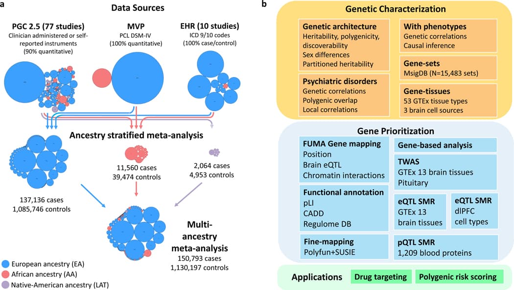
Figure 1 |. Data sources and analyses in PTSD Freeze 3.

Results

Data collection and GWAS

The PGC-PTSD Freeze 3 data collection includes 1,307,247 individuals from 88 studies (Supplementary Table 1). Data in this freeze were assembled from three primary sources (Fig. 1a): PTSD studies based on clinician administered or self-reported instruments (Freeze 2.5 plus subsequently collected studies), MVP release 3 GWASs utilizing the Posttraumatic Stress Disorder Checklist (PCL for DSM-IV), and 10 biobank studies with electronic health record (EHR)-derived PTSD status. We included 95 GWASs, including EA (n = 1,222,882; effective sample size (neff) = 641,533), AA (n = 51,034; neff = 42,804) and LAT (n = 7,017; neff = 6,530) participants (Supplementary Table 2).

European ancestry PTSD GWAS

Population, screening, and case ascertainment differences between datasets led to the assumption that there would be substantial cross-dataset variation in PTSD genetic signal. We investigated this possibility using the software MiXeR. Overall, we found no evidence for subset-specific genetic causal variation (see Supplementary Note, Supplementary Tables 3 and 4 and Extended Data Fig. 1 for further details). Given the similarities of the PTSD subsets, we performed a sample-size weighted fixed-effects meta-analysis of GWAS. For the EA meta-analysis (137,136 cases and 1,085,746 controls), the GC lambda was 1.55, the LDSC intercept was 1.0524 (s.e. = 0.0097) (Supplementary Table 5), and the attenuation ratio was 0.0729 (s.e. = 0.0134), indicating that 92.7% of the observed inflation in test-statistics was due to polygenic signal; thus, artifacts produced only minimal inflation.

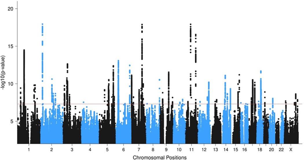
The EA meta-analysis identified 81 independent genome-wide significant (GWS) loci, including 5 GWS loci on the X chromosome (Extended Data Fig. 2, Supplementary Figs. 1 and 2, Supplementary Table 6, regional association plots in Supplementary Data 1, forest plots in Supplementary Data 2, and Supplementary Note). Relative to recent prior PTSD GWAS, 67 loci are novel (Supplementary Table 7). No region exhibited significant effect size heterogeneity (Supplementary Fig. 3).

We next sought to gain insights into whether loci harbor multiple independent variants. While FUMA annotations reported independent lead SNPs within risk loci based on pair-wise LD (Supplementary Table 8), COJO analysis of each locus conditional on the leading variants suggested that only one locus carried a conditionally independent GWS SNP (rs3132388 on chromosome 6, P = 2.86 × 10⁻⁹). This locus however, is in the MHC region, whose complicated linkage disequilibrium (LD) structure may not be accurately captured by reference panels.

African and Native American ancestry PTSD GWAS meta-analyses

The AA meta-analysis included 51,034 predominantly admixed individuals (n = 11,560 cases and 39,474 controls). There was minimal inflation of test statistics, with GC lambda = 1.031. No GWS loci were identified (Supplementary Fig. 4). The LAT meta-analysis was performed in 7,017 individuals (n = 2,064 cases and 4,953 controls). There was minimal inflation of test statistics (GC lambda = 0.993) and no GWS loci were identified (Supplementary Fig. 5).

Multi-ancestry GWAS meta-analysis

A multi-ancestry fixed-effects meta-analysis of EA, AA, and LAT GWAS (150,793 cases, 1,130,197 controls) identified 85 GWS loci. Compared to the EA meta-analysis, 10 loci lost GWS, while 14 previously suggestive loci (P < 5 × 10⁻⁷) became GWS (Fig. 2). In total, the present study identified 95 unique GWS PTSD loci between the EA and multi-ancestry meta-analyses (Table 1). Due to the complex local ancestry structure in AA and LAT individuals, which complicates LD modeling, we focused subsequent fine-mapping analyses (Fig. 1b) on data from the EA GWAS.
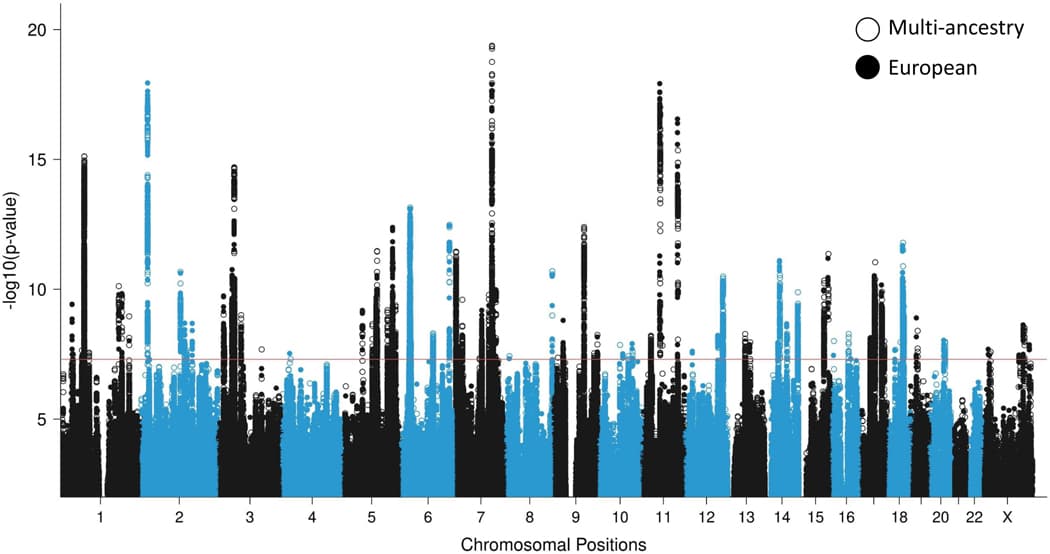
Figure 2 |. GWAS meta-analyses in European and multi-ancestry individuals identify a total of 95 PTSD risk loci.

Table 1 |. Genome-wide significant loci associated with PTSD in the multi-ancestry and European PGC-PTSD Freeze 3 data.

Table 1

Gene-mapping

To link GWS SNPs to relevant protein coding genes, we applied three gene mapping approaches implemented in FUMA: positional mapping, expression quantitative trait loci (eQTL), and chromatin interaction mapping (Supplementary Table 9). GWS SNPs within the 81 EA loci mapped to 415 protein coding genes under at least one mapping strategy. A total of 230 genes (55%) were mapped by two or more strategies, and 85 (20%) genes were mapped by all three strategies (Supplementary Fig. 6). Notably, some genes were implicated across independent risk loci by chromatin interactions/eQTL mapping, including EFNA5, GRIA1, FOXP2, MDFIC, WSB2, VSIG10, PEBP1, and C17orf58. Chromatin interaction plots are shown in Supplementary Data 3.

Functional annotation and fine-mapping of risk loci

Functional annotations were used to gain insights into the functional role of SNPs within the 81 risk loci (Supplementary Table 10): 72 loci contained at least one SNP with Combined Annotation Dependent Depletion (CADD) scores suggestive of deleteriousness to gene function (≥12.37), 43 loci contained GWS SNPs with Regulome DB scores likely to affect binding, and 23 loci contained at least one SNP in the exon region of a gene.

To narrow the credible window of risk loci and identify potentially causal SNPs, we fine-mapped loci using PolyFun+SUSIE, which identified a credible set for 67 loci. Credible set window lengths were on average 62% of the original set lengths (Supplementary Table 11) and contained a median of 23 credible SNPs (range 1–252). Only one contained a SNP with posterior inclusion probability > 0.95, a missense SNP in the exon of ANAPC4 (rs34811474, R[CGA]>Q[CAA]; Supplementary Table 12).

Gene-based, gene-set, and gene-tissue analyses

As an alternative approach to SNP-based association analysis, we tested the joint association of markers within genes using a gene-based association analysis in MAGMA, which is a 2-stage method that first maps SNPs to genes and then tests whether a gene is significantly associated with PTSD. The gene-based analysis identified 175 GWS genes (Supplementary Table 13 and Supplementary Fig. 7). Of these, 52 were distinct from the genes implicated by the gene-mapping of individual SNPs within GWS loci. These notably include DRD2, which has been thoroughly investigated in the context of psychiatric disorders and is a significant GWAS locus for multiple psychiatric disorders including schizophrenia (see Supplementary Note and Supplementary Table 14 for further investigation of conditionally independent SNPs within these 52 genes).

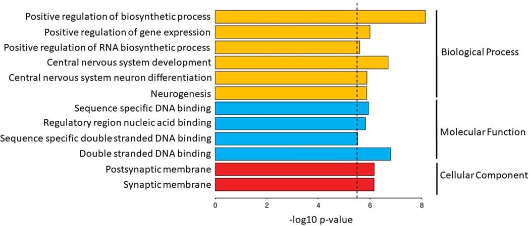
MAGMA gene-set analysis of 15,483 pathways and gene ontology (GO) terms from MSigDB identified 12 significant GO terms. Significant terms were related to the development and differentiation of neurons (e.g. go_central_nervous_system_development, P = 2.0 × 10⁻⁷), the synaptic membrane (e.g. go_postsynaptic_membrane, P = 6.9 × 10⁻⁷), gene regulation (go_positive_regulation_of_gene_expression, P = 1.0 × 10⁻⁶), and nucleic acid binding (P = 1.52 × 10⁻⁶) (Extended Data Fig. 3 and Supplementary Table 15).

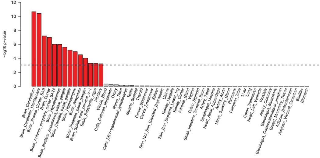
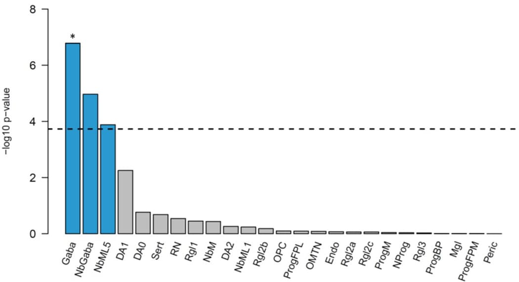
MAGMA gene-tissue analysis of 54 tissue types showed PTSD gene enrichment in the brain (most notably in cerebellum, but also cortex, hypothalamus, hippocampus and amygdala) and in the pituitary, with enrichment found across all 13 examined brain regions (Extended Data Fig. 4). Cell type analysis conducted in midbrain tissue data identified GABAergic neurons, GABA neuroblasts, and mediolateral neuroblast type 5 cell types as having enriched associations above other brain cell types tested (P < 0.05/268) (Extended Data Fig. 5). GABAergic neurons remained significant (P = 4.4 × 10⁻⁵) after stepwise conditional analysis of other significant cell types.

Multi-omic investigation of PTSD

To gain insights into which particular genes in enriched brain tissues were contributing to PTSD, we conducted a combination of a transcriptome-wide association study (TWAS) and summary based Mendelian randomization (SMR) analyses using GTEx brain tissue data based on the EA GWAS summary data. TWAS identified 25 genes within 9 loci with Bonferroni-significantly different genetically regulated expression levels between PTSD cases and controls (P < 0.05/14,935 unique genes tested) (Fig. 3a, Supplementary Fig. 8, and Supplementary Table 16). SMR identified 26 genes within 4 loci whose expression levels were putatively causally associated with PTSD (P < 0.05/9,003 unique genes tested) (Fig. 3b and Supplementary Table 17). Many of these genes have been previously implicated in PTSD and other psychiatric disorders (e.g., CACNA1E, CRHR1, FOXP2, MAPT, WNT3). Notably, the 3p21.31 (including RBM6, RNF123, MST1R, GMPPB, INKA1), 6p22.1 (including ZCAN9 and HCG17) and 17q21.31 (including ARHGAP27, ARL17A, CRHR1, MAPT, FAM215B, LRRC37A2, PLEKHM1, and SPPL2C) regions contained >10 putative causal genes each.
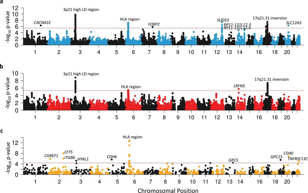
Figure 3 |. Manhattan plots of PTSD associations in multi-omic analyses.

Among the GTEx tissues with the most TWAS and SMR signals was the dorsolateral prefrontal cortex (dlPFC). To gain insight into cell type resolution, we conducted MAGMA for cell-type-specific markers of dlPFC and cell-type-specific SMR. MAGMA showed a significant enrichment of dlPFC inhibitory and excitatory neurons, but also of oligodendrocytes and oligodendrocyte precursor cells (Supplementary Table 18), while the SMR analyses identified cell-type-specific signals for 8 genes (KANSL1, ARL17B, LINC02210-CRHR1, LRRC37A2, ENSG00000262633, MAPT, ENSG00000273919, PLEKHM1) over 3 loci (6 out of 8 from 17q21.31) and all cell types (P < 0.05/1,885 unique genes tested) whose expression levels were potentially causally associated with PTSD (Supplementary Table 19). The top gene, KANSL1, was significant in all cell types.

Given previously reported associations between blood-based protein levels and PTSD, we performed protein quantitative trait loci (pQTL) SMR analysis for PTSD using data from the UK Biobank Pharma Proteomics Project (n = 54,306 samples and n = 1,209 proteins). We identified 16 genes within 9 loci whose protein levels were significantly associated with PTSD (P < 0.05/1,209 and PHEIDI > 0.05) (Fig. 3c and Supplementary Table 20), including members of the TNF superfamily (e.g., CD40, TNFRSF13C), implicating TNF-related immune activation in PTSD.

Gene prioritization

One research objective was to identify the genes with the greatest evidence of being responsible for the associations observed at each identified PTSD locus. Following recent research methods, we prioritized genes based on weighted sum of evidence scores taken across the functional annotation and post-GWAS analyses (Fig. 1b). Based on the absolute and relative scores of genes within risk loci, we ranked genes into Tier 1 (greater likelihood of being the causal risk gene) and Tier 2 (prioritized over other GWAS-implicated genes, but lower likelihood than Tier 1 of being the causal gene). 75% of loci contained prioritized genes (Tier 1 or Tier 2); the remaining loci did not contain any genes over the minimum threshold of evidence (score ≥ 4) to suggest prioritization. The prioritized genes for the top 20% of loci (ranked by locus P-value) are shown in Figure 4. A complete list of scores and rankings for all 415 protein coding genes mapped to risk loci is available in Supplementary Data 4.
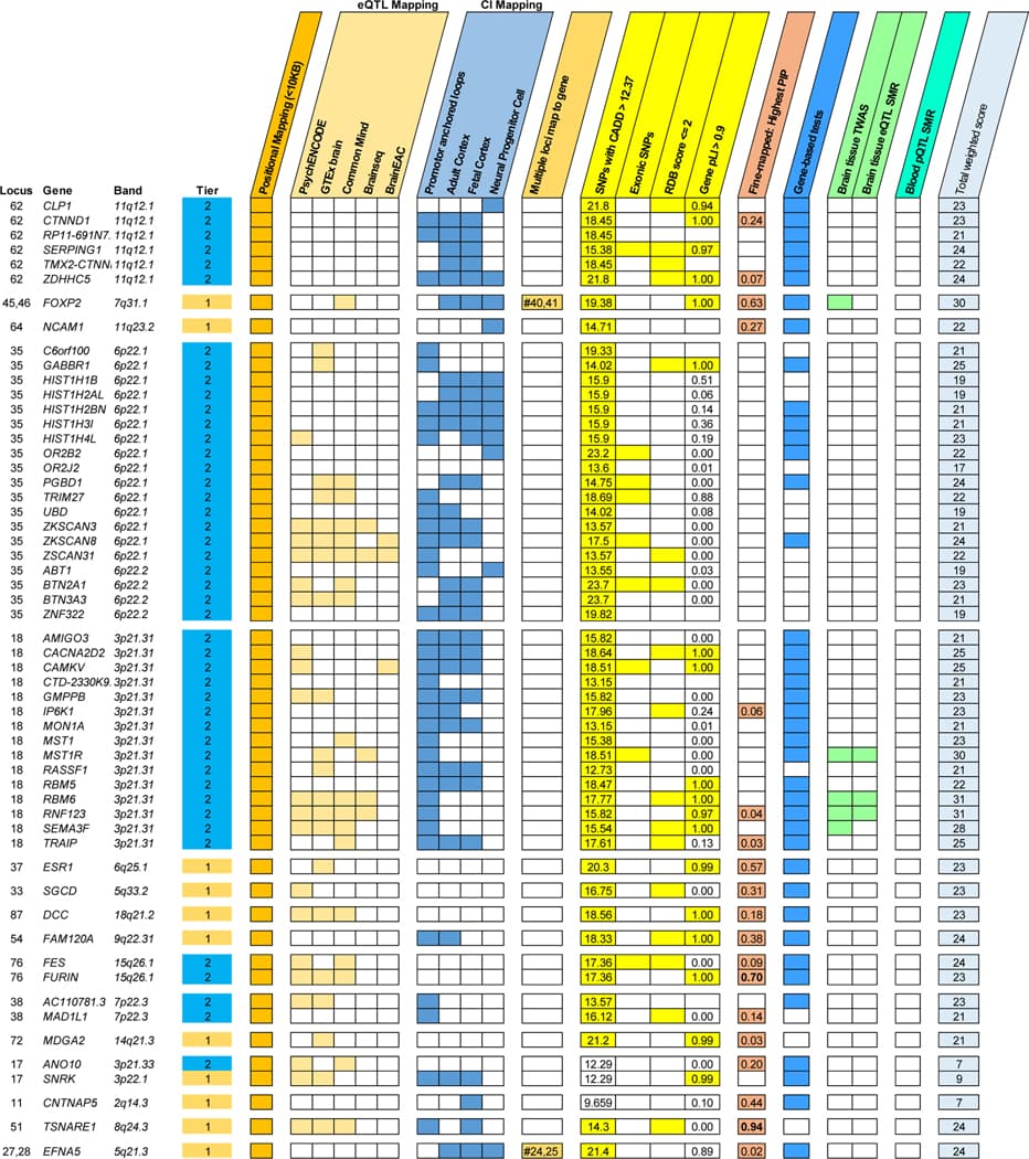
Figure 4 |. Gene prioritization in PTSD loci.

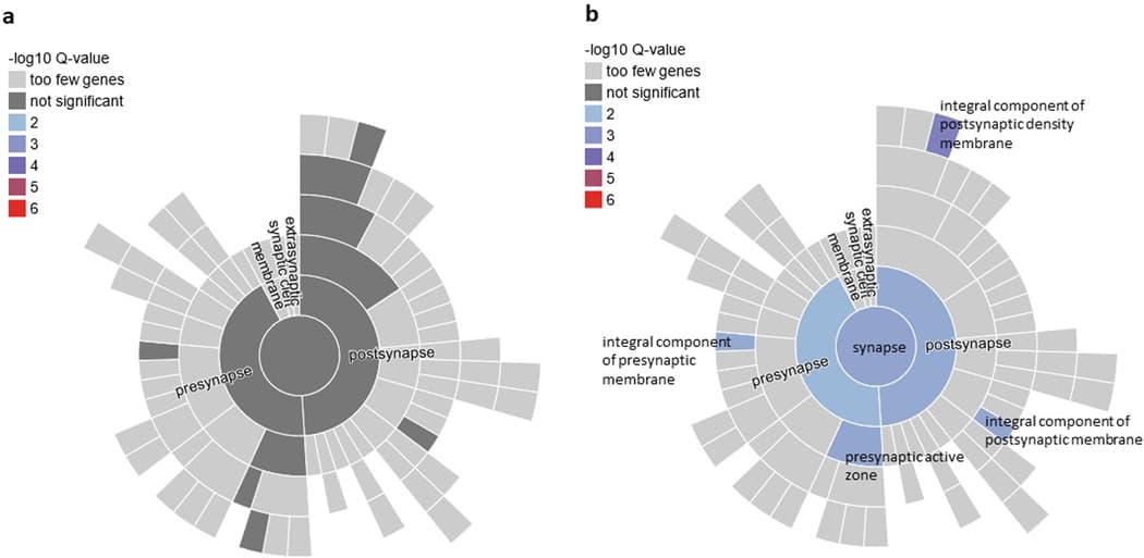
We performed pathway enrichment analysis of the Tier 1 genes in SynGO. From Tier 1, 11 genes mapped to the set of SynGO annotated genes (CACNA1E, DCC, EFNA5, GRIA1, GRM8, LRFN5, MDGA2, NCAM1, OLFM1, PCLO, and SORCS3). Relative to other brain-expressed genes, Tier 1 genes were significantly overrepresented in the synapse (P = 0.0009, qFDR = 0.003), pre- and post-synapse (P = 0.0086, qFDR = 0.0086 and P = 0.003, qFDR = 0.004, respectively), and four subcategories (Extended Data Fig. 6). By contrast, there was no significant overrepresentation of genes when we applied this test to the entire set of 415 protein coding genes. Other notable Tier 1 genes included PDE4B related to synaptic function and TNF-related immune-regulatory genes, including TANK and TRAF3.

Genetic architecture of PTSD

SNP-based heritability (h2SNP) estimated by LDSC was 0.053 (s.e. = 0.002, P = 6.8 × 10−156). Whereas previous reports suggested sex-specific differences in PTSD11, no significant differences were found (P = 0.13), and rg between male and female subsets was high (rg = 0.98, s.e. = 0.05, P = 1.2 × 10−98; Supplementary Table 5). MiXeR estimated 10,863 (s.e. = 377) influential variants and a discoverability of 7.4 × 10⁻⁶ (s.e. = 2.2 × 10⁻⁷) (Supplementary Table 3), indicating a genetic architecture comparable to other psychiatric disorders.

Partitioned heritability across 28 functional categories identified enrichment in histone markers (H3K9ac peaks: 6.3-fold enrichment, s.e. = 1.12, P = 3.11 × 10⁻⁶; H3K4me1: 1.5-fold enrichment, s.e. = 0.14, P = 3.3 × 10⁻⁴; Supplementary Table 21), and in evolutionary constrained regions across 29 Eutherians (18.37-fold enrichment, s.e. = 1.18, P = 1.29 × 10⁻¹⁷). This is consistent with findings for multiple other psychiatric disorders, but has not been previously identified in PTSD.

Contextualization of PTSD among psychiatric disorders

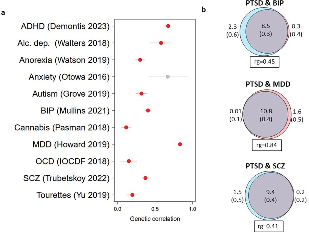
We measured the genetic overlap between PTSD and other psychiatric disorders using the most recent available datasets. We observed moderate to high positive rg between PTSD and other psychiatric disorders (Extended Data Fig. 7a). To gain further insights into this overlap, we used MiXeR to quantify the genetic overlap in causal variation between PTSD and bipolar disorder (BPD), MDD, and schizophrenia (SCZ) (Extended Data Fig. 7b). The strong majority (79–99%) of the variation influencing PTSD risk also influenced these disorders (Extended Data Fig. 7b and Supplementary Tables 22 and 23). Similar to rg, PTSD had the highest fraction of concordant effect directions with MDD (among the shared variation) (87% concordant, s.e. = 2%), significantly higher than the directional concordance with BPD (67%, s.e. = 1%) and SCZ (65%, s.e. = 0.5%).

While our results indicate an overall strong rg between PTSD and MDD (rg = 0.85, s.e. = 0.008, P < 2 × 10⁻¹⁶), the correlation between PTSD and MDD varied significantly across PTSD subsets, with the most homogeneously assessed subset, MVP, showing the lowest correlation, and the biobank subset being most strongly associated (Supplementary Table 24). Further, to evaluate if specific genetic regions differ substantially from genome-wide estimates, we used LAVA to estimate the local h2SNP and rg of PTSD and MDD across the genome, as partitioned into 2,495 approximately independent regions (Supplementary Table 25). Local h2SNP was significant (P < 0.05/2,495) for both PTSD and MDD in 141 regions. Of these, local rg was significant (P < 0.05/141) in 40 regions, all in the positive effect direction, where the mean local rg was 0.57 (s.d. = 0.24). In addition, we assessed the local rg between PTSD and MDD specifically for the 76 autosomal GWS EA loci (Supplementary Table 26). While LAVA identified 20 significantly correlated loci (rg < 6.58 × 10⁻⁴), there was also evidence for PTSD loci lacking evidence for correlation with MDD (Supplementary Figs. 9 and 10 showcase 6 selected loci with low and high rg).

Contextualization of PTSD across other phenotype domains

Considering all 1,114 traits with SNP-based heritability z > 6 available from the Pan-UKB analysis, we observed Bonferroni-significant rg of PTSD with 73% of them (Supplementary Table 27). Examining the extremes of estimates observed, the top positive rg was with sertraline prescription (rg = 0.88, P = 3.25 × 10⁻²⁰), a medication frequently prescribed for PTSD and other internalizing disorders. Other leading associations included medication poisonings (e.g. “Poisoning by psychotropic agents” rg = 0.88, P = 3.92 × 10⁻²⁰), which could support a link with accidental poisonings or self-harm behaviors. Converging with epidemiologic studies, there were correlations with gastrointestinal symptoms (e.g., “Nausea and vomiting” rg = 0.80, P = 2.39 × 10⁻¹⁶), mental health comorbidities (e.g., “Probable Recurrent major depression (severe)” rg = 0.87, P = 1.18 × 10⁻¹⁸; “Recent restlessness” rg = 0.86, P = 4.21 × 10−54), chronic pain (multi-site chronic pain rg = 0.63, P = 7.5 × 10−301) and reduced longevity (“Mother’s age at death” rg = −0.51, P = 7.6 × 10⁻²⁷).

Drug target and class analysis

We extended MAGMA gene-set analysis to investigate 1,530 gene sets comprising known drug targets (Supplementary Table 28). We identified one drug (stanozolol, an anabolic steroid) significantly enriched for targets associated with PTSD (P = 1.62 × 10⁻⁵). However, stanozolol has only two target genes in our analyses (ESR1, JUN), and likely reflects the strong association of ESR1 with PTSD in gene-level analyses (P = 8.94 × 10⁻¹²).

We further examined whether high-ranking drug targets were enriched for 159 drug classes defined by Anatomical Therapeutic Chemical (ATC) codes. We identified two broad classes where drugs were significantly enriched for association in drug target analyses (Supplementary Table 29). These were opioid drugs (ATC code N02A, P = 2.75 × 10⁻⁴), and psycholeptics (ATC code N05, P = 3.62 × 10⁻⁵), particularly antipsychotics (ATC code N05A, P = 3.55 × 10⁻⁷). However, sensitivity analyses limited to drugs with 10 or more targets identified no significant drug target sets nor drug classes.

Polygenic predictive scoring

We evaluated the predictive accuracy of PRS based on PTSD Freeze 3 in a set of MVP holdout samples (Fig. 5). In EA holdouts, risk was significantly different across the range of PTSD PRS. For example, individuals in the highest quintile of PTSD PRS had 2.4 times the relative risk of PTSD (log relative risk s.e. = 0.032; 95%CI = [2.25, 2.56]; P = 1.16 × 10−167) than individuals in the lowest quintile. PRS explained 6.6% of the phenotypic variation in PTSD (Nagelkerke’s R2 transformed to the liability scale at 15% population and sample prevalence), representing a major improvement over PRS based on Freeze 2. In contrast, among AA holdout samples, PRS explained only 0.9% (liability scale) of the variation in PTSD, consistent with previous work suggesting that AA PRS based on EA data lag behind in prediction.
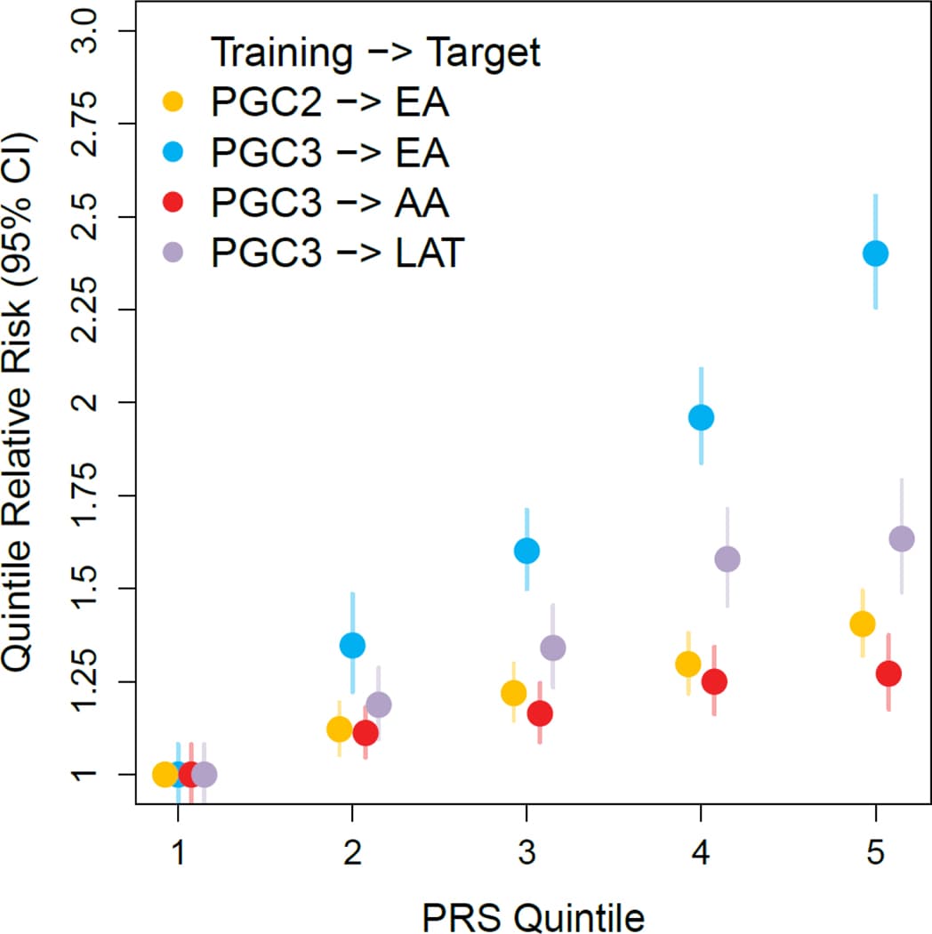
Figure 5 |. Polygenic risk score analysis for PTSD across different data sets and ancestries.

Discussion

In the largest PTSD GWAS to date, we analyzed data from over one million individuals and identified a total of 95 independent risk loci across analyses, a five-fold increase over the most recent PTSD GWAS. Compared to previous PTSD GWAS, we confirmed 14 out of 24 loci and identified 80 novel PTSD loci. Variant discovery in psychiatric GWAS follows a sigmoid curve, rapidly increasing once sample size passes a given threshold. This analysis passes that inflection point in PTSD, thus representing a major milestone in PTSD genetics. Moreover, by leveraging complementary research methodologies, our findings provide new functional insights and a deeper characterization of the genetic architecture of PTSD.

Tissue and cell-type enrichments revealed involvement of cerebellum, in addition to other traditionally PTSD-associated brain regions, and interneurons in PTSD risk. Structural alterations in the cerebellum are associated with PTSD, and large postmortem transcriptomic studies of PTSD consistently reveal differential expression of interneuron markers in prefrontal cortical tissue and amygdala nuclei. We used a combination of TWAS and SMR to probe the causal genes operating within the enriched tissues and cell types with brain transcriptomic data. The identified signals were concentrated in some GWAS loci like 17q21.31 whose inversion region is associated with a range of psychiatric phenotypes and linked to changes in brain structure and function. KANSL1, ARL17B, LINC02210-CRHR1 (encoding a fusion protein with CRHR1) and LRRC37A2 were the top causal genes in both neuronal and non-neuronal cell-types. KANSL1 plays a critical role in brain development. Furthermore, the first single cell transcriptomic study of PTSD confirmed neuronal, excitatory and inhibitory, alterations in 17q21.31 with top alterations in ARL17B, LINC02210-CRHR1 and LRRC37A2, while also emphasizing the involvement of immune and glucocorticoid response in neurons.

Notably, although PTSD risk in epidemiological studies is higher in women than men, here we found no sex differences in heritability. Five loci on the X chromosome associated with the disorder. Our finding that the estrogen receptor (ESR1) gene was identified in GWAS, as well as observations of differential effects of estrogen levels on a variety PTSD symptoms, suggests the importance of further analyses of ESR1 as a potential mediator of observed sex differences.

Our analyses prioritized 43 genes as Tier 1 (likely causal) based on weighted sum of evidence scores taken across the functional annotation and post-GWAS analyses. These genes can broadly be classified as neurotransmitter and ion channel synaptic plasticity modulators (e.g., GRIA1, GRM8, CACNA1E), developmental, axon guidance and transcription factors (e.g., FOXP2, EFNA5, DCC), synaptic structure and function genes (e.g., PCLO, NCAM1, PDE4B), and endocrine and immune regulators (e.g., ESR1, TRAF3, TANK). Furthermore, many additional genes with known function in related pathways were genome-wide significant and met Tier 2 prioritization criteria (e.g., GABBR1, CACNA2D2, SLC12A5, CAMKV, SEMA3F, CTNND1, and CD40). Together, these top genes show a remarkable convergence with neural network, synaptic plasticity and immune processes implicated in psychiatric disease. Furthermore, CRHR1, WNT3, and FOXP2, among other genes, are implicated in preclinical and clinical work related to stress, fear and threat-processing brain regions thought to underlie the neurobiology of PTSD. These findings largely support existing mechanistic hypotheses, and it will be important to examine how these genes and pathways function in already identified stress-related neural circuits and biological systems. Furthermore, while some of the prioritized genes are largely within pathways currently indicated in PTSD, many of the specific genes and encoded proteins were not previously established and warrant further investigation. Additionally, many genes and noncoding RNAs were not previously identified in any psychiatric or stress-related disorder, and offer an important road map for determining next steps in understanding new mechanisms of vulnerability for posttraumatic psychopathology. Future mechanistic research in preclinical models should examine whether targeting combinations of these genes, for example via polygenic targeting, epigenetic, or knockdown approaches, would have increased power in regulating stress, fear, cognitive dysfunction or other symptoms and behaviors seen in PTSD.

We observed highly shared polygenicity between PTSD and other psychiatric disorders, albeit with effect discordance across the shared variation. In particular, in some cases we found that the genetic correlation of PTSD with MDD is as high or higher than genetic correlations between different cohorts, with different measures, of PTSD. Thus, our findings corroborate the hypothesis that psychiatric disorders share a substantial amount of risk variation but are differentiated by disorder-specific effect sizes. Across the disorders we assessed, the correlation between PTSD and MDD was highest, in agreement with existing genetic multi-factor models of psychopathology that consistently cluster these disorders together and concordant with their epidemiologic co-morbidity. Evaluation of local patterns of heritability and genetic correlation however indicates disorder-specific risk variation, which will serve as targets for follow-up in cross-disorder investigations. We note that as GWAS of psychiatric traits grow in size and power, the field is seeing relatively strong genetic correlations among these traits, as well as with other behavioral and medical traits. This likely reflects, in part, the reality that there is substantial shared genetic variance among these traits, while not excluding the consistent observations that: (1) these traits do vary considerably in the magnitude of their genetic correlations, and (2) local genetic correlations reveal even greater genetic heterogeneity among these traits than global genetic correlations alone would lead us to believe. Finally, while PTSD is the most well-understood psychiatric outcome of trauma exposure, it is well documented that trauma is a risk factor for many different psychiatric disorders, with perhaps depression as the highest risk. Thus, these shared areas of overlap may represent general trauma vulnerability as well.

Despite the high level of overall correlation between PTSD and depression, we also note certain areas of clear distinction. When we examined local genetic correlations between PTSD and depression within all significant loci from the EA PTSD GWAS, we found that there were some regions with significant local heritability for PTSD but not depression, suggestive of PTSD-specific signals. In contrast, we also find other regions with clear shared signals showing local correlation across depression and PTSD, indicating that we have the power to detect shared and distinct local heritability. Together these findings suggest several PTSD-specific loci worthy of further investigation.

Further identification of PTSD genetic loci will provide therapeutic insights. We explored whether genes targeted by specific drugs (and drug classes) were enriched for GWAS signal. These analyses provided tentative support for antipsychotics and opioid drugs – known psychiatric drug classes – and were driven by gene-wise associations with DRD2 (antipsychotics) and CYP2D6 (opioids). Atypical antipsychotics may have efficacy in treating severe PTSD, but otherwise their use is not supported. Similarly, whereas some observational studies find that chronic opioid use worsens PTSD outcomes, there is preclinical work motivating the further study of opioid subtype-specific targeting (e.g., partial MOR1 agonism, κ-type opioid receptor [KOR1] antagonism) in the treatment of comorbid PTSD and opioid use disorders. Analyses in better-powered datasets may identify drug repositioning opportunities and could use the predicted effect of associated variants on gene expression to indicate whether drug candidates would be beneficial or contraindicated in people with PTSD.

In summary, we report 81 loci associated with PTSD in EA meta-analysis and 85 loci when expanding to cross-ancestry analyses. While these results represent a milestone in PTSD genetics and point to exciting potential target genes, further investment into data collection from underrepresented populations of diverse ancestries is needed for identification of additional risk variants and to generate equitable and more robust PRS.

Methods

Participants and studies

PTSD assessment and DNA collection for GWAS analysis were performed by each study following their protocols. A description of the studies included and the phenotypic and genotyping methods for each study are provided in the Supplementary Note and Supplementary Table 1. We complied with relevant ethical regulations for human research. All participants provided written informed consent, and studies were approved by the relevant institutional review boards and the UCSD IRB (protocol #16097×).

EHR studies

A total of 10 EHR-based cohorts (not including the MVP, which also contributed data) provided GWAS summary statistics. These cohorts consisted of four US-based sites (Vanderbilt University Medical Center’s BioVu, the Mass General Brigham Biobank, Mount Sinai’s BioMe, and Mayo Clinic’s MayoGC) and six non-US sites (iPSYCH from Denmark, FinnGen, HUNT Study from Norway, STR-STAGE from Sweden, UK Biobank, and Estonia Biobank). More details on procedures at each site are provided in the Supplementary Note. At each site, a broad definition of PTSD cases was defined based on patients having at least 1 PTSD or other stress disorder code (see Supplementary Note for the list of corresponding ICD-9 and 10 codes). All other patients without such a code were defined as controls. From a total of 817,181 participants across all cohorts, this case definition resulted in 78,687 cases based on the broad definition (9.6%).

Data assimilation

Subjects were genotyped on Illumina (n = 84 studies) or Affymetrix genotyping arrays (n = 5 studies) (Supplementary Table 1). Studies that provided direct access to pre-quality control genotype data (n = 64 studies) were deposited on the LISA server for central processing and analysis by the PGC-PTSD analyst. Studies with data sharing restrictions (n = 24 studies) were processed and analyzed following their own site-specific protocols (Supplementary Table 28) and shared GWAS summary statistics for inclusion in meta-analysis.

Genotype quality control and imputation

Genotype data was processed separately by study. For genotype data processed by the PGC-PTSD analyst, quality control was performed using a uniform set of criteria, as implemented in the RICOPILI pipeline version 2019_Oct_15.001. Modifications were made to the pipeline to allow for ancestrally diverse data and are noted where applicable. Quality control: using SNPs with call rates >95%, samples were excluded with call rates <98%, deviation from expected inbreeding coefficient (fhet < −0.2 or >0.2), or a sex discrepancy between reported and estimated sex based on inbreeding coefficients calculated from SNPs on X chromosomes. SNPs were excluded for call rates <98%, a > 2% difference in missing genotypes between cases and controls, or being monomorphic. Hardy-Weinberg equilibrium was calculated within only in the largest homogenous ancestry group found in the data. SNPs with a Hardy-Weinberg equilibrium P-value < 1 × 10⁻⁶ in controls were excluded.

After quality control, datasets were lifted over to the GRCh37/hg19 human genome reference build. SNP name inconsistencies were corrected, and genotypes were aligned to the strand of the imputation reference panel. Markers with non-matching allele codes or with excessive MAF difference (> 0.15) with the selected corresponding population in the reference data were removed. The pipeline was modified so that only the largest homogenous ancestry group in the data was used for the calculation of MAF. For ambiguous markers, strand was matched by comparing allele frequencies: if a strand flip resulted in a lower MAF difference between the study and the reference data, the strand was flipped. Ambiguous markers with high MAF (> 0.4) were removed. The genome was broken into 132 approximately equally sized chunks. For each chunk, genotypes were phased using Eagle v2.3.5 and phased genotypes were imputed into the Haplotype Reference Consortium panel86 using minimac3. Imputed datasets were deposited with the PGC DAC and are available for approved requests.

Studies with data sharing restrictions followed similar criteria for quality control, as detailed in Supplementary Table 28 and in the references in the supplemental material. Studies were imputed to either the 1000G phase 3, HRC, SISu panel, or a composite panel. GWAS summary data were lifted to the GRCh37 reference build where required. As differences in the imputation panels and genome reference build can result in SNP-level discrepancies between datasets, each set of summary data was examined for correspondence to the centrally imputed data. Multi-allelic SNPs and SNPs with non-matching allele codes were excluded. Strand ambiguous SNPs with high MAF difference (>20%) from the average frequency calculated in the PGC-PTSD data were flagged and examined for strand correspondence.

Ancestry determination

For studies where the PGC analyst had genotype data access, ancestry was determined using a global reference panel using SNPweights. The ancestry pipeline was shared with external sites to be utilized where possible. Participants were placed into three large groupings: European and European Americans (EA; individuals with ≥90% European ancestry), African and African-Americans (AA; individuals with ≥5% African ancestry, <90% European ancestry, <5% East Asian, Native American, Oceanian, and Central-South Asian ancestry; and individuals with ≥50% African ancestry, <5% Native American, Oceanian, and <1% Asian ancestry), and Latin Americans (LAT; individuals with ≥5% Native American ancestry, <90% European, <5% African, East Asian, Oceanian, and Central-South Asian ancestry). Native Americans (individuals with ≥60% Native American ancestry, <20% East Asian, <15% Central-South Asian, and <5% African and Oceanian ancestry) were grouped together with LAT. All other individuals were excluded from the current analyses. For the MVP cohort, ancestry was determined using standard principal components analysis approach where MVP samples were projected onto a PC space made from 1000 Genomes Phase 3 (KGP3) samples with known population origins (EUR, AFR, EAS, SAS, and AMR populations). EHR cohorts followed their own site-specific ancestry classification protocols.

GWAS

GWAS was performed with stratification by ancestry group and study. Strata were only analyzed if they had a minimum of 50 cases and 50 controls, or alternatively 200 participants total. Where noted (Supplementary Table 2), small studies of similar composition were jointly genotyped so they could be analyzed together as a single unit. For GWAS, the association between each SNP and PTSD was tested under an additive genetic model, using a regression model appropriate to the data structure. The statistical model, covariates, and analysis software used to analyze each study is detailed in Supplementary Table 30. In brief, studies of unrelated individuals with continuous (case/control) measures of PTSD were analyzed using PLINK 1.9 using a linear (logistic) regression model that included 5 PCs as covariates. For studies that retained related individuals, analyses were performed using methods that account for relatedness. QIMR was analyzed using GEMMA v0.96, including the first five PCs as covariates. RCOG was analyzed using the generalized disequilibrium test. UKBB was analyzed using BOLT-LMM including 6 PCs, and batch and center indicator variables as covariates. VETS was analyzed using BOLT-LMM including 5 PCs as covariates. EHR-based studies that included related individuals were analyzed using saddle point approximation methods to account for case/control imbalances. AGDS and QIM2 were analyzed using SAIGE including 4 PCs and study-specific covariates. BIOV was analyzed using SAIGE including 10 PCs and age of record. ESBB, FING, HUNT, and SWED were analyzed using SAIGE including 5 PCs. UKB2 was analyzed using REGENIE including 6 PCs, assessment center, and genotyping batch covariates. GWAS was additionally performed stratified by sex. For the X chromosome analysis, sex was added as a covariate.

Meta-analysis

Sample-size weighted fixed-effects meta-analysis was performed with METAL. Within each dataset and ancestry group, summary statistics were filtered to MAF ≥ 1% and imputation information score ≥ 0.6. Meta-analyses were performed within the EA, AA, and LAT ancestry groups. A multi-ancestry meta-analysis was performed as the meta-analysis of the three meta-analyses. Genome-wide significance was declared at P < 5 × 10⁻⁸. Heterogeneity between datasets was tested with the Cochran test. Markers with summary statistics in less than 80% of the total effective sample size were removed from meta-analyses. LDSC intercept was used to estimate inflation of test statistics related to artifacts rather than genetic signal. The proportion of inflation of test statistics due to the actual polygenic signal (rather than other causes such as population stratification) was estimated as 1 − (LDSC intercept − 1)/(mean observed Chi-square − 1).

Regional association plots

Regional association plots were generated using LocusZoom with 1.5-Mb windows around the index variant (unless the locus region was wider than 1.5 Mb, in which case it was the locus region plotted plus an additional buffer to include data up to the recombination region). The LD patterns plotted were based on the 1000 Genomes Phase 3 reference data, where a sample ancestry appropriate subpopulation (EUR, AFR, or AMR) was used.

Conditional analysis of significant loci

To determine if there were independent significant SNPs within risk loci, GCTA Conditional and Joint Analysis was performed. Stepwise selection was performed using the --cojo-slct option and default parameters, where UKBB European genotype data was used to model LD structure.

SNP heritability

h2SNP of PTSD was estimated using LDSC. LD scores calculated within KGP3 European populations (https://data.broadinstitute.org/alkesgroup/LDSCORE/) were used for the input. Analyses were limited to HapMap 3 SNPs, with the MHC region excluded (chr6: 26–34 million base pairs). SNP-based heritability was also calculated as partitioned across 28 functional annotation categories (https://data.broadinstitute.org/alkesgroup/LDSCORE/) using stratified LDSC.

Comparisons of genetic architecture

We used univariate MiXeR (version 1.3)to contrast the genetic architecture of phenotypes. MiXeR estimates SNP-based heritability and two components that are proportional to heritability: the proportion of non-null SNPs (polygenicity), and the variance of effect sizes of non-null SNPs (discoverability). MiXeR was applied to GWAS summary statistics under the default settings with the supplied European ancestry LD reference panel. The results reported for the number of influential variants reflects the number of SNPs necessary to explain 90% of SNP-based heritability. Bivariate MiXeR was used to estimate phenotype-specific polygenicity and the shared polygenicity between phenotypes. Goodness of fit of the MiXeR model relative to simpler models of polygenic overlap was assessed using AIC values. Heritability, polygenicity and discoverability estimates were contrasted between datasets using the z-test.

Local genetic correlation analyses

Local h2SNP and rg between PTSD and MDD were estimated using LAVA. KGP3 European data were used as the LD reference. Local h2SNP and rg were evaluated across the genome, as partitioned into 2,495 approximately equally sized LD blocks. Local rg was only evaluated for loci where local heritability was significant (P < 0.05/2,495) in both phenotypes. Significance of local rg was based on Bonferroni adjustment for the number of rg evaluated.

Polygenic risk scores (PRS)

PRS were calculated in ancestry-stratified MVP holdout samples, based on the EA Freeze 3 PTSD GWAS. GWAS summary statistics were filtered to common (MAF > 1%), well-imputed variants (INFO > 0.8). Indels and ambiguous SNPs were removed. PRS-CS was used to infer posterior effect sizes of SNPs, using the KGP3 EUR based LD reference panel supplied with the program, with the global shrinkage parameter set to 0.01, 1,000 MCMC iterations with 500 burn-in iterations, and the Markov chain thinning factor set to 5. PRS were calculated using the --score option in PLINK 1.9, using the best-guess genotype data of target samples, where for each SNP the risk score was estimated as the posterior effect size multiplied by the number of copies of the risk allele. PRS was estimated as the sum of risk scores over all SNPs. PRS were used to predict PTSD status under logistic regression, adjusting for 5 PCs. The proportion of variance explained by PRS for each study was estimated as the difference in Nagelkerke’s R2 between a model containing PRS plus covariates and a model with only covariates.

Functional mapping and annotation

We used the SNP2GENE module in FUMA v1.4.1 (https://fuma.ctglab.nl) to annotate and visualize GWAS results. The complete set of parameters used for FUMA analysis are shown in the Supplementary Note. Independent genomic risk loci were identified (r2 < 0.6, calculated using ancestry-appropriate KGP3 reference genotypes). SNPs within risk loci were mapped to protein coding genes using positional mapping (10-kb window), eQTL mapping (GTEx v8 brain tissue, BRAINEAC, and CommonMind data sources), and chromatin interaction mapping (PsychENCODE and HiC of brain tissue types) methods. Chromatin interactions and eQTLs were plotted in circos plots. SNPs were annotated to functional annotation databases including ANNOVAR, CADD, and RegulomeDB.

Novelty of risk loci

The start and stop positions of independent risk loci were assessed for positional overlap with existing PTSD loci11–13. Loci were declared novel if their boundaries did not overlap with a variant reported significant in prior GWAS.

MAGMA gene-based and gene-set analyses

Gene-based association analyses were conducted using MAGMA v1.08. SNPs were positionally mapped (0-kb window) to 19,106 protein-coding genes. The SNP-wide mean model was used to derive gene-level P-values, with an ancestry appropriate KGP3 reference panel was used to model LD. Significance was declared based on Bonferroni adjustment for the number of genes tested. Gene-based association statistics were used in MAGMA for gene-set and gene-property analyses. Gene-set analysis used the MsigDB version 7.0 including 15,483 curated gene-sets and gene-ontology (GO) terms. Gene-property analysis of tissues and tissue subtypes was performed using GTEx v8 expression data, with adjustment for the average expression of all tissues in the dataset. To evaluate cell type specific enrichment, the FUMA cell type module was used, selecting 12 datasets related to the brain (full list in Supplementary Note). Finally, MAGMA was used to estimate the enrichment of dlPFC cell types in PTSD risk based on the DER21 marker gene list from PsychEncode Consortium Phase 1 resource release.

GWAS fine-mapping

Polygenic functionally informed fine-mapping (PolyFun) software was used to annotate our results data with per-SNP heritabilities, as derived from a meta-analysis of 15 UK Biobank traits. PTSD risk loci were fine-mapped using SUSIE, with these per SNP heritabilities used as priors, pre-computed UKB-based summary LD information used as the LD reference, and locus start and end positions as determined by FUMA. The SUSIE model assumed a maximum of two causal variants.

Expression quantitative trait loci (eQTL) and blood protein quantitative trait loci (pQTL) analyses

To test for a joint association between GWAS summary statistics SNPs and eQTL, the SMR method, a Mendelian randomization approach, was used. SMR software (version 1.03) was run using the default settings. The European samples of the 1000G were used as a reference panel. Bonferroni multiple-testing correction was applied on SMR P-value (PSMR). Moreover, a post-filtering step was applied by conducting heterogeneity in dependent instruments (HEIDI) test. The HEIDI test distinguishes the causality and pleiotropy models from the linkage model by considering the pattern of associations using all SNPs significantly associated with gene expression in the cis-eQTL region. The null hypothesis is that a single variant is associated with both trait and gene expression, while the alternative hypothesis is that trait and gene expression are associated with two distinct variants. Finally, gene-trait associations based on SMR-HEIDΙ were defined as the ones for which PSMR met the Bonferroni significance threshold and had PHEIDI > 0.05. We conducted a combination of SMR and HEIDI based on GTEx project latest (version 8) multi-tissue cis-eQTL databases from 13 brain regions and pituitary tissue that showed significant enrichment in MAGMA/FUMA analyses (see above). We also used cell-type-specific eQTLs in dlPFC for SMR analyses. Finally, we used a blood UK Biobank pQTLs database of 1,463 plasma proteins relying on a very large population (54,306) for SMR/HEIDI analysis to evaluate biomarker potential.

Brain focused TWAS

JEPEGMIX2-P software with default settings was used to conduct TWAS on 13 brain regions and pituitary tissue that showed significant enrichment in MAGMA/FUMA analyses using our PEC-DLPFC GReX model. JEPEGMIX2-P was applied on GWAS summary statistics to estimate gene-trait associations. This method was preferable since it relied on a covariance matrix based on 33k samples compared to other TWAS methods which use less than 3k samples. To determine significance, a Bonferroni correction threshold for the unique number of genes tested was applied (P < 0.05/14,935). As a less conservative approach, we also applied FDR at a q-value threshold of 0.05.

Gene prioritization

Genes within risk loci were prioritized following the general approach previously described. Genes were given prioritization scores based on the weighted sum of evidence across all evidence categories: FUMA positional, eQTL, and CI mapping, variant and gene annotation scores (CADD, predicted loss of impact [pLI], and RDB scores), positional overlap in fine-mapping, significance in gene-based analyses, brain tissue TWAS, eQTL SMR, and pQTL SMR. Weights for each evidence category are provided in Supplementary Table 31. Within a given locus, the evidence scores were compared across genes to identify the most likely causal gene. Genes with scores ≥ 4 were ranked as either Tier 1 (greater likelihood of being the causal risk gene) or Tier 2 (lower likelihood of being the causal risk gene) and genes with scores < 4 were left unranked. The ranking algorithm is as follows. For a given locus, if there was a gene whose evidence score ≥ 4 and this gene’s score was > 20% higher than all other genes in the locus, it was ranked as a Tier 1 gene (greater likelihood of being the causal risk gene). Within a locus with a Tier 1 gene, other genes with scores between 20% and 50% lower than the Tier 1 gene were labeled as Tier 2. For loci without a Tier 1 gene, all genes with scores ≥ 4 that were within 50% of the leading gene were ranked as Tier 2.

SynGO

PTSD-related genes were tested for overrepresentation among genes related to synaptic terms in the SynGO web interface (https://www.syngoportal.org/). Brain expressed genes were selected as the background list for the overrepresentation tests. SynGO terms with FDR q < 0.05 were considered as being overrepresented.

Drug targeting analyses

Following a previously described approach, we analyzed the enrichment of gene-level associations with PTSD in genes targeted by individual drugs. We then examined the enrichment of specific drug classes among these drug target associations. We obtained gene-level associations using MAGMA v1.08. Variant-level associations were converted to gene-level associations using the “multi=snp-wise” model, which aggregates Z scores derived from the lowest and the mean variant-level P value within the gene boundary. We set gene boundaries 35 kb upstream and 10 kb downstream of the transcribed regions from build 37 reference data (National Center for Biotechnology Information, available at https://ctg.cncr.nl/software/magma).

We performed drug target analysis using competitive gene-set tests implemented in MAGMA. Drug target sets were defined as the targets of each drug from: the Drug–Gene Interaction database DGIdb v.4.2.0, the Psychoactive Drug Screening Database Ki DB, ChEMBL v27, the Target Central Resource Database v6.7.0, and DSigDB v1.0, all downloaded in October 2020. We additionally used the drug target sets to identify targets of drugs of interest from gene-based analyses.

We grouped drugs according to the Anatomical Therapeutic Chemical class of the drug. Results from the drug target analysis were ranked, and the enrichment of each class in the drug target analysis was assessed with enrichment curves. We calculated the area under the enrichment curve and compared the ranks of drugs within the class to those outside the class using the Wilcoxon Mann-Whitney test. Multiple testing was controlled using a Bonferroni-corrected significance threshold of P < 3.27 × 10⁻⁵ for drug target analysis and P < 4.42 × 10⁻⁴ for drug class analysis, accounting for 1,530 drug sets and 113 drug classes tested.

We initially limited drug target analyses to drugs with two or more targets. However, results suggested this low limit may lead to false positive findings. As a sensitivity analysis, we further limited these analyses to drugs with 10 or more targets. Multiple testing was controlled using a Bonferroni-corrected significance threshold of P < 5.42 × 10⁻⁵ for drug target analysis and P < 7.94 × 10⁻⁴ for drug class analysis, accounting for 923 drug sets and 63 drug classes tested.

Genetic correlations and causal associations with other phenotypes

Using LDSC, we assessed the rg of PTSD derived from the PGC meta-analysis conducted in EUR cohorts with traits available from the Pan-UKB analysis conducted in EUR samples. Details regarding the Pan-UKB analysis are available at https://pan.ukbb.broadinstitute.org/. Briefly, Pan-UKB genome-wide association statistics were generated using the SAIGE and including a kinship matrix as a random effect and covariates as fixed effects. The covariates included age, sex, age × sex, age2, age2 × sex, and the top-10 within-ancestry principal components. We limited our analysis to data derived from UKB participants of European descent (n = 420,531) because of the limited sample size available in the other ancestry groups. Initially, we calculated SNP-based heritability of phenotypes available from Pan-UKB, retaining only those with SNP-based heritability z > 6 (Supplementary Table 25) as recommended by the developers of LDSC. To define traits genetically correlated with PTSD, we applied a Bonferroni correction accounting for the number of tests performed.

Extended Data
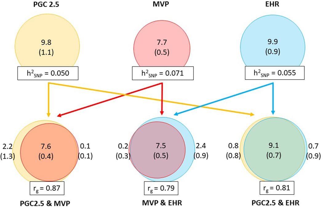
Extended data Figure 1: Comparison of the genetic architecture of PTSD in the three main data sources.Extended data Figure 2: Manhattan plot of the PTSD GWAS meta-analysis in individuals of European ancestry (EA).Extended Data Figure 3: Significant PTSD gene-sets.Extended Data Figure 4: MAGMA tissue enrichment analysis.Extended Data Figure 5: MAGMA cell-type enrichment analysis in midbrain.Extended Data Figure 6: PTSD genes in SynGO.Extended Data Figure 7: Genetic correlations and polygenic overlap between PTSD and other psychiatric disorders.

Supplementary Material

Supplementary Tables

NIHMS2015499-supplement-Supplementary_Tables.xlsx (5MB, xlsx)

Supplementary Information

NIHMS2015499-supplement-Supplementary_Information.docx (8.8MB, docx)

Supplementary Data 2

NIHMS2015499-supplement-Supplementary_Data_2.pdf (559KB, pdf)

Supplementary Data 1

NIHMS2015499-supplement-Supplementary_Data_1.pdf (3.5MB, pdf)

Supplementary Data 4

NIHMS2015499-supplement-Supplementary_Data_4.xlsx (61.4KB, xlsx)

Supplementary Data 3

NIHMS2015499-supplement-Supplementary_Data_3.pdf (8.1MB, pdf)

Supplementary Methods

NIHMS2015499-supplement-Supplementary_Methods.docx (338.1KB, docx)

Open Article as PDF

Abstract

Post-traumatic stress disorder (PTSD) genetics are characterized by lower discoverability than most other psychiatric disorders. The contribution to biological understanding from previous genetic studies has thus been limited. We performed a multi-ancestry meta-analysis of genome-wide association studies across 1,222,882 individuals of European ancestry (137,136 cases) and 58,051 admixed individuals with African and Native American ancestry (13,624 cases). We identified 95 genome-wide significant loci (80 new). Convergent multi-omic approaches identified 43 potential causal genes, broadly classified as neurotransmitter and ion channel synaptic modulators (for example, GRIA1, GRM8 and CACNA1E), developmental, axon guidance and transcription factors (for example, FOXP2, EFNA5 and DCC), synaptic structure and function genes (for example, PCLO, NCAM1 and PDE4B) and endocrine or immune regulators (for example, ESR1, TRAF3 and TANK). Additional top genes influence stress, immune, fear and threat-related processes, previously hypothesized to underlie PTSD neurobiology. These findings strengthen our understanding of neurobiological systems relevant to PTSD pathophysiology, while also opening new areas for investigation.

Introduction

Posttraumatic stress disorder (PTSD) involves intrusive thoughts, heightened reactions, avoidance, and negative changes in thinking and mood that can last a long time after a traumatic event. Globally, about 5.6% of adults exposed to trauma experience PTSD in their lifetime, with higher rates seen in combat survivors and assault victims. For many, PTSD is a long-lasting condition that significantly impacts their quality of life and places a burden on society.

Understanding the biological basis of PTSD has advanced significantly through studies, many of which focus on the brain's fear systems and are being applied to human research. Brain imaging studies suggest problems in brain circuits involved in fear, specifically issues with how regulatory areas like the anterior cingulate and ventromedial prefrontal cortex control the amygdala. Hormone studies have also found issues with the HPA axis and gene expression related to stress hormones in PTSD development and persistence. However, many questions about PTSD's underlying biology remain, and new targets are needed for prevention and treatment.

Studies of twins and genetics show that the risk of developing PTSD after trauma is partly influenced by inherited factors. The specific genetic makeup of PTSD is now becoming clearer as large-scale genetic studies become available. Recent research has shown that PTSD is a complex disorder influenced by many genes. Even with studies involving over 200,000 individuals, only a limited number of specific genetic locations linked to PTSD risk have been found, and these were not consistent across all datasets, indicating the need for even larger studies. Also, these studies often did not examine the X chromosome, which makes up 5% of the human genome and might be especially important given differences in PTSD rates between sexes.

Current genetic studies have also had limited success in identifying promising treatment options. PTSD frequently occurs with and shares genetic links with other mental health conditions, like major depressive disorder and attention deficit hyperactivity disorder, as well as physical health issues, such as cardiovascular disease and obesity. However, existing studies are limited in their ability to distinguish genetic factors unique to PTSD from those shared with other conditions and connect them to underlying biological systems. Importantly, previous genetic studies struggle to apply their findings to people of non-European ancestry. Advances in polygenic risk scores for PTSD show potential for research but have limited applicability across different populations. Without expanding research to other ancestries, there is a risk that genetic discoveries in PTSD could worsen existing research and treatment disparities, which is particularly concerning in the US where populations of African, Native, and Latin American origin face a disproportionately high burden of trauma and PTSD.

The current analysis combined data from 88 studies to perform a large-scale genetic meta-analysis across individuals of European, African, and Native American ancestries, including analysis of the X chromosome. Researchers then investigated these genetic findings to understand overall and localized genetic influences, infer the involvement of brain regions and neuronal systems, describe shared genetic effects with co-occurring conditions, and identify a set of potentially causal genes using multiple types of biological data. Finally, this information was used to pinpoint potential pathways for future PTSD treatment studies, representing significant progress toward understanding the biology of trauma and stress-related disorders and informing future interventions.

Results

The data collection for this study included over one million individuals from 88 different studies. Genetic information was gathered from various sources, including studies using clinician assessments or self-reports, a program for veterans, and biobank studies that used electronic health records to identify PTSD cases. These genetic analyses included participants of European, African, and Native American ancestries.

In the analysis of European ancestry participants, researchers investigated whether there were substantial differences in genetic signals across various datasets. No evidence was found for specific genetic differences between subsets. A meta-analysis of European ancestry data identified 81 independent genetic locations significantly linked to PTSD, including 5 on the X chromosome. A substantial number of these locations, 67, were newly discovered compared to previous PTSD genetic studies. Further detailed analysis suggested that most of these locations likely contain only one primary genetic variant contributing to risk.

Genetic analyses of African ancestry and Native American ancestry populations, although smaller, showed minimal statistical inflation, and no genetic locations reached genome-wide significance in these groups. However, a combined meta-analysis of all ancestries (European, African, and Native American) identified a total of 85 significant genetic locations. This multi-ancestry analysis revealed some changes compared to the European-only analysis, with some locations losing significance and others becoming significant. Overall, the study identified 95 unique genetic locations linked to PTSD across both European and multi-ancestry analyses.

To connect these significant genetic locations to specific genes, three mapping approaches were used, identifying 415 protein-coding genes in European ancestry individuals. A large portion of these genes were identified by multiple mapping strategies, and some genes were linked across several independent risk locations. Functional annotations provided insights into the role of these genetic variants, with many suggesting an impact on gene function or binding. Fine-mapping techniques were used to narrow down the specific genetic variants most likely to be causal within these locations, identifying credible sets of variants for most locations.

Beyond individual genetic variants, gene-based analyses identified 175 genes significantly associated with PTSD, including some not found through the direct mapping of individual variants, such as DRD2, which is well-known in psychiatric research. Gene-set analysis revealed that significant genetic pathways were related to neuron development and differentiation, synaptic membranes, gene regulation, and nucleic acid binding. Gene-tissue analysis showed PTSD gene enrichment primarily in brain regions, particularly the cerebellum, but also the cortex, hypothalamus, hippocampus, and amygdala. Cell type analysis of midbrain tissue further pinpointed GABAergic neurons and neuroblasts as having enriched associations.

A multi-omic investigation, combining genetic and gene expression data, aimed to identify genes in enriched brain tissues contributing to PTSD. This led to the identification of several genes whose expression levels were significantly different or causally linked to PTSD, many of which have been previously implicated in PTSD and other psychiatric disorders (e.g., CACNA1E, CRHR1, FOXP2, MAPT, WNT3). Specific chromosomal regions contained multiple such causal genes. Analysis of the dorsolateral prefrontal cortex, a key brain area, showed enrichment in inhibitory and excitatory neurons, as well as oligodendrocytes. Furthermore, analysis of blood protein levels identified genes, including members of the TNF superfamily, suggesting a role for immune activation in PTSD.

A key objective was to prioritize genes most likely responsible for the observed genetic associations. By combining evidence from functional annotation and various genetic analyses, 43 genes were prioritized as "Tier 1" (highly likely causal) and other genes as "Tier 2." The Tier 1 genes were found to be significantly overrepresented in the synapse and related structures, playing roles as neurotransmitter and ion channel modulators, developmental factors, and immune regulators.

Regarding the genetic architecture of PTSD, analyses estimated that common genetic variants explain a portion of the disorder's variability. No significant sex differences were found in this heritability. The genetic architecture was found to be comparable to other psychiatric disorders, involving many influential genetic variants. Analysis of partitioned heritability identified enrichment in specific histone markers and evolutionarily conserved regions, consistent with findings for other psychiatric disorders.

To understand PTSD in the context of other conditions, genetic overlap was measured with various psychiatric disorders, including bipolar disorder, major depressive disorder (MDD), and schizophrenia. Moderate to high positive genetic correlations were observed. A significant portion of the genetic variation influencing PTSD also influenced these other disorders, with the highest concordance in effect directions found with MDD. While there was a strong overall genetic correlation between PTSD and MDD, this correlation varied across different PTSD datasets. Localized genetic correlation analyses also indicated both shared and disorder-specific genetic influences across the genome. Beyond psychiatric disorders, PTSD showed significant genetic correlations with many other traits, including medication use, poisonings, gastrointestinal symptoms, other mental health issues, chronic pain, and reduced lifespan.

Investigations into drug targets and classes revealed limited but notable associations. One anabolic steroid, stanozolol, showed enrichment for targets associated with PTSD, largely driven by the estrogen receptor gene ESR1. Broader drug class analyses suggested enrichment for opioid drugs and psycholeptics, particularly antipsychotics. However, sensitivity analyses with stricter criteria for the number of drug targets did not yield significant results, highlighting the need for more powerful datasets.

Finally, polygenic risk scores (PRS) were evaluated for their ability to predict PTSD. In European ancestry individuals, those with the highest PRS had a significantly increased risk of PTSD, and PRS explained a notable portion of the phenotypic variation, marking a significant improvement over previous studies. In contrast, among African ancestry individuals, PRS explained a much smaller portion of the variation, consistent with previous observations of limited transferability of PRS based on European data to other populations.

Discussion

This study, the largest genetic analysis of PTSD to date, included over one million individuals and identified a total of 95 independent genetic risk locations, a five-fold increase compared to earlier studies. This marks a significant achievement in PTSD genetics, as the analysis reached a sample size threshold where variant discovery rapidly accelerates. The findings also offer new functional insights and a deeper understanding of PTSD's genetic makeup.

Analyses of tissue and cell types revealed that the cerebellum, along with other brain regions traditionally linked to PTSD, and specific types of interneurons are involved in PTSD risk. Structural changes in the cerebellum are known to be associated with PTSD, and large postmortem studies consistently show differences in interneuron markers in the prefrontal cortex and amygdala in PTSD. Combining genetic and expression data helped pinpoint causal genes operating within these brain tissues and cell types. Significant signals were concentrated in certain genetic regions, like 17q21.31, which has been linked to various psychiatric conditions and changes in brain structure and function. Genes such as KANSL1, ARL17B, and CRHR1 were identified as top causal candidates in both neuronal and non-neuronal cell types, with KANSL1 being crucial for brain development.

Despite epidemiological studies showing higher PTSD risk in women, this study found no sex differences in overall heritability. However, five genetic locations on the X chromosome were associated with the disorder. The identification of the estrogen receptor gene ESR1 in genetic analyses, along with observations of estrogen's varying effects on PTSD symptoms, suggests ESR1 is important for further investigating observed sex differences.

The study prioritized 43 genes as highly likely causal based on a weighted sum of evidence. These genes generally fall into categories such as modulators of neurotransmitters and ion channel synaptic plasticity, developmental and axon guidance factors, genes involved in synaptic structure and function, and regulators of the endocrine and immune systems. Many other genes with known functions in related pathways also met prioritization criteria. These top genes strongly align with neural network, synaptic plasticity, and immune processes already implicated in psychiatric disease. Furthermore, several identified genes are supported by preclinical and clinical research related to stress, fear, and threat-processing brain regions thought to be central to PTSD neurobiology. These findings largely support existing biological hypotheses, and it is crucial to examine how these genes and pathways function within known stress-related neural circuits and biological systems. While some prioritized genes are within already recognized PTSD pathways, many specific genes and proteins were not previously established and warrant further investigation. Moreover, many genes and noncoding RNAs were not previously linked to any psychiatric or stress-related disorder, providing an important roadmap for understanding new mechanisms of vulnerability for posttraumatic mental health issues. Future research in models should explore whether targeting combinations of these genes, perhaps through polygenic or epigenetic approaches, could be more effective in regulating stress, fear, cognitive dysfunction, or other symptoms seen in PTSD.

A high degree of shared genetic influence was observed between PTSD and other psychiatric disorders, although with differences in how these effects manifest. In some cases, the genetic correlation between PTSD and major depressive disorder (MDD) was as strong as or stronger than correlations between different groups or measures of PTSD itself. These findings support the idea that psychiatric disorders share many risk factors but are distinguished by specific genetic effects. Among the disorders assessed, PTSD and MDD showed the highest correlation, consistent with models of mental illness that group these disorders together and with their frequent co-occurrence. However, evaluating local patterns of genetic influence indicated specific risk variations for each disorder, which can be targets for future cross-disorder studies. It is noted that as genetic studies of psychiatric traits grow, strong genetic correlations between these traits, and with other behavioral and medical traits, are increasingly observed. This reflects significant shared genetic variance, while also showing that traits vary considerably in the strength of their genetic correlations, and local genetic correlations reveal even greater genetic diversity than global correlations alone suggest. Additionally, while PTSD is a well-understood outcome of trauma, trauma is a known risk factor for many psychiatric disorders, with depression often being a high-risk outcome. Thus, these shared genetic overlaps may represent a general vulnerability to trauma.

Despite the strong overall correlation between PTSD and depression, distinct areas were also noted. When local genetic correlations were examined within significant PTSD genetic locations, some regions showed significant local heritability for PTSD but not depression, pointing to PTSD-specific genetic signals. Conversely, other regions showed clear shared signals with local correlations across both depression and PTSD, indicating the ability to detect both shared and distinct local genetic influences. These findings suggest several PTSD-specific genetic locations warranting further investigation.

Further identifying PTSD genetic locations can provide insights for new treatments. The study explored whether genes targeted by specific drugs or drug classes were enriched for genetic signals. These analyses provided tentative support for antipsychotics and opioid drugs, known psychiatric drug classes. These associations were driven by gene-level links with DRD2 (for antipsychotics) and CYP2D6 (for opioids). Atypical antipsychotics may be effective for severe PTSD, but their general use is not widely supported. Similarly, while some studies suggest chronic opioid use worsens PTSD outcomes, preclinical research encourages further investigation into specific opioid targeting for co-occurring PTSD and opioid use disorders. Analyses in larger, more powerful datasets may reveal opportunities for repositioning existing drugs and could use predicted genetic effects on gene expression to determine whether drug candidates would be beneficial or harmful for individuals with PTSD.

In summary, this study identified 81 genetic locations associated with PTSD in a meta-analysis of European ancestry individuals, expanding to 85 locations with cross-ancestry analyses. These results represent a significant milestone in PTSD genetics and highlight exciting potential target genes. However, continued investment in collecting data from diverse and underrepresented populations is essential for identifying additional genetic risk variants and developing fair and more accurate polygenic risk scores.

Methods

This study involved participants from various research projects, with PTSD assessed through clinical evaluations or self-report measures. Genetic material was collected and processed according to established protocols. Ethical regulations for human research were followed, and all participants provided informed consent. Ten cohorts based on electronic health records also contributed data, defining PTSD cases broadly based on diagnostic codes.

Genetic data was processed carefully for each study, undergoing quality control to ensure data reliability, including checking for consistent call rates, inbreeding coefficients, and sex information. Genetic markers were also assessed for quality before being prepared for imputation, a process that estimates missing genetic information based on reference panels. Ancestry was determined for participants, categorizing them into European, African, and Latin American groups, with only the largest homogeneous ancestry groups used for certain calculations.

Genetic association studies were performed for each ancestry group, testing the association between each genetic variant and PTSD status. Statistical models adjusted for factors like population structure and relatedness among individuals. Meta-analyses were then conducted to combine results across studies within each ancestry group, and then a multi-ancestry meta-analysis combined these results. Genome-wide significance was set at a very low probability threshold to identify robust associations.

Further analyses included mapping significant genetic variants to specific protein-coding genes, using methods based on physical proximity, gene expression associations (eQTL), and chromatin interactions within brain tissues. Functional annotations described the potential impact of these genetic variants on gene function. Fine-mapping techniques were applied to pinpoint the most likely causal genetic variants within identified risk regions. Gene-based analyses examined the combined effect of multiple genetic markers within genes, identifying genes significantly associated with PTSD. Gene-set and gene-tissue analyses investigated whether specific biological pathways or brain regions were enriched for PTSD genetic signals.

Multi-omic investigations combined genetic data with gene expression data (transcriptome-wide association studies, TWAS) and blood protein level data (protein quantitative trait loci, pQTL) to identify genes whose expression or protein levels might be causally linked to PTSD. Genes were prioritized based on a weighted sum of evidence from these various analyses, categorizing them as highly likely or lower likelihood causal risk genes. Overrepresentation analyses explored whether prioritized genes were significantly linked to synaptic functions. Genetic correlations were calculated to understand the shared genetic basis between PTSD and other psychiatric disorders, as well as a wide range of other physical and behavioral traits. Finally, polygenic risk scores (PRS) were calculated in independent groups of participants to evaluate how well the identified genetic variants could predict PTSD risk, assessing predictive accuracy across different ancestries. Investigations into drug targets and classes explored whether genes targeted by specific medications were enriched for PTSD genetic associations.

Open Article as PDF

Abstract

Post-traumatic stress disorder (PTSD) genetics are characterized by lower discoverability than most other psychiatric disorders. The contribution to biological understanding from previous genetic studies has thus been limited. We performed a multi-ancestry meta-analysis of genome-wide association studies across 1,222,882 individuals of European ancestry (137,136 cases) and 58,051 admixed individuals with African and Native American ancestry (13,624 cases). We identified 95 genome-wide significant loci (80 new). Convergent multi-omic approaches identified 43 potential causal genes, broadly classified as neurotransmitter and ion channel synaptic modulators (for example, GRIA1, GRM8 and CACNA1E), developmental, axon guidance and transcription factors (for example, FOXP2, EFNA5 and DCC), synaptic structure and function genes (for example, PCLO, NCAM1 and PDE4B) and endocrine or immune regulators (for example, ESR1, TRAF3 and TANK). Additional top genes influence stress, immune, fear and threat-related processes, previously hypothesized to underlie PTSD neurobiology. These findings strengthen our understanding of neurobiological systems relevant to PTSD pathophysiology, while also opening new areas for investigation.

Introduction

Posttraumatic stress disorder (PTSD) involves troubling thoughts, heightened reactions, avoidance behaviors, and negative changes in thinking and mood. These symptoms can become long-lasting for some individuals after they experience a traumatic event. Approximately 5.6% of adults worldwide who have experienced trauma develop PTSD during their lives. The rates are even higher for those who have faced severe or specific types of trauma, such as combat survivors or victims of assault. For many, PTSD is a chronic condition that significantly impacts their quality of life and creates a substantial burden on both individuals and society.

Considerable progress is being made in understanding the biological mechanisms of PTSD through studies in laboratory settings, many of which focus on the brain's fear systems. Some of these findings are now being applied to human studies of PTSD. Brain imaging studies in humans point to potential problems in the brain circuits related to fear. These problems include difficulties in the brain's higher-level control centers, such as the anterior cingulate and ventromedial prefrontal cortex, in regulating the amygdala, a region involved in processing emotions. Studies of hormones have also found issues in the HPA axis and changes in gene expression related to stress hormones, which contribute to the development and persistence of PTSD. However, many questions about the physical processes of PTSD remain unanswered, and new targets for prevention and treatment are still needed.

Twin and genetic studies have shown that a person's risk of developing PTSD after trauma is partly influenced by their genes. The specific genetic makeup of PTSD is only now starting to become clear, thanks to very large meta-analyses of genome-wide association studies (GWAS). Recent research from groups like the Psychiatric Genomic Consortium for PTSD (PGC-PTSD) and the VA Million Veteran Program (MVP) has increased our understanding of PTSD's genetic complexity, showing it is a highly polygenic disorder, meaning many genes contribute. Despite examining over 200,000 individuals, these studies identified at most 16 genetic locations linked to PTSD risk, and these locations were not consistently found across different datasets, indicating a need for even larger sample sizes. Also, these studies did not examine the X chromosome, which makes up 5% of the human genome and may be especially important given that PTSD is more common in women.

Previous GWAS efforts have also had limited success in identifying promising candidates for treatment. PTSD is often present with other mental health conditions, such as major depressive disorder (MDD) and attention deficit hyperactivity disorder, and physical health conditions, like cardiovascular disease and obesity. However, earlier studies were limited in their ability to separate shared genetic factors from those specific to PTSD and link them to underlying biological systems. Importantly, prior GWAS have been significantly limited in applying their findings to people of non-European ancestries. Recent work on polygenic risk scores (PRS) in PTSD shows their potential for research but also highlights their limited transferability across different populations. Without expanding research to include other ancestries, there is a risk that advances in PTSD genetics could worsen existing research and treatment disparities. This inequality is particularly concerning in the United States, given the disproportionately high burden of trauma and PTSD faced by populations of African, Native, and Latin American origin.

In the current study, data from 88 studies were combined to perform a multi-ancestry meta-analysis of GWAS data. This included samples from individuals of European ancestry (over 1.2 million people), African ancestry (over 50,000 people), and Native American ancestry (over 7,000 people), as well as analyses of the X chromosome. Researchers followed up on the GWAS findings to examine overall and local genetic heritability, infer the involvement of specific brain regions and neuronal systems using genetic expression data, describe shared genetic effects with co-occurring conditions, and use multiple types of genetic and biological data to prioritize a set of 43 potentially causal genes. Finally, this information was used to identify potential pathways for future PTSD treatment studies. Taken together, these findings represent significant progress toward uncovering the underlying biology of trauma and stress-related disorders and can inform future treatment approaches for PTSD and related conditions.

Results

The data collection for this study, known as PGC-PTSD Freeze 3, involved over 1.3 million individuals from 88 different studies. This extensive dataset was gathered from three main sources: existing PTSD studies based on clinician assessments or self-reports, GWAS using a PTSD checklist from the VA Million Veteran Program, and ten biobank studies that identified PTSD status from electronic health records. The analysis included 95 GWAS, covering participants of European, African, and Native American ancestries. For the European ancestry analysis, which included over 1.2 million participants, 81 distinct genetic locations linked to PTSD were identified, with 5 of these found on the X chromosome. Many of these identified locations were new discoveries compared to earlier PTSD GWAS. However, for the African and Native American ancestry groups, which had smaller sample sizes, no genetic locations reached genome-wide significance in their individual analyses.

A multi-ancestry meta-analysis, combining data from European, African, and Native American ancestries (totaling over 1.2 million individuals), identified 85 significant genetic locations for PTSD. This brought the total number of unique significant locations identified across both the European and multi-ancestry analyses to 95. To understand which protein-coding genes were affected by these genetic locations, three gene mapping methods were used, linking the significant genetic variations to 415 protein-coding genes. Functional annotations provided insights into the roles of these genetic variations, indicating that many had characteristics suggesting they could be damaging to gene function or affect gene regulation. Further detailed analysis, called fine-mapping, helped to narrow down the potential causal genetic variations within 67 of these locations, with one specific variant in the ANAPC4 gene showing a high probability of being causal.

Beyond looking at individual genetic variations, gene-based analyses identified 175 genes significantly associated with PTSD, including genes like DRD2, which is known to be involved in other mental health disorders. Analyses of gene sets revealed that significant terms were related to the development and differentiation of neurons, the synaptic membrane (where nerve cells communicate), gene regulation, and nucleic acid binding. Gene-tissue analyses showed that PTSD-related genes were particularly active in the brain, especially in the cerebellum, but also in the cortex, hypothalamus, hippocampus, and amygdala, as well as the pituitary gland. Further cell type analysis identified GABAergic neurons, which are inhibitory nerve cells, as having especially strong associations.

To better understand which genes in these brain tissues contributed to PTSD, a combination of transcriptome-wide association studies (TWAS) and Mendelian randomization (SMR) analyses were conducted using brain tissue data. These analyses identified a number of genes whose expression levels were significantly linked to PTSD, with many previously implicated in PTSD or other psychiatric disorders. Notably, specific regions on chromosomes 3, 6, and 17 contained multiple genes identified as potentially causal. Further cell-type-specific analyses in the prefrontal cortex pointed to inhibitory and excitatory neurons, along with other brain cells, and identified genes like KANSL1, ARL17B, and CRHR1 as potentially causal. In addition, analyses of blood protein levels identified 16 genes, including members of the TNF superfamily, suggesting immune system involvement in PTSD. Based on a scoring system that combined evidence from these various analyses, 43 genes were prioritized as Tier 1 (likely causal), broadly categorized into those affecting synaptic plasticity, development, synaptic structure, and endocrine/immune regulation. These Tier 1 genes were significantly overrepresented in synaptic functions.

Regarding the overall genetic makeup of PTSD, analyses estimated a modest but significant SNP-based heritability. Importantly, no significant differences in heritability were found between men and women, and the genetic correlation between male and female subsets was very high. The genetic architecture was found to be comparable to other psychiatric disorders, involving over 10,000 influential genetic variants. Studies of genetic overlap showed moderate to high positive genetic correlations between PTSD and other psychiatric disorders like bipolar disorder, major depressive disorder (MDD), and schizophrenia, with the highest correlation found with MDD. However, local differences in genetic correlation between PTSD and MDD were also observed. PTSD also showed significant genetic correlations with many other health conditions, including those related to prescribed medications (like sertraline), medication poisonings, gastrointestinal symptoms, mental health comorbidities, chronic pain, and reduced lifespan. An analysis of drug targets tentatively supported antipsychotics and opioid drugs, driven by specific gene associations. Lastly, polygenic risk scores demonstrated improved predictive accuracy for PTSD in European ancestry individuals but showed limited prediction in African ancestry individuals.

Discussion

This study represents the largest genome-wide association study (GWAS) for PTSD to date, analyzing data from over one million individuals. It successfully identified a total of 95 independent genetic locations linked to PTSD, which is a five-fold increase compared to previous GWAS efforts. This significant leap in variant discovery means the field has passed a critical point in understanding the genetics of PTSD. Beyond simply identifying these genetic locations, the study used complementary research methods to provide new functional insights and a more detailed understanding of the genetic architecture of PTSD.

The analyses revealed that PTSD risk is linked to specific brain tissues and cell types, including the cerebellum, in addition to brain regions traditionally associated with PTSD, and interneurons (a type of nerve cell). Changes in the cerebellum are known to be linked to PTSD, and postmortem brain studies of PTSD consistently show differences in interneuron markers in the prefrontal cortex and amygdala. Through a combination of analyses, the study pinpointed causal genes operating within these brain tissues and cell types. These signals were concentrated in certain GWAS locations, such as 17q21.31, a region associated with various psychiatric conditions and changes in brain structure and function. Genes like KANSL1, ARL17B, and CRHR1 were identified as top causal genes across different neuronal and non-neuronal cell types. While epidemiological studies show higher PTSD risk in women, this study found no sex differences in heritability. However, the identification of the estrogen receptor (ESR1) gene in the GWAS suggests that further research into ESR1 could help explain observed sex differences.

The study prioritized 43 genes as "Tier 1" (most likely causal) based on a weighted sum of evidence from various analyses. These genes broadly fall into categories related to neurotransmitter and ion channel synaptic plasticity, developmental and transcription factors, synaptic structure and function, and endocrine and immune regulators. Many other genes, known to function in related pathways, also showed significant associations. These top genes strongly align with known neural network, synaptic plasticity, and immune processes involved in psychiatric diseases. Specific genes like CRHR1, WNT3, and FOXP2 have been implicated in stress, fear, and threat-processing brain regions thought to underpin PTSD neurobiology. While many of the prioritized genes support existing theories, many specific genes and proteins identified were not previously known to be involved and warrant further investigation. They provide a crucial roadmap for future research into new mechanisms of vulnerability for posttraumatic mental health issues.

A high degree of shared genetic influence was observed between PTSD and other psychiatric disorders, though with differences in the direction of their effects among the shared genetic variations. The findings support the idea that psychiatric disorders share a substantial amount of genetic risk but are also distinguished by disorder-specific effects. Among the disorders assessed, the genetic correlation between PTSD and major depressive disorder (MDD) was the highest, aligning with genetic models that consistently group these disorders together and consistent with their common co-occurrence. However, evaluating local patterns of genetic influence indicated specific genetic variations unique to each disorder, which can be targets for further cross-disorder investigations. It is important to note that trauma is a risk factor for many psychiatric disorders, with depression often being a highly associated risk. Thus, these shared genetic overlaps may partly represent a general vulnerability to trauma.

Despite the strong overall genetic correlation between PTSD and depression, distinct areas were also noted. When local genetic correlations were examined within significant PTSD genetic locations, some regions showed significant local heritability for PTSD but not depression, suggesting PTSD-specific signals. Conversely, other regions displayed clear shared signals, with local correlations across both depression and PTSD. These findings highlight that the study had the power to detect both shared and distinct local genetic influences. This suggests that several PTSD-specific genetic locations warrant further investigation.

Further identification of genetic locations linked to PTSD will offer therapeutic insights. The study investigated whether genes targeted by specific drugs or drug classes were enriched for GWAS signals. These analyses tentatively supported antipsychotics and opioid drugs, which are known psychiatric drug classes, with specific gene associations for DRD2 (antipsychotics) and CYP2D6 (opioids). While some antipsychotics may help treat severe PTSD, their general use is not widely supported. Similarly, some studies suggest chronic opioid use can worsen PTSD, but preclinical research encourages further investigation into specific opioid targeting for co-occurring PTSD and opioid use disorders. Future analyses with more powerful datasets could identify opportunities for repurposing drugs and use predicted effects of genetic variants on gene expression to determine if drug candidates would be beneficial or harmful for individuals with PTSD. In conclusion, this study identified 81 genetic locations associated with PTSD in the European ancestry analysis and 85 locations in the multi-ancestry analysis. These results mark a significant achievement in PTSD genetics and point to exciting potential target genes. However, continued investment in collecting data from underrepresented populations of diverse ancestries is essential to identify even more risk variants and to develop more equitable and robust polygenic risk scores.

Methods

This research included over one million participants from 88 studies. PTSD status and DNA samples for GWAS analysis were collected by each study according to their specific protocols, with all studies adhering to ethical regulations for human research. All participants provided written informed consent, and studies were approved by relevant institutional review boards. PTSD diagnoses were determined through various methods, including clinician-administered assessments, self-reported instruments, and electronic health record data, which broadly defined cases based on specific diagnostic codes.

Genotype data was processed separately for each study. For many studies, quality control was performed using a standardized pipeline, which included removing samples or genetic markers that did not meet specific quality criteria, such as low call rates or deviations from expected genetic patterns. Ancestry was determined for participants using a global reference panel, classifying individuals into European, African, and Latin American groups. These classifications were crucial for conducting ancestry-stratified analyses.

Genome-wide association studies (GWAS) were conducted to test the association between each genetic marker (SNP) and PTSD under an additive genetic model. These analyses used regression models appropriate for the data structure, including covariates such as principal components to account for population stratification. For studies with related individuals, methods accounting for familial relationships were employed. The results from individual GWAS were then combined using a sample-size weighted fixed-effects meta-analysis tool, conducted separately for European, African, and Latin American ancestry groups, and then a combined multi-ancestry meta-analysis. Genetic locations with a p-value less than 5 x 10^-8 were considered genome-wide significant.

To understand the function of the identified genetic variations, SNPs within risk locations were mapped to protein-coding genes using positional, gene expression, and chromatin interaction mapping methods. Genes showing significant associations were further analyzed using gene-based and gene-set analyses to identify pathways and biological processes associated with PTSD, such as neuron development and synaptic function. Gene-tissue analyses explored which tissues and cell types showed enrichment for PTSD-related genes, highlighting various brain regions and specific neuron types. Detailed fine-mapping techniques were also used to narrow down the credible sets of potentially causal SNPs within the identified risk locations.

A multi-omic approach was used to investigate PTSD further, combining transcriptome-wide association studies (TWAS) and Mendelian randomization (SMR) analyses with brain tissue and blood protein data. These methods helped to identify genes whose expression or protein levels were putatively causally associated with PTSD. Genes were prioritized based on a weighted sum of evidence from various functional annotations and post-GWAS analyses, categorizing them into tiers based on their likelihood of being causal. The genetic overlap between PTSD and other psychiatric disorders, as well as with a wide range of other health conditions, was assessed using several analytical methods. Finally, polygenic risk scores (PRS) were calculated and tested in holdout samples to evaluate their predictive accuracy for PTSD. Drug target analyses were also performed to explore potential therapeutic insights by examining the enrichment of gene-level associations with PTSD in known drug targets and drug classes.

Open Article as PDF

Abstract

Post-traumatic stress disorder (PTSD) genetics are characterized by lower discoverability than most other psychiatric disorders. The contribution to biological understanding from previous genetic studies has thus been limited. We performed a multi-ancestry meta-analysis of genome-wide association studies across 1,222,882 individuals of European ancestry (137,136 cases) and 58,051 admixed individuals with African and Native American ancestry (13,624 cases). We identified 95 genome-wide significant loci (80 new). Convergent multi-omic approaches identified 43 potential causal genes, broadly classified as neurotransmitter and ion channel synaptic modulators (for example, GRIA1, GRM8 and CACNA1E), developmental, axon guidance and transcription factors (for example, FOXP2, EFNA5 and DCC), synaptic structure and function genes (for example, PCLO, NCAM1 and PDE4B) and endocrine or immune regulators (for example, ESR1, TRAF3 and TANK). Additional top genes influence stress, immune, fear and threat-related processes, previously hypothesized to underlie PTSD neurobiology. These findings strengthen our understanding of neurobiological systems relevant to PTSD pathophysiology, while also opening new areas for investigation.

Introduction

Posttraumatic stress disorder (PTSD) involves intrusive thoughts, heightened alertness, avoidance, and changes in thinking and mood that can last a long time after a traumatic event. About 5.6% of adults exposed to trauma worldwide develop PTSD during their lives. For many, PTSD is a long-lasting condition that significantly affects quality of life. Research is advancing the understanding of PTSD's biology, especially concerning fear systems in the brain. Brain imaging studies suggest problems in brain circuits related to fear, and hormone studies have found issues with the HPA axis and how certain genes are expressed. Despite these advances, new approaches for prevention and treatment are still needed.

The risk of developing PTSD after trauma is partly due to genetic factors. However, understanding the specific genetic makeup of PTSD has been challenging. Previous large genetic studies, involving over 200,000 people, identified only a few genetic markers for PTSD, and these findings were not always consistent. Earlier studies also did not examine the X chromosome, which might be important given sex differences in PTSD rates. A major limitation has been the inability of past genetic studies to apply findings to people of non-European ancestry, risking wider health inequalities, particularly for populations of African, Native, and Latin American origin who face a higher burden of trauma and PTSD.

The current study combines genetic data from over one million individuals from 88 studies, including those of European, African, and Native American ancestry, and analyzes the X chromosome. Researchers used this extensive data to understand how genetic traits are passed down, identify involved brain regions and nerve systems, describe shared genetic effects with other conditions, and pinpoint 43 genes likely to be important in PTSD. This information helps identify potential targets for future PTSD treatments, marking significant progress in understanding trauma and stress-related disorders.

Results

This study included genetic data from over 1.3 million individuals across 88 studies. The analysis of European ancestry individuals identified 81 distinct genetic markers, or loci, associated with PTSD, including 5 on the X chromosome. Sixty-seven of these loci were newly identified. While individual analyses of African and Native American ancestries did not find significant loci, a combined multi-ancestry analysis of European, African, and Native American data identified 85 significant loci. In total, the study found 95 unique genetic markers linked to PTSD across all analyses.

Researchers further explored these genetic findings to understand how genes relate to PTSD. This involved identifying 415 protein-coding genes linked to the genetic markers. Detailed analysis showed these genes play roles in how neurons function and communicate, and in brain development. The study found that genes associated with PTSD are particularly active in various brain regions, including the cerebellum, cortex, hypothalamus, hippocampus, and amygdala, as well as in specific brain cells called GABAergic neurons.

Using additional biological data, researchers identified 43 genes that are highly likely to directly cause PTSD. Many of these genes are involved in regulating neurotransmitters, which are chemicals that transmit signals in the brain, as well as in brain development, synaptic function (communication between brain cells), and the body's immune and hormonal systems. Some of these genes, such as CRHR1 and FOXP2, have already been linked to stress and fear processing.

The genetic makeup of PTSD, known as its genetic architecture, was found to be similar to that of other psychiatric disorders. No significant genetic differences were observed between men and women regarding the overall heritability of PTSD. The study also examined PTSD's genetic overlap with other conditions, finding moderate to high genetic correlations with other psychiatric disorders like major depressive disorder, bipolar disorder, and schizophrenia. Strong genetic links were also found with various physical health conditions and traits, including chronic pain, gastrointestinal symptoms, and certain medication prescriptions.

Analysis of drug targets tentatively suggested that certain antipsychotic and opioid medications might be relevant to PTSD. Additionally, the study evaluated polygenic risk scores (PRS), which combine many genetic markers to predict risk. For individuals of European ancestry, PRS successfully predicted PTSD risk, with those in the highest risk group having 2.4 times the risk compared to the lowest. However, PRS were much less effective for individuals of African ancestry, highlighting the ongoing challenge of applying genetic predictions across different populations.

Discussion

This study, the largest genetic analysis of PTSD to date, included over one million individuals and identified 95 independent genetic markers linked to PTSD. Eighty of these markers were new discoveries, marking a significant achievement in PTSD genetics. These extensive findings provide new insights into the condition's biological basis and genetic structure.

The research revealed that the cerebellum, a brain region, is involved in PTSD risk, in addition to other known areas. Certain brain cells called interneurons also showed involvement. Specific genetic signals were concentrated in a region known as 17q21.31, which is linked to various psychiatric conditions. Key genes identified as likely causes, such as KANSL1, ARL17B, and LINC02210-CRHR1, are important for brain development and cell function. Despite PTSD being more common in women, this study found no genetic differences in how the trait is inherited between sexes, though the identification of the estrogen receptor (ESR1) gene suggests it may play a role in the observed sex differences in PTSD symptoms.

The study pinpointed 43 genes that are very likely to be direct causes of PTSD. These genes fall into categories related to brain cell communication, brain development, and the body's immune and hormonal systems. Many of these top genes align with processes already known to be important in psychiatric disorders. Some, like CRHR1 and FOXP2, are linked to how the brain processes stress and fear. These findings support current theories and also highlight many specific genes that were not previously known to be involved in PTSD, opening new avenues for understanding vulnerability and developing treatments.

A significant genetic overlap was confirmed between PTSD and other psychiatric disorders, particularly major depressive disorder (MDD). The genetic link between PTSD and MDD was notably strong. This supports the idea that many psychiatric disorders share common genetic risk factors. While much of this shared genetic risk might reflect a general vulnerability to trauma, the analysis also identified specific genetic regions that appear unique to PTSD, providing targets for future research into what makes PTSD distinct from other conditions.

The study also explored whether genetic findings could point to potential drug targets. It found tentative links to antipsychotics and opioid drugs, though more research is needed to confirm these connections and understand their implications for treatment. In conclusion, this research marks a major step in PTSD genetics, identifying many new genetic markers and providing new biological insights. However, to ensure fair and accurate genetic predictions and treatments for all, continued efforts are crucial to collect data from diverse, underrepresented populations.

Methods

Genetic data was collected from over 1.3 million individuals across 88 studies. These participants included those from clinical studies, the VA Million Veteran Program, and large biobanks that use electronic health records. Rigorous quality control procedures were applied to the genetic data to ensure accuracy and consistency. Participants were categorized into European, African, and Latin American ancestry groups for analysis.

Researchers performed genetic association studies (GWAS) and then combined these results in a large-scale meta-analysis across the different ancestry groups. Advanced computational methods were used to link genetic markers to specific genes, predict their functions, and identify how they are expressed in various brain regions and cell types. The study also examined the overall genetic makeup of PTSD, explored its genetic connections with other psychiatric and physical health conditions, and investigated potential drug targets. Lastly, polygenic risk scores were developed to assess how well genetics could predict an individual's risk for PTSD.

Open Article as PDF

Abstract

Post-traumatic stress disorder (PTSD) genetics are characterized by lower discoverability than most other psychiatric disorders. The contribution to biological understanding from previous genetic studies has thus been limited. We performed a multi-ancestry meta-analysis of genome-wide association studies across 1,222,882 individuals of European ancestry (137,136 cases) and 58,051 admixed individuals with African and Native American ancestry (13,624 cases). We identified 95 genome-wide significant loci (80 new). Convergent multi-omic approaches identified 43 potential causal genes, broadly classified as neurotransmitter and ion channel synaptic modulators (for example, GRIA1, GRM8 and CACNA1E), developmental, axon guidance and transcription factors (for example, FOXP2, EFNA5 and DCC), synaptic structure and function genes (for example, PCLO, NCAM1 and PDE4B) and endocrine or immune regulators (for example, ESR1, TRAF3 and TANK). Additional top genes influence stress, immune, fear and threat-related processes, previously hypothesized to underlie PTSD neurobiology. These findings strengthen our understanding of neurobiological systems relevant to PTSD pathophysiology, while also opening new areas for investigation.

Introduction

Posttraumatic stress disorder, or PTSD, involves bad memories, feeling jumpy, avoiding things, and having sad or angry thoughts. These feelings can last a long time after a person goes through a traumatic event. About 5.6 out of every 100 adults who have experienced trauma around the world will have PTSD at some point in their lives. This number is higher for people who have been through certain traumas, such as soldiers in combat or victims of assault. For many, PTSD is a long-lasting condition that greatly affects their quality of life and costs a lot for both individuals and society.

Scientists are learning more about what causes PTSD in the body, especially through studies on how the brain's fear system works. Some of these studies are now being done with people. Brain imaging studies show problems in the brain's fear circuits, including issues with how certain parts of the brain control the amygdala, which handles emotions. Studies on hormones also show differences in the body's stress response system in people with PTSD. However, there are still many questions about how PTSD affects the body, and new ways to prevent and treat it are needed.

Studies on families have shown that the risk of getting PTSD after a trauma is partly passed down through genes. But scientists are only just starting to understand the full genetic picture of PTSD through very large studies of many genes. Recent work from groups like the Psychiatric Genomic Consortium for PTSD and the VA Million Veteran Program has shown that PTSD is very complex. Many genes play a part in it. Even with studies involving over 200,000 people, only a few gene spots linked to PTSD risk were found, and these were not always the same in different studies. This means even bigger studies are needed. Also, previous studies did not look at the X chromosome, which makes up 5 out of every 100 human genes and might be important because more women than men get PTSD.

Past gene studies have also not been very good at finding new ways to treat PTSD. PTSD often occurs with other mental health issues, like depression, and physical health problems, like heart disease. But earlier studies couldn't tell which gene spots were shared between conditions and which were unique to PTSD. Importantly, older gene studies were not good at finding results that apply to people from non-European backgrounds. This lack of diversity means that new genetic tools for PTSD might not work well for everyone. This is a big problem in places like the United States, where groups of African, Native, and Latin American origin carry a much higher load of trauma and PTSD.

In this study, researchers looked at information from 88 different studies to combine gene data from people of European, African, and Native American backgrounds. This included looking at the X chromosome. They then looked into what these gene findings mean for brain areas and systems. The study also explored shared gene effects with other health problems and used different kinds of data to find 43 genes that are likely to be involved in causing PTSD. Finally, this information was used to find possible pathways for future PTSD treatments. These findings are a big step toward understanding how trauma and stress affect the body and brain, which can help guide new ways to help people with PTSD.

Results

Data collection and GWAS

This research collected information from over 1.3 million people across 88 studies. This data came from three main places: studies where doctors diagnosed PTSD, a program for military veterans using PTSD surveys, and ten health systems using electronic health records to identify PTSD. In total, 95 gene studies were included, covering people of European, African, and Native American backgrounds.

European ancestry PTSD GWAS

It was thought that the gene findings for PTSD might be very different across various studies. However, researchers found that the gene signals were similar, so they combined the data. When combining data from 137,136 people with PTSD and over one million without, 81 new gene "spots" were found that are linked to PTSD. Five of these spots were on the X chromosome. Many of these 81 gene spots had not been found in earlier PTSD gene studies. Most of these spots seemed to have only one main gene change that affects PTSD.

African and Native American ancestry PTSD GWAS meta-analyses

When looking at gene data from people of African background, no clear gene spots linked to PTSD were found. The same was true for people of Native American background. This means more research is needed for these groups.

Multi-ancestry GWAS meta-analysis

Combining gene data from people of European, African, and Native American backgrounds together, 85 gene spots linked to PTSD were found. This means that 95 unique gene spots for PTSD were found in total across all analyses in this study. For deeper studies, the researchers mainly focused on the data from people of European background because gene structures in other groups are more complex.

Gene-mapping

To understand which genes are connected to these important gene spots, researchers used three ways to link them. The 81 gene spots found in people of European background were linked to 415 genes that make proteins. More than half of these genes were linked by two or more methods, and some were linked by all three. It was also noted that some genes were linked to more than one of the independent risk spots.

Functional annotation and fine-mapping of risk loci

Researchers looked at what these 81 gene spots might do. Many spots contained gene changes that could harm how a gene works. Other spots contained gene changes that likely affect how genes bind to other things. Some spots even contained gene changes within the active part of a gene. To find the most likely genes causing risk, researchers narrowed down the gene spots. This process helped them identify the best candidates for causing PTSD.

Gene-based, gene-set, and gene-tissue analyses

Another way to look at gene connections was to test how groups of genes work together. This showed 175 genes linked to PTSD. Of these, 52 were new and had not been found by looking at single gene spots. These included a gene called DRD2, which is well-known in studies of mental health problems. Groups of genes related to how brain cells grow and develop, parts of brain cells that send signals, and how genes are controlled were also found. When looking at different body tissues, the study found that PTSD genes are more active in the brain, especially in parts like the cerebellum, as well as the pituitary gland. All 13 brain regions studied showed this pattern. Specific brain cells were also found to be more linked to PTSD.

Multi-omic investigation of PTSD

To understand which genes in the brain are most involved in PTSD, researchers looked at gene activity in brain tissues. This showed 25 genes where the way they are turned on or off was different in people with PTSD. Many of these genes have been linked to PTSD or other mental health problems before, such as CACNA1E and FOXP2. Certain regions on chromosomes, like 17q21.31, contained many genes that were likely involved. Specific brain areas, like the dorsolateral prefrontal cortex, were also highlighted, along with specific brain cell types. The study also found that levels of 16 different proteins in the blood were linked to PTSD, suggesting that the body's immune system might play a role in the disorder.

Gene prioritization

A main goal of this research was to find the genes most likely responsible for the links seen in each PTSD gene spot. Researchers gave scores to genes based on how much evidence pointed to them. They then ranked 43 genes as "Tier 1," meaning they are most likely to be the genes causing risk. Other genes were ranked "Tier 2," meaning they are still important but less likely to be the primary cause than Tier 1 genes. Many of the Tier 1 genes were found to be important for how brain cells connect and communicate, and also for how the immune system works.

Genetic architecture of PTSD

Studies showed that about 5.3 out of every 100 gene differences contribute to PTSD. Interestingly, the study found no real difference in how genes affect PTSD risk between men and women. It was found that over 10,000 gene changes work together to influence PTSD, similar to other mental health problems. The study also showed that gene changes linked to PTSD are found in parts of our DNA that help turn genes on or off, and in parts of the genome that have stayed the same over a long time in evolution. This is also similar to findings for many other mental health conditions.

Contextualization of PTSD among psychiatric disorders

Researchers looked at how PTSD's genetic risks compare to those of other mental health conditions. They found that PTSD shares some genetic risks with bipolar disorder, major depressive disorder (MDD), and schizophrenia. Most of the genetic changes that influence PTSD also influence these other conditions. Among the shared genetic changes, PTSD had the most similar effects with MDD. While PTSD and MDD share a lot of genetic overlap, the study also found some unique genetic areas for PTSD, meaning some gene risks are specific to PTSD. These shared genetic areas might point to general risks that appear after trauma, which can lead to many different mental health problems, not just PTSD.

Contextualization of PTSD across other phenotype domains

Looking at many other health conditions, the study found that PTSD shares genetic links with a large number of them. For example, the genes for PTSD were strongly linked to being prescribed sertraline, a common medicine for PTSD and anxiety. Other strong links were found with accidental poisonings, stomach problems, other mental health conditions, long-term pain, and even a shorter lifespan. These genetic links match what doctors and scientists already know about how PTSD is connected to many other health issues.

Drug target and class analysis

Researchers also looked at whether genes targeted by certain medicines were linked to PTSD. One drug, stanozolol (an anabolic steroid), was found to target genes linked to PTSD. However, this might be mostly due to a strong link with one specific gene. When looking at types of drugs, opioid drugs and medicines used for mental health problems (like antipsychotics) showed links to PTSD genes. However, more focused studies are needed to confirm these findings and see if these drugs could be used to treat PTSD.

Polygenic predictive scoring

The study tested how well gene scores could predict a person's risk of PTSD. For people of European background, individuals with the highest gene scores for PTSD had 2.4 times higher risk of having PTSD than those with the lowest scores. These scores were much better than in previous studies. However, for people of African background, these gene scores did not work as well, explaining only a small part of their PTSD risk. This shows that gene scores based on data from one group may not work well for others.

Discussion

This study analyzed gene data from over a million people, making it the largest PTSD gene study to date. It identified 95 new genetic risk spots for PTSD, which is five times more than previous studies. This marks a major step forward in understanding the genetics of PTSD. The findings give new insights into how PTSD affects the body's systems and the overall genetic makeup of the disorder.

The study showed that brain areas like the cerebellum, along with other known PTSD-related brain regions, and specific types of brain cells are involved in PTSD risk. Researchers used different methods to find the genes that cause changes in these brain tissues and cells. Key signals were found in gene spots like 17q21.31, which is linked to many mental health problems and changes in brain structure. Genes like KANSL1, ARL17B, and LRRC37A2 were identified as important. These genes play a key role in brain development, and some also suggest that the immune system and stress hormone response in brain cells are involved.

Even though more women than men get PTSD in general, this study found no differences in how genes contribute to PTSD risk between the sexes. However, five gene spots on the X chromosome were linked to PTSD. The estrogen receptor gene (ESR1) was also identified, which suggests that further study of this gene could help explain why PTSD rates differ between men and women.

The study picked out 43 genes as "Tier 1" genes, meaning they are very likely to cause PTSD. These genes can be grouped by their functions: they help brain cells connect and send signals, guide brain development, form brain cell structures, and control hormones and the immune system. Many of these top genes show clear links to brain networks, how brain cells change and adapt, and immune processes that are already known to be involved in mental health problems. Some genes, like CRHR1, WNT3, and FOXP2, are already part of research on stress and fear in the brain related to PTSD. These findings largely support existing ideas about what causes PTSD, but many specific genes and proteins found were not previously known and need more research. These new discoveries offer important clues for understanding how some people are more likely to develop mental health issues after trauma.

The study found a lot of shared genetic risk between PTSD and other mental health problems, even though the specific effects might differ. For example, PTSD and major depressive disorder (MDD) had a very high genetic overlap. This supports the idea that many mental health conditions share a lot of genetic risk, but also have unique features. The high genetic link between PTSD and MDD matches what is known about how often these two conditions occur together. However, looking at specific gene regions, the study also found genetic risks unique to PTSD, which can help guide future research to find ways to help people with PTSD specifically. These shared genetic areas might also point to general genetic risks that make someone more likely to develop any mental health problem after trauma.

Finding more PTSD gene spots can lead to better treatments. The study looked at whether genes targeted by specific drugs or drug types were linked to the genetic signals for PTSD. There was some evidence linking antipsychotic and opioid drugs. This was driven by connections with genes like DRD2 (for antipsychotics) and CYP2D6 (for opioids). Atypical antipsychotics might help with severe PTSD, but generally are not recommended. While some studies suggest that long-term opioid use can worsen PTSD, other research suggests that targeting specific opioid receptors could help with PTSD and opioid addiction. Future studies with more data might find new ways to use existing drugs for PTSD.

In short, this study found 81 gene spots linked to PTSD in people of European background and 85 spots when including other backgrounds. These results are a big step in understanding PTSD genetics and point to exciting new target genes. However, more research is needed, especially collecting data from diverse populations, to find more risk genes and to create gene-based tools that work fairly and effectively for everyone.

Methods

Participants and studies

Information for this study came from many different groups and studies. Each study followed its own rules for checking for PTSD and collecting DNA. All people who took part gave their written permission, and the studies were approved by ethical review boards.

EHR studies

Ten groups that use electronic health records (EHRs) gave gene data. These groups included health centers in the US and other countries. For this study, PTSD cases were defined as patients who had at least one medical code for PTSD or another stress disorder in their records. All other patients without such a code were considered controls. From over 800,000 participants, about 78,000 were identified with PTSD using this broad definition.

Data assimilation

People's genetic information was collected using special chips. For most studies, this information was sent to a central processing center. For other studies with privacy rules, the data was processed on site and only the summary results were shared for the main analysis. All genetic data was checked to make sure it met certain quality standards.

Genotype quality control and imputation

The genetic data was checked for quality in each study. This involved removing data that was incomplete or showed problems. Then, the genetic data was aligned to a standard human gene map. Any markers that did not match or had big differences were removed. The genome was divided into small sections, and missing genetic information was filled in using a large reference database. This process helped make sure the data was accurate for the analysis.

Ancestry determination

For studies where the researchers had direct access to genetic data, people's backgrounds (ancestry) were figured out using a special tool. People were put into three main groups: European, African, and Latin American. People with Native American ancestry were grouped with Latin Americans. The aim was to ensure that the genetic analysis was done within groups of similar ancestry. Other studies followed their own rules for grouping by ancestry.

GWAS

Gene studies (GWAS) were done for each ancestry group and study. A study was only included if it had enough participants. For each gene marker, researchers looked for connections between the marker and PTSD. This was done using computer models that also took into account other factors like age or sex. For the X chromosome, sex was also included as a factor.

Meta-analysis

To combine the results, researchers used a method that gives more weight to larger studies. This was done separately for the European, African, and Latin American ancestry groups. Then, a final combined analysis was done using the results from these three groups. Any gene marker with a very low chance of being random was considered important.

Regional association plots

Special graphs were made to show the gene markers found in important areas of the genome. These graphs showed how the gene markers were linked based on data from the 1000 Genomes Project, using information from people of the same ancestry.

Conditional analysis of significant loci

To find out if there were several important gene changes within one gene spot, researchers performed an additional analysis. This helped determine if more than one gene change in a certain area was linked to PTSD.

SNP heritability

The study looked at how much of PTSD risk comes from small changes in genes. This was done using a special method that only included certain gene markers and excluded a complex region on chromosome 6.

Comparisons of genetic architecture

Researchers used a tool to compare the overall genetic makeup of PTSD with other conditions. This tool estimated how much of the genetic risk is due to many small gene changes and how easy it is to find these changes. These estimates were compared between different datasets.

Local genetic correlation analyses

The study looked at specific parts of the genome to see how much genetic risk for PTSD and major depressive disorder (MDD) overlapped in those areas. This helped identify regions where the genetic link between the two conditions was strong, or where they had separate genetic influences.

Polygenic risk scores (PRS)

Researchers created "gene scores" to predict PTSD risk. These scores were based on the gene findings from people of European background. The scores were then tested in a separate group of veterans. For each person, the score added up the risk from many different gene changes. These scores were used to predict PTSD, after taking into account other factors.

Functional mapping and annotation

A special software was used to describe and display the gene study results. This helped identify important gene regions and link gene markers to the genes that make proteins. This included looking at how genes are expressed in different brain tissues and how genes interact with each other. The gene markers were also described based on how they might affect gene function.

Novelty of risk loci

Researchers checked if the newly found gene risk spots for PTSD had been reported in earlier studies. Any spot that did not overlap with previous findings was considered new.

MAGMA gene-based and gene-set analyses

Another method was used to look at groups of gene markers within genes. This helped find genes that were strongly linked to PTSD. These gene findings were then used to look at groups of genes and different types of tissues. For example, they looked at which brain tissues and cell types showed a stronger link to PTSD.

GWAS fine-mapping

A special computer program was used to narrow down the important gene spots even further. This helped find the most likely gene changes that cause PTSD.

Expression quantitative trait loci (eQTL) and blood protein quantitative trait loci (pQTL) analyses

To see how gene changes might affect gene activity and protein levels, researchers used specific analysis methods. This looked at how gene changes might influence how much genes are turned on in different brain tissues and how much of certain proteins are found in the blood. This helped find genes whose activity or protein levels might be linked to PTSD.

Brain focused TWAS

Another computer tool was used to look at how genes are expressed (turned on) in brain regions. This helped estimate how gene activity in the brain is linked to PTSD.

Gene prioritization

Genes within the important risk spots were given scores based on all the evidence. Genes with high scores were ranked as either "Tier 1" (most likely to be the cause) or "Tier 2" (also important, but less likely to be the primary cause).

SynGO

Genes linked to PTSD were checked to see if they were overly common among genes related to brain cell connections. This helped understand if brain cell communication is a key area affected in PTSD.

Drug targeting analyses

Researchers looked at whether genes that are targeted by specific drugs or drug types were also strongly linked to PTSD. This involved converting gene-level associations into drug target associations. They then grouped drugs by their known medical classes. This helped to see if certain types of drugs might be effective for PTSD. To make sure the results were reliable, they also did a careful check by focusing only on drugs that target many genes.

Genetic correlations and causal associations with other phenotypes

The study looked at how much PTSD's genetic risk overlapped with over a thousand other health traits. This helped identify other conditions that share genetic influences with PTSD. Only traits with clear genetic links were included in this analysis.

Open Article as PDF

Footnotes and Citation

Cite

Nievergelt, C. M., Maihofer, A. X., Atkinson, E. G., Chen, C. Y., Choi, K. W., Coleman, J. R. I., Daskalakis, N. P., Duncan, L. E., Polimanti, R., Aaronson, C., Amstadter, A. B., Andersen, S. B., Andreassen, O. A., Arbisi, P. A., Ashley-Koch, A. E., Austin, S. B., Avdibegoviç, E., Babić, D., Bacanu, S. A., Baker, D. G., … Koenen, K. C. (2024). Genome-wide association analyses identify 95 risk loci and provide insights into the neurobiology of post-traumatic stress disorder. Nature genetics, 56(5), 792–808. https://doi.org/10.1038/s41588-024-01707-9

    Highlights