Early Life Stress and Risks for Opioid Misuse: Review of Data Supporting Neurobiological Underpinnings
Lynn Oswald
Kelly Dunn
David Seminowicz
Carla Storr
SimpleOriginal

Summary

Early life stress may alter brain circuits and opioid sensitivity, increasing OUD risk. Changes in dopamine and opioid systems may help explain misuse, overdose, and poor treatment outcomes, pointing to new prevention targets.

2021

Early Life Stress and Risks for Opioid Misuse: Review of Data Supporting Neurobiological Underpinnings

Keywords early life stress; opioid use disorder; opioid sensitivity; mesocorticolimbic; endogenous opioid; dopamine; emotion processing; reward processing

Abstract

A robust body of research has shown that traumatic experiences occurring during critical developmental periods of childhood when neuronal plasticity is high increase risks for a spectrum of physical and mental health problems in adulthood, including substance use disorders. However, until recently, relatively few studies had specifically examined the relationships between early life stress (ELS) and opioid use disorder (OUD). Associations with opioid use initiation, injection drug use, overdose, and poor treatment outcome have now been demonstrated. In rodents, ELS has also been shown to increase the euphoric and decrease antinociceptive effects of opioids, but little is known about these processes in humans or about the neurobiological mechanisms that may underlie these relationships. This review aims to establish a theoretical model that highlights the mechanisms by which ELS may alter opioid sensitivity, thereby contributing to future risks for OUD. Alterations induced by ELS in mesocorticolimbic brain circuits, and endogenous opioid and dopamine neurotransmitter systems are described. The limited but provocative evidence linking these alterations with opioid sensitivity and risks for OUD is presented. Overall, the findings suggest that better understanding of these mechanisms holds promise for reducing vulnerability, improving prevention strategies, and prescribing guidelines for high-risk individuals.

1. Introduction

1.1. Prevalence of Early Life Stress (ELS)

In 2018, United States (US) Child Protective Service (CPS) agencies received an estimated 4.3 million referrals involving approximately 7.8 million children [1]. Approximately one-fifth of the children investigated were determined to be victims of maltreatment (abuse or neglect). The youngest and most vulnerable children (aged birth to one-year) have the highest rates of victimization at 26.7 per 1000. While these numbers are appalling, they probably represent only the tip of the iceberg as they do not include cases that go unreported or are unverified or cases that involve other forms of traumatic experiences. Findings from one of the largest and most diverse studies on adverse childhood experiences conducted to date revealed that 62% of the 248,934 non-institutionalized adults sampled across the US reported at least one traumatic experience during childhood and about a quarter reported at least three [2].

Adverse childhood experiences, also referred to as early life stress (ELS), may include events such as physical, sexual, or emotional abuse or neglect; exposure to domestic violence; family dysfunction; divorce; or death of a parent [3], which are outside the control of the child, have the potential to impair normal development, and may impair the child’s physical and/or psychological well-being [4]. The events often reflect either a threat involving harm to the physical integrity of the child or deprivation involving the absence of expected support, nurturance, or environmental enrichments [5]. Risk factors for maltreatment include caregivers with alcohol or drug problems, which are present in 12.3% and 30.7% of cases reported to the CPS, respectively. The rates of both of these risk factors have increased in the past several years, which is believed to be at least partially related to the increasing misuse of both prescription and non-prescription opioids [1].

1.2. Consequences of ELS

A robust body of research has now demonstrated that ELS exposure during early development when neuronal plasticity is high seems to have particularly damaging effects on an individual’s well-being. A history of severe or prolonged ELS is associated with increased likelihood of engaging in health risk behaviors, such as smoking, drug taking, and suicide attempts, as well as heightened risks for an array of emotional and physical health problems across their lifespan [6,7,8,9]. Indeed, findings of a growing number of studies, beginning with the landmark Adverse Childhood Experiences (ACE) study [10], suggest that there is a “dose–response” relationship between ELS and adult pathology, such that greater trauma is associated with a greater likelihood of such problems and with worse prognosis [11,12,13,14,15,16,17].

Epidemiological studies have identified positive associations between ELS and substance use disorders (SUDs) in both adolescents and adults [18,19,20]. While the majority of these findings have not been specific to opioid use disorder (

Figure 1.

OUD), the number of studies targeting opioids has grown in recent years. Consistent with the relationships observed between ELS and other types of SUDS, the findings show that ELS is associated with increased risks for opioid use initiation, injection drug use, overdose, use disorder, and poor treatment outcome [21,22,23,24,25,26,27]. Nevertheless, empirical studies elucidating the neurobiological mechanisms linking ELS with vulnerability for OUD are lacking.

1.3. Aim of this Review

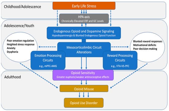
There is substantial and well-recognized variation in opioid sensitivity across individuals [28,29,30], which has been proposed to confer different levels of risk for opioid misuse and eventual OUD [31]. This review aims to establish a theoretical model (Figure 1) that summarizes the potential neurobiological mechanisms by which ELS alters opioid sensitivity, thereby contributing to future risks for OUD. This review first describes individual variation in risks for opioid misuse, the public health implications of opioid misuse/OUD, and the relationships between ELS and OUD. Next, the effects of ELS on mesocorticolimbic circuits that underlie emotion and reward processing and on the endogenous opioid and dopamine (DA) neurotransmitter circuits that play fundamental roles in these processes are discussed. Evidence linking these changes with individual differences in opioid sensitivity and risks for OUD is presented in each of the sections. The overarching hypothesis is that ELS-induced derangements in mesocorticolimbic brain regions lead to altered opioid sensitivity, which increases the abuse liability of these drugs and represents a preexisting vulnerability phenotype for OUD.

2. Individual Differences Confer Differential Risk for OUD

2.1. Opioid Sensitivity

Animal studies examining characteristics of drug intake repeatedly find that a subset of animals display a strong preference for opioids over other reinforcers and that preference is opioid-specific and does not represent a general sensitivity to reinforcing substances. For instance, rats given the opportunity to self-administer heroin versus other drug (e.g., cocaine) or non-drug (e.g., saccharin) reinforcers frequently develop an immediate preference for one but not all substances; this preference predicts future heroin-administration and development of “heroin addiction” [32,33,34,35,36]. Such individual differences in preference are also evident in nonhuman primates; one study found that nonhuman primates exposed to five different psychoactive substances (cocaine, remifentanil, methohexital, ethanol, and ketamine) developed strong individual preferences for a single drug class in a manner similar to what was observed in rats [37]. Although there are limited data to inform of this effect in humans, empirical support is growing. One human laboratory study found that a given individual generally experienced the same level of effect (positive or negative) when administered the opioids heroin and hydromorphone but that the level of effect differed markedly across individuals, suggesting that humans also display pronounced individual differences in risk for opioid misuse [38]. Moreover, retrospective human cohort studies have repeatedly found that patient self-reported experiences of their first opioid exposure varied widely across individuals and that the subset of participants who experienced an initial euphoric effect were more likely to develop OUD [39,40], suggesting that experiencing euphoria during the first opioid exposure was not a universal response but did signal a risk for opioid misuse [41].

2.2. Mechanisms Underlying Opioid Sensitivity

The mechanisms underlying variations in opioid effects have not yet been clearly elucidated but are hypothesized to include an array of pharmacokinetic, pharmacodynamic, and pharmacogenetic contributions [42]. For instance, rats that were tested with four different mu agonists displayed strong within-subject consistency but substantial dose variability (30- to 300-fold) across animals with regard to analgesia [43], suggesting a strong biological mechanism underlying differences in opioid sensitivity. An examination of opioid effects across different animal strains revealed that the opioid dose needed to produce equivalent levels of conditioned place preference (CPP, a measure of drug reward) was substantially and consistently higher among Wistar versus Sprague Dawley rats and corresponded to differential levels of DA release in the nucleus accumbens (NAc) [44]. Such a variability is also evident within the same animal strain; one study observed a continuum of responses in Sprague Dawley rats ranging from the 10% of animals showing a preference for morphine over food after 4 days to another 10% of the sample not developing a morphine preference even after 38 days of access [45]. A final study observed pronounced differences in the rate of heroin and oxycodone intake and preference across the four different sub-strains of the 129 mouse [46]. These strain-based differences suggest a pharmacogenetic contribution to individual differences; the most frequently explored gene in the context of opioid use has been OPRM1, which codes for the mu opioid receptor. OPRM1 appears to produce clinically meaningful differences in human opioid sensitivity [47,48,49,50] and to modulate human cortisol stress response [51], thus providing a putative pathway by which ELS may confer a unique risk for opioid misuse and OUD.

Stress has been independently associated with opioid sensitivity, with several studies observing evidence of greater OUD risk (e.g., increased opioid self-administration and drug reinstatement) [52,53,54] among animals exposed to stress from restraint [55], foot-shock [52,56], and intermittent swim [57] assays. Stressful stimuli have also been shown to produce a hyporesponsive state of the endogenous opioid system in animals [58], and corticosterone release following a stressful stimulus has been causally and independently linked to increased opioid self-administration [57]. Early life stress appears to be particularly destructive because it produces enduring conformational changes in the endogenous opioid system that impact drug use behavior [59,60]. The deleterious impact of ELS can be observed as far back as gestational exposure, wherein pups whose mothers were exposed to stress during gestation were more likely to acquire morphine CPP during their adolescence [61]. Converging evidence indicates that ELS in animals reduces opioid (particularly mu) receptor availability [62,63,64] and decreases downstream dopamine signaling [63,65] in a way that may bolster the reinforcing effects of opioids. Such an effect is also evident in behavioral tests where young animals exposed to stress display more CPP for µ- versus κ-opioid agonists [66]. Altogether, these data suggest that ELS may produce conformational changes in the opioid system that enhance opioid sensitivity, which may serve as the basis for the pronounced behavioral effects observed among adults.

3. Societal Impact of Opioid Misuse/Use Disorder

3.1. Epidemiological Findings

Not all persons who are exposed to opioids develop OUD, but those who do can experience devastating consequences. Opioids are considered essential medicines for acute and cancer pain, palliative care, and treatment of opioid dependence by the World Health Organization [67] and are used for medicinal purposes worldwide. Although the availability and consumption of opioid analgesics are considered inadequate to provide sufficient pain relief in some regions [68], in high-income countries, such as the US and Canada, increased prescribing and availability, aggressive promotion, and under-regulation of these drugs from the 1990s to around 2011 has led to a serious national crisis and spiraling needs for healthcare services. In 2018, 57.8 million people globally were estimated to have used opioids in the past year, with almost half of them misusing pharmaceutical opioids [69]. In fact, prescription pain reliever misuse in the US is second only to marijuana as the first illicit substance that people try, with approximately 4400 new initiates each day [70].

Examination of trends in opioid analgesic abuse from 2002 to 2011 showed that approximately 75% of US heroin users reported being introduced to opioids through prescription drug use [71,72]. However, despite evidence that misuse of prescription pain relievers has declined (from 4.7% in 2015 to 3.5% in 2019), heroin use has remained relatively stable over the past decade. It is estimated that 1.6 million people in the US suffered from OUD in 2019 [70]. These problems are accompanied by school dropout, unemployment, poor quality of life, cooccurring psychiatric disorders, and problems with the criminal justice system [73], which represent not only tremendous burdens to individuals and families but also a US “economic burden” of roughly $78.5 billion a year [74]. Two out of three drug overdose deaths in the US in 2018 involved an opioid, which can be attributed to the wider availability of high potency opioid analgesics and synthetic opioids since about 2013 [73,75,76].

3.2. Risk Factors

Risk factors for opioid misuse and OUD include both genetic and environmental determinants. Findings of twin studies suggest that genetic variance explains approximately half of the liability for OUD, although some of this variance is likely a genetic disposition to drug use disorders in general [77,78,79]. However, environmental factors also play a prominent role, including drug availability, peer pressure, other substance use, adverse childhood experiences, family history of alcohol and drug use disorder, and other comorbid mental health problems [71,73,80,81,82,83,84]. Predictors of prolonged opioid use in patients with musculoskeletal problems include past or current substance use problems, higher initially prescribed doses, mood disorders, and depression [71,85,86]. Among chronic pain patients, predictors of misuse include anxiety, anger, pain intensity, and depression [87,88,89] as well as measures of distress intolerance [90], pain catastrophizing [91], and difficulties in emotion regulation [92].

4. Evidence of ELS and OUD Associations

4.1. ELS Is Highly Prevalent among Persons with OUD

An extensive body of research has shown that ELS exposure profoundly increases risks for the development of alcohol, cocaine, marijuana, and nicotine use disorders [18,93,94,95,96,97], but only recently has it become evident that childhood adversity is also common among individuals with OUD [22,98,99,100,101]. A recent meta-analysis found that 41% of women and 16% of men with OUD reported a history sexual abuse and that 38–42% reported other types of mistreatment before the age of 18 [100]. Early life stress has also been linked to higher rates of relapse, suicidal ideation, overdose, and a more rapid transition from misuse to OUD in these individuals [21,22,102,103].

Data from the National Longitudinal Study of Adolescent to Adult Health study (n = 12,288) revealed a dose–response relationship between ELS and adulthood prescription pain reliever misuse that increased in strength from young to middle adulthood [104]. There is also evidence that the odds of prescription opioid/pain reliever misuse increase as a function of the number of ELS events experienced [26,104,105,106] and that other behaviors, such as age of first opioid use and injection drug use, are also associated with the number of early life stressors endorsed [25,104,106]. Although the aggregate number of events seems to have a cumulative effect [22,107], the type of early life stressor may also impact the outcomes. For example, in one study, neglect, emotional abuse, and parental incarceration were associated with 25–55% increased odds of prescription pain reliever misuse in young adults while sexual abuse and witnessing violence were associated with nearly three and five times the odds of injection drug use [104]. Chronicity, severity, and developmental timing of these experiences are also differentially related to drug use outcomes [18].

4.2. Persons with ELS and/or OUD Exhibit Similar Pathologies

Early life stress is associated with deficits in emotion regulation and neuroendocrine responses to stress that persist into adulthood and are associated with the development of several forms of psychopathology [5,108,109,110,111,112,113,114,115,116,117,118]. Recently, it has been suggested that such deficits may be intermediaries in the pathway that links ELS with the development of OUD [90,103,119,120,121]. For instance, emotional responses to neutral and unpleasant stimuli were positively associated with childhood neglect and severity of addiction in heroin users, suggesting that ELS impairs the ability to modulate emotions and to cope with stress. These deficits then give rise to a range of maladaptive behaviors that can predispose to OUD [122]. Similar findings were reported by Ghorbani et al. [123], who showed that ELS was indirectly related to heroin craving via a limited ability to regulate emotions. Internalizing and externalizing symptoms have been shown to partially mediate the association between ELS and prescription opioid misuse [24,106]. These findings suggest that self-medication may play a role in generating and maintaining both recreational and prescription opioid misuse in ELS-exposed individuals [124,125]. In addition to their euphoric and analgesic effects, opioids relieve stress through their inhibitory actions on the hypothalamic-pituitary-adrenal (HPA) axis [120,126,127], which can be highly negatively reinforcing [128] to individuals with such affective vulnerabilities.

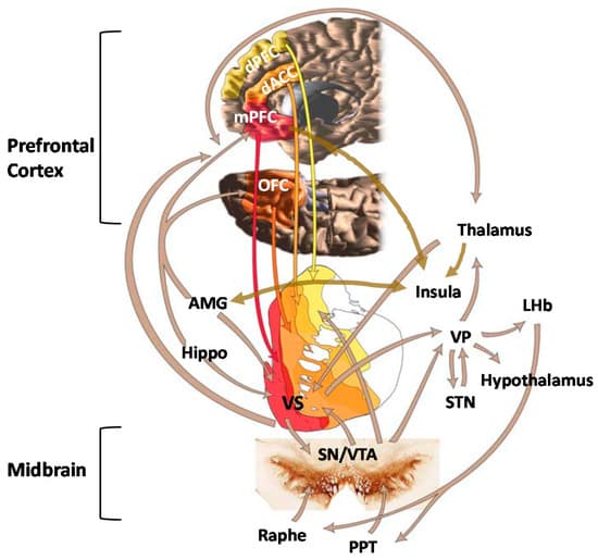
5. Role of Mesocorticolimbic Emotion Processing Circuits in ELS and OUD

5.1. Effects of ELS on Emotion Processing Circuits

Mesocorticolimbic brain circuits include several cortical and subcortical structures that are integrally involved in stress regulation and emotion and reward processing [129] (Figure 1). Two regions that seem to be particularly impacted by ELS are the amygdala (AMG) and medial prefrontal cortex (mPFC) [130,131,132,133]. These structures have extensive bidirectional connections and normally work together to integrate the expression of fundamental aspects of emotional learning, memory, and behavior as well as play prominent roles in stress responsivity [134,135,136]. As maturation of these regions occurs throughout juvenile and adolescent periods, perturbations that occur during childhood can alter their normal neurodevelopmental trajectories, which may negatively impact psychological function later in life [135]. Alterations in AMG volume [137,138] and task-related hyper-responsivity [133,139,140,141,142,143] have been observed in both adults and children with a history of ELS. Oshri et al. [144] showed that ELS-related changes in AMG volume were associated with increased anxiety, depressive symptoms, and alcohol use. Heightened threat-related AMG reactivity has been shown to predict internalizing symptoms [145] and risks for alcohol use disorder [146,147]. In contrast, ELS has been associated with reduced mPFC volume [148,149] and lower mPFC activation during both resting state and cognitive tasks [150,151,152].

Normally, the mPFC, particularly the ventral mPFC, modulates fear behavior by providing top-down control of AMG and HPA-axis responses to stress [153,154]. Dysfunction in AMG-mPFC connectivity has been implicated in several psychiatric disorders, including depression and schizophrenia [135], and there is evidence that top-down control may be altered in individuals with a history of ELS. While valence (positive or negative) and regional specificity vary somewhat across studies [136], atypical patterns of connectivity have been observed between the AMG and PFC in both youth and adults with a history of ELS. Findings in children and adolescents include evidence of weakened left AMG–anterior cingulate cortex (ACC) resting state functional connectivity (rs-FC), which was associated with higher levels of current anxiety in one study and mediated the relationship between ELS and internalizing symptoms in another [155,156,157]. Gee et al. [140] reported precocious maturation of the mPFC–AMG pathway in previously institutionalized youth, which reflected a developmental shift from positive to negative coupling and seemed to confer some degree of reduced anxiety. These somewhat counterintuitive findings suggested that accelerated maturation of these connections may serve to facilitate coping with environmental insults in the short term but lead to less efficient stress regulation and increased vulnerability for psychopathology in adulthood [135]. Associations between AMG–PFC connectivity and cortisol levels have also been reported in humans [158] and nonhuman primates [159], suggesting that ELS-related changes in neuroendocrine function contribute to the neural deficits and problems with emotion regulation that are observed in these individuals in later life.

Similar ELS-related findings have been reported in adults, including weakened AMG–pregenual ACC rs-FC, which predicted elevated state anxiety [160], and reduced AMG–ventral mPFC rs-FC, which predicted increased levels of pro-inflammatory cytokines [161]. However, inconsistent findings have also been reported, which may be related to variations in network configurations between resting state and task-related co-activations, task-specific engagement, or other methodological differences among studies. In contrast to the findings during resting state scans, Jedd et al. [162] reported increased AMG–PFC during an emotion processing task in ELS-exposed adults. Kaiser et al. [163] found that ELS was associated with greater negative AMG–dorsolateral PFC rs-FC, which mediated the relationship between ELS severity and blunted cortisol response to acute stress, and elevated dynamic AMG–rostral ACC rs-FC, which was associated with reduced negative mood following a social evaluation stress challenge. These findings suggested that ELS may be associated with both maladaptive and compensatory changes in mesocorticolimbic circuits. Though most studies in this area of research are cross-sectional and preclude determinations of causality, overall, these findings suggest that neuroplastic aberrations incurred as a result of ELS may persist decades later into adulthood, leading to alterations in physiological and emotional responses to stress.

There has been growing evidence that the effects of ELS on mesocorticolimbic brain structures may be partially related to repeated or chronically high levels of corticotropin-releasing factor (CRF) and glucocorticoids (GCs) [164,165,166,167,168,169] that occur during early stages of ELS [170,171,172]. Although other mechanisms may be involved, composite findings from several lines of research indicate that epigenetic processes involving HPA-axis regulation and GC signaling underlie many of these effects [173,174]. Glucocorticoid receptors (GR) are densely located throughout the brain, including stress-sensitive regions such as the mPFC, AMG, hippocampus, NAc, and hypothalamus [175,176]. Regional differences in methylation and both increases and decreases in brain GR expression have been observed in rodents and monkeys exposed to ELS [177,178,179,180]. Findings of one postmortem study showed decreased levels of GR mRNA in the hippocampus of suicide victims with a history of ELS compared to those without this history [181]. Greater GC receptor methylation has also been found in the peripheral cells of ELS-exposed humans [182], which was recently shown to moderate associations between ELS and cortisol stress reactivity [183].

5.2. Emotion Processing Circuit Interface between ELS and OUD

Although OUD is underrepresented in the neuroimaging literature on addiction compared to other SUDs [184], findings from a limited number of human fMRI studies have implicated brain circuits involved in stress and emotion regulation in the perpetuation of this disorder. For example, the AMG has been shown to be activated in response to heroin drug cues [185,186,187] and plays a central role in the generation of cue-elicited craving in persons with OUD [188]. Schmidt et al. [189] found that patients with OUD displayed higher left AMG response to fearful faces than healthy controls during acute withdrawal, which correlated with levels of state anxiety, ACTH, and cortisol levels in all subjects and with heroin craving in patients. However, AMG connectivity was reduced to levels that did not differ from those of healthy controls after acute heroin maintenance treatment, suggesting that the results were driven by the drug-related attenuation of stress hormone release [189]. Alterations have also been found in mPFC activation and connectivity in abstinent, currently using, and methadone-maintained persons with OUD during resting state and cue reactivity, inhibitory control, and emotion processing tasks [190,191,192,193]. Administration of the opioid antagonist naltrexone has been shown to decrease AMG and to increase PFC responses to drug cues in abstinent heroin users, which suggests that naltrexone’s clinical effects may result in part from its ability to increase the capacity for conscious self-regulation [186]. Wang et al. [194] further showed that greater pretreatment mPFC response to heroin-related cues predicted greater adherence to naltrexone in detoxified heroin-dependent individuals, demonstrating that lower mPFC activity may contribute to increased craving, negative affect, and reduced treatment compliance. In general, the imaging data suggest that OUD is associated with reduced PFC monitoring, weak inhibitory controls, dysfunctional stress responses, problems with emotion regulation, and heightened negative affective states.

Nevertheless, in spite of the commonalities in behavioral and neural deficits that have been observed between ELS-exposed individuals and persons with OUD, to our knowledge, relationships between ELS-induced changes in brain function and opioid misuse have never been examined. Findings from several lines of research support speculations that ELS-related changes in circuits that underlie stress and emotion processing may represent an underlying opioid vulnerability pathway. For instance, impairments in AMG connectivity have been shown to mediate associations between ELS and internalizing symptoms in adolescents [155] while internalizing and externalizing symptoms partially mediate associations between ELS and opioid misuse [24,106]. These findings suggest that ELS may lead to changes in brain function that impair the ability to modulate emotions and to cope with stress, which may then lead to a range of maladaptive behaviors that can predispose to opioid misuse. Poor coping and emotion regulation profiles have previously been shown to predict initiation of opioids at an earlier age, past 90-day heroin use, increased probability of injecting a drug, and less likelihood of heroin abstinence after treatment, which may all be related to a need for self-medication [195].

It is important to note that these findings do not specifically explain the preference for opioids in these individuals as poor coping and emotion regulation have also been associated with misuse of other substances [195,196]. However, findings of a growing body of research suggest that ELS-related changes in emotion processing regions may lead to altered opioid sensitivity. Prefrontal and NAc activation are partially mediated by the endogenous opioid system [31,197], which is altered in ELS-exposed individuals. In one study, opioid antagonist naltrexone modulated activation of the mPFC during negative emotional processing as a function of ELS in both healthy controls and individuals with alcohol, cocaine, and/or opioid use disorders [198]. Specifically, the greater the severity of abuse, the greater the sensitivity of the mPFC to the effects of naltrexone, suggesting that ELS-related changes in mPFC function may underlie individual differences in responsiveness to the drug. Persons with AUD/SUD also reported higher levels of depression, anxiety, and stress sensitivity than the control group, which was somewhat ameliorated by naltrexone. These data are consistent with preclinical evidence showing that naltrexone reduces ethanol consumption in rats that experienced prolonged separation but not short absences of the dam [199]. Collectively, the data support hypotheses that ELS-induced derangements in endogenous opioid function in stress and emotion processing circuits may underlie part of the variability detected in opioid sensitivity across individuals. However, significant gaps still remain in our understanding of how ELS-induced changes in these circuits influence the course of opioid addiction and why alterations in function are associated with maladaptive behaviors in some individuals but not others.

6. Role of Mesocorticolimbic Reward Processing Circuits in ELS and OUD

6.1. Effects of ELS on Reward Processing Circuits

Reward functions underlying addiction are generally thought to involve the ventral tegmental area (VTA) and mesolimbic DA neurons that project from the midbrain VTA to the ventral striatum (VS) or, more specifically, the NAc, which is considered the center of reward learning. It is well-established that ELS increases feelings of dysphoria and anhedonia in humans and leads to an attenuation of behavioral responses to primary and conditioned rewards in animals [200,201,202,203,204,205]. Several investigators have hypothesized that these outcomes may be the result of ELS-induced alterations in NAc function. Goff et al. [206] reported hypoactivation of the NAc during an emotional faces task in adolescents with a history of ELS, which was associated with higher levels of depression. Between-subject comparisons of children ages 5–10 yrs. and adolescents 11–15 yrs. old further indicated that the ELS group failed to show the developmentally typical rise in NAc reactivity shown by the comparison group. The findings suggested that ELS impacts the development of the VS, resulting in hypoactivity, which leads to dysfunctional reward and motivational processing. Other investigators have shown similar findings consistent with notions of reduced activation of striatal structures and dampened behavioral responses to reward in ELS-exposed individuals [203,207,208]. Given prior evidence that blunted reward responsivity may be a marker of motivational mechanisms underlying addiction vulnerability [209,210,211,212,213], these findings suggest that alterations in vs. function may be one mechanism that underlies increased susceptibility for addiction in ELS-exposed individuals.

Several cross-sectional fMRI studies have examined associations between ELS-induced changes in reward circuitry and subjective measures of reward [214,215,216]. For example, Dillon et al. [203] found that ELS-exposed young adults reported elevated depressive and anhedonic symptoms, rated reward cues less positively, and exhibited decreased anticipatory reward activity in left basal ganglia regions relative to controls during completion of a monetary reward task. Corral-Frias et al. [217] showed that blunted vs. reactivity to reward was associated with increased anhedonic symptoms, which indirectly predicted other depressive symptoms and problematic alcohol use in ELS-exposed young adults. Marusak et al. [218] examined blood-oxygen-level-dependent (BOLD) responses to an emotional conflict task, showing that greater conflict-related AMG reactivity was also associated with diminished trait reward sensitivity in adolescents with a history of ELS. Overall, these findings are consistent with behavioral evidence from preclinical studies showing that ELS impairs motivation to work for rewards in animals, suggesting a downregulation of reward functions [219,220,221,222].

Longitudinal studies are somewhat rare in this area of investigation. However, Birn and colleagues [223] found that young adults who experienced a high level of stress as children showed deficits in decision-making during a reward processing task and reported more real-life risk-taking behaviors than individuals without this history. They also displayed alterations in reward processing regions, including the middle temporal gyrus, precuneus, putamen, insula, and left inferior frontal gyrus, some of which mediated relationships between ELS and reward processing or self-reported risk-taking behavior. Casement et al. [224] showed that cumulative life stress from ages 15–18 years was associated with decreased mPFC response during both anticipation and receipt of monetary reward at age 20 in adult males. The blunted mPFC response to reward predicted greater symptoms of alcohol dependence and mediated the relationship between life stress and alcohol use. Boecker et al. [225] found that ELS assessed at 3 months after birth and between the ages of 2 and 15 years was associated with hyporesponsiveness in reward circuits during reward anticipation (i.e., VS, putamen, and thalamus) and hyperresponsiveness during reward delivery (i.e., insula, pallidum, substantia nigra, and right posterior hippocampus) in healthy young adults who had been followed prospectively over 25 years.

To date, only a small number of studies have examined the influence of ELS on functional connectivity in reward processing regions. Fareri et al. [226] found that resting state coupling between the VS and mPFC was stronger in previously institutionalized youth than in youth who were raised by their biological parents; the fMRI findings mediated differences in social problems between the two groups. Blunted maturation of VTA–mPFC resting state connections [227] and increased connectivity of the insula to salience network seed regions have also been observed in trauma-exposed youth, which was associated with diminished reward sensitivity [216]. Hanson et al. [228] found elevated VS–mPFC functional connectivity during a monetary reward task in college-age adults with a history of both ELS and higher levels of recent life stress, which suggested that the deficits observed in youth persist into young adulthood. Although further prospective research is needed, collectively, the findings suggest that blunted VS activity and elevated functional connectivity in reward processing regions may represent neurobiological markers that serve as indicators of diathesis for psychological dysfunction in adults with a history of ELS. Overall, the findings are consistent with those of preclinical research showing broad changes in connectivity of the limbic and reward networks of adult rats exposed to ELS [229] as well as deficits in reward responsiveness and approach motivation [220].

6.2. Reward Processing Circuit Interface between ELS and OUD

In recent years, fMRI has been used to evaluate abnormalities in activation and functional connectivity of reward regions in abstinent and currently using heroin-dependent individuals using a variety of paradigms [184,230]. In general, brain activation has been shown to be upregulated in the reward and salience network brain regions of persons with OUD in response to drug-related cues [231]. Dysfunctional connectivity has also been reported using a variety of methods, including at rest, in response to heroin-related cues, and while performing decision-making or response inhibition tasks in regions that include the mPFC, orbitofrontal cortex (OFC), dorsolateral PFC, ACC, posterior cingulate cortex (PCC), and NAc [191,192,232,233,234,235]. In one study, rs-FC between regions involved in reward and motivation (e.g., VS-ACC and VS-OFC) was increased and connectivity between regions involved in cognitive control (e.g., PFC-ACC) was decreased in chronic heroin users, most of whom were being treated with methadone [236]. Functional connections between the AMG and mPFC have also been shown to be critical for the processing of opioid rewards [237,238,239]. Engagement of the reward networks is associated with craving, addiction severity, duration of use, and/or relapse in persons with OUD [191,240,241].

Preclinical studies have demonstrated that chronic opioid use produces abnormalities in mesocorticolimbic reward circuits that contribute to opioid misuse and OUD [191,239,242,243,244,245,246]. However, genetic and environmental factors, such as stress, may also lead to functional deficits in these circuits that underlie increased susceptibility for drug misuse and dysregulated responses to opioids. As previously described, ELS-related derangements in reactivity and connectivity of reward circuits have been associated with problematic alcohol use and risks for alcohol use disorder as well as well-established intermediate phenotypes for SUDs (e.g., anhedonia, deficits in decision-making, and problems with reward-based learning). However, virtually nothing is known about how ELS-induced alterations in reward circuits influence opioid sensitivity or about the role that such alterations play in the transition from opioid use to misuse. Similarities between ELS-induced alterations in connectivity and those observed in persons with OUD suggest that ELS-related derangements may predate and increase vulnerability for this disorder. This relationship is also suggested by evidence that ELS-induces changes in DA and endogenous opioid neurotransmission, which mediate the reward functions of these regions [247,248]. However, further research is needed to test these hypotheses.

7. Role of Endogenous Opioid Neurotransmitter System in ELS and OUD

7.1. Effects of ELS on Endogenous Opioid Function

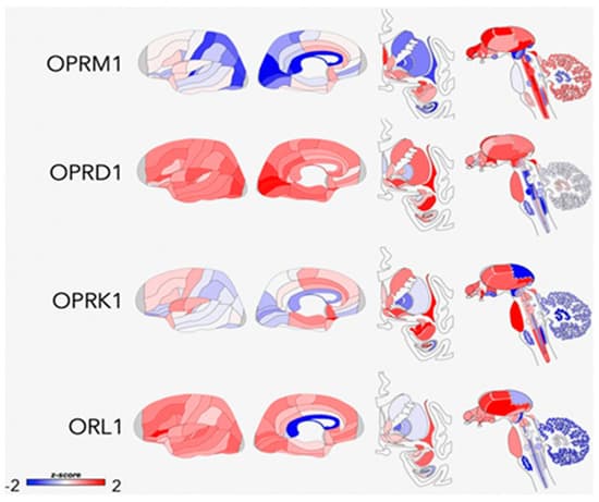
Opioid peptides and receptors are widely distributed throughout the central nervous system and are thought to modulate many aspects of human behavior, including reward, affective states, pain responses, and other physiological functions [249] (Figure 2). Considerable variability in responses to opioid drugs has been noted in the general population, which are associated with differences in therapeutic response to treatment as well as risks for drug misuse [41,47,250,251]. Such differences may be due to both heritable factors and environmental influences that interact with the genome through epigenetic or transcription mechanisms to produce long-term alterations in the endogenous opioid system [252]. Endogenous opioid peptides are found throughout the peripheral and central nervous systems, where they play a role in many different types of functions, including nociception and analgesia, stress responses, physiological functions, social behavior, mood, and reinforcement [127,253,254,255,256,257]. Normally, this system is activated by acute stress, leading to the release of endogenous opioids at multiple sites in the brain. The release of opioids generally serves to attenuate stress responses by actions that include modulating the release of CRF, which returns the systems to pre-perturbation levels. However, repetitive stress exposure (which is characteristic of ELS) leads to an imbalance between CRF and opioids such that opioid inhibitory tone is favored [258]. Although the relationships require further testing, there is some evidence that chronically high tonic levels of endogenous opioids may trigger downregulation or reduced affinity of µ-opioid receptors (MOR) and lower phasic release of opioid peptides, resulting in hypoactivity of this system [259,260].

Fig. 2

Conformational changes that have been specifically associated with ELS in preclinical studies include changes in opioid peptide levels [63,261], kappa receptor signaling [262], and variations in mu- and kappa-receptor (KOR) gene expression [62,64,65,263,264] in brain regions that include the hypothalamus, PFC, periaqueductal gray (PAG), AMG, NAc, rostral ventromedial medulla, and lateral habenula. Nylander and Roman [251] concluded that the most pronounced effect of ELS on opioid peptides is on Met-inkephalinArg6Phe7 (MEAP) levels, which are reduced in ELS-exposed animals. Rats with lower MEAP levels exhibit altered risk-taking behavior and a propensity for high ethanol intake, consistent with theories that an inherent opioid deficiency leads to increased susceptibility for addiction. Given the large number of physiological functions that are regulated by the endogenous opioid system, it is reasonable to speculate that hypofunction of this system could also lead to dysregulation of stress responses, altered pain-processing, and an array of stress-related disorders.

The first direct evidence of ELS effects on opioid neurotransmission in humans was reported by Lutz et al. [92], who conducted a postmortem study showing that ELS was associated with the downregulation of kappa receptors in the anterior insula of both depressed individuals who died by suicide and controls who died suddenly from accidental causes. Cortisol response to naltrexone has also been found to be blunted in high vs. low ELS women, which similarly linked ELS with downregulation of endogenous opioid activity [265]. The authors posited that these effects may reflect an adaptation of the central opioid system that shapes how a person responds to motivationally significant stimuli. The findings of a recent study by Garland et al. [266] are consistent with these notions, showing that ELS was associated with blunted heart rate variability (HRV) and increased cue-elicited drug craving during a task involving negative emotions in female opioid-treated chronic pain patients. In theory, the reduced capacity to respond to negative emotional stimuli could be the result of reduced opioid function. However, opioid function was not specifically examined and the cross-sectional nature of the study makes it impossible to know whether the deficits predated or were a result of chronic opioid use. It is also unclear whether the findings would be replicated in an opioid-naïve sample without prior history of chronic pain. Nevertheless, in spite of the limitations, aggregate findings from this small body of human studies are consistent with the preponderance of preclinical evidence suggesting that ELS leads to a deficiency in opioid neurotransmission.

7.2. Interface between ELS and OUD via Endogenous Opioid Function

Much of the current research on the neurobiology of OUD is focused on gaining a better understanding of the molecular and cellular aspects of opioid receptor function that contribute to vulnerability for this disorder [267] or on characterizing how genetic influences on receptor function translate to abuse liability and treatment outcomes [268,269,270]. Although this research holds promise for explaining some of the variability in clinical responses to opioids, the evidence that heritability estimates for OUD are only about 23–54% [271] suggests that there is also a need for better understanding of the effects of environmental factors that help to shape the behavioral and molecular profiles of individuals with this disorder.

Recent preclinical findings have provided initial evidence that ELS-induced changes in endogenous opioid function may alter opioid agonist and antagonist sensitivity. In one study, ELS-exposed rats showed greater place preference for the µ-agonist morphine but lesser aversion to the k-receptor agonist spiradoline [66], suggesting that ELS may enhance opioid abuse vulnerability by both increasing reward sensitivity and by decreasing the aversive effects at k-receptors [272]. Vazquez et al. [273] showed that maternal deprivation in rat pups was associated with hypersensitivity to the reinforcing effects of morphine and development of morphine dependence in adulthood, which was likely a result of basal hypoactivity of the nucleus accumbens (NAc) enkephalinergic system. Nakamoto et al. [64] found that ELS-exposed mice displayed decreased µ- and k-opioid receptor messenger mRNA expression in the PAG and increased k-opioid receptor expression in the AMG. A lack of morphine antinociception was observed in stressed mice in adulthood but not immediately after ELS exposure. Bruehl et al. [41] recently extended these findings to humans, showing that endogenous opioid function assessed by naloxone administration was inversely associated with euphoric effects of a single dose of 0.09 mg/kg morphine sulfate in patients with low back pain. According to reinforcement theory [274], either hypersensitivity to the reinforcing effects or diminished sensitivity to the antinociceptive effects of opioids could lead to misuse of these drugs, through positive or negative reinforcement, respectively. There is also evidence that ELS-induced changes in endogenous opioid function may lead to dysfunctions in DA neurotransmission in rats [63,275]. In one study, ELS increased KOR-mediated inhibition of baseline and stimulated DA release, which contributed to a hypodopaminergic state and escalated ethanol intake. Taken together, these findings provide support for hypotheses that ELS-induced conformational changes in the endogenous opioid system [58,273] may alter the effects of opioid drugs in ways that increase risks for their misuse [59,276,277,278].

Although not specific to OUD, the findings from human positron-emission tomography (PET) imaging studies have found associations between endogenous opioid function, and subjective and behavioral responses to both drugs of abuse and pain. For example, k-opioid receptor availability is associated with stress-induced cocaine self-administration in subjects with cocaine-use disorder [279], altered vs. binding potential for the µ-receptor agonist [11C]carfentanil is associated with alcohol craving and relapse risk in abstinent alcoholics [280,281,282], the µ-receptor binding potential correlates with nicotine dependence and reward in smokers [283], and alterations in endogenous opioids and µ-receptors in patients with chronic nonspecific back pain are associated with both sensory and affective elements of the pain experience [284]. To date, we are not aware of any neuroimaging studies that have evaluated the effects of ELS on opioid neurotransmission in humans or any controlled human laboratory studies that have examined ELS contribution to human opioid sensitivity. Better understanding of the role that individual differences in sensitivity play as mediators in the transition from prescription or recreational opioid use to opioid misuse and risky-drug related behaviors may help to inform the development of more effective interventions for ELS-exposed individuals.

8. Role of Dopamine Neurotransmitter System in ELS and OUD

8.1. Effects of ELS on Dopamine Function

Another neurotransmitter system that plays a fundamental role in stress responses and emotional-motivational activation of reward seeking is the midbrain DA system [285,286,287,288]. The DA mesolimbic pathway projects from the VTA to the VS, AMG, and hippocampus, and the mesocortical pathway projects from the VTA to cortical regions such as the ACC, OFC, mPFC, and insula. Considerable preclinical evidence has emerged showing that ELS may lead to profound and long-term derangements in brain DA neurotransmission. Abnormalities that have been observed include but are not limited to altered D1, D2, and D3 receptor mRNA expression; decreased density of DA transporters; increased DA metabolites in the striatum and/or NAc [289,290,291,292,293,294]; and reduced rates of DA clearance in the mPFC [295]. Both enhanced [290,296,297] and blunted [290,296,297,298] striatal DA responses to stress have been observed in adult rodents exposed to ELS. It has been suggested that, in general, ELS induces a hypodopaminergic state with an associated enhancement of DA system responses to salient stimuli [63]. There is also a general consensus that the effects of ELS on DA neurotransmission has broad-based clinical implications for the development of psychopathological conditions, such as schizophrenia and addiction, which are known to be associated with malfunctions in DA neurotransmission [96,299,300,301]. It is hypothesized that these effects may be the result of excessive exposure to glucocorticoids during early life, which impacts the organization and epigenetic control of midbrain DA systems [302,303,304,305].

Functional changes that occur in the DA system as a result of ELS may also be associated with altered neurochemical and behavioral responses to drug abuse. Early life stress may lead to enhanced DA and behavioral responses to psychostimulants and changes in drug consumption patterns that reflect greater vulnerability for drug abuse in later life in animals [275,290,297,298,305,306,307,308,309]. The first evidence that some of these findings may translate to humans was provided by findings of an [11C]raclopride positron emission tomography (PET) study conducted by Pruessner and colleagues [310], who found that persons who reported low maternal care had greater VS DA release in response to stress than individuals who reported high maternal care. Oswald et al. [311] extended this line of research by showing that ELS is also associated with enhanced VS DA responses to amphetamine in healthy young adults. The relationship between ELS and DA response was partially mediated by current levels of perceived stress, which suggested that ELS may not directly influence DA function in some individuals unless accompanied by elevated levels of psychological stress in adulthood.

8.2. Interface between ELS and OUD via Dopamine Function

It has been proposed that the reinforcing effects of opioid agonists are, at least, partially dependent on their actions on mesolimbic DA circuits [312,313,314,315]. Interactions between the DA and endogenous opioid systems are well-established within mesocorticolimbic brain circuits [316,317,318,319,320] and play a role in behavioral responses to opioids and in relief of pain in animals [321,322,323]. It is reasonable to posit that extensive ELS-related dysfunctions in DA circuits could, therefore, influence sensitivity to opioids and vulnerability for OUD. Blum and colleagues [210] hypothesized that addiction results from an underlying reward deficiency state characterized by hypodopaminergia. This condition may be innate or acquired and is clinically manifested as anhedonia, numbing, apathy, or decreased motivation for natural reinforcers [324,325]. According to the hypothesis, opioid/endorphin deficiency increases a person’s vulnerability for OUD by disrupting interactions between midbrain opioid and DA neurotransmitter systems [326]. This has been supported by evidence that ELS alters µ- and k-receptor mRNA levels [62,63,64] and decreases downstream DA signaling [63,65] in a way that may alter the reinforcing and antinociceptive effects of opioids. Dopamine-deficient mice display decreased sensitivity to the analgesic effects of morphine [313] and mice lacking DA D2 receptors fail to self-administer morphine [327], which also provides evidence that individual differences in DA function may contribute to differential opioid sensitivity.

9. Conclusions

Epidemiological research has shown that ELS is highly prevalent in persons with OUD and is associated with opioid use initiation, injection drug use, overdose, and poor treatment outcome. However, despite evidence that ELS has a profound impact on mesocorticolimbic brain circuits implicated in OUD, the role that these alterations play in vulnerability for and severity of this disorder has yet to be elucidated. Figure 3 outlines a theoretical model based on the extant literature reviewed here, wherein ELS initiates a cascade of neurobiological changes that lead to altered opioid sensitivity and increased risks for OUD. This model is not meant to be exhaustive but to provide a foundation to support focused research in this area. The data suggest that ELS leads to chronically elevated CRF and GC levels in stress-sensitive mesocorticolimbic brain circuits during early exposure, which trigger changes in neurochemistry, activation patterns, and connectivity that persist into adulthood. The consequences include conformational changes in the endogenous opioid system that engender an endorphinergic deficiency and alterations in the DA neurotransmitter system that lead to hypodopaminergia. The presence of a vulnerability pathway involving opioid sensitivity is most strongly supported by preclinical evidence that ELS-induced changes in endogenous opioid function are associated with increased drug-seeking behavior and altered sensitivity to the reinforcing and antinociceptive effects of opioids and that DA-deficient mice exhibit decreased sensitivity to the analgesic effects of morphine, suggesting that DA function may also contribute to the ELS-exposed behavioral phenotype. Derangements in functional activation and connectivity in mesocorticolimbic circuits have been associated with emotion regulation and reward processing deficits in ELS-exposed humans and may reflect the underlying neurochemical imbalances in these regions. Such deficits may predispose an individual to a range of behaviors that independently increase opioid use and misuse. Once opioid use is initiated, ELS-induced neurochemical changes may be manifested as altered opioid sensitivity, which may facilitate the transition from use to misuse and ultimately OUD. Additional research that evaluates the effects of ELS exposure on opioid sensitivity and establishes the degree to which ELS-induced changes in endogenous opioid or DA neurotransmitter systems are present and/or contribute to these processes is warranted. This research could delineate key mechanisms underlying substantial individual variation in opioid risk and could lead to improved prevention strategies and prescribing guidelines for high-risk individuals.

Fig. 3

Open Article as PDF

Abstract

A robust body of research has shown that traumatic experiences occurring during critical developmental periods of childhood when neuronal plasticity is high increase risks for a spectrum of physical and mental health problems in adulthood, including substance use disorders. However, until recently, relatively few studies had specifically examined the relationships between early life stress (ELS) and opioid use disorder (OUD). Associations with opioid use initiation, injection drug use, overdose, and poor treatment outcome have now been demonstrated. In rodents, ELS has also been shown to increase the euphoric and decrease antinociceptive effects of opioids, but little is known about these processes in humans or about the neurobiological mechanisms that may underlie these relationships. This review aims to establish a theoretical model that highlights the mechanisms by which ELS may alter opioid sensitivity, thereby contributing to future risks for OUD. Alterations induced by ELS in mesocorticolimbic brain circuits, and endogenous opioid and dopamine neurotransmitter systems are described. The limited but provocative evidence linking these alterations with opioid sensitivity and risks for OUD is presented. Overall, the findings suggest that better understanding of these mechanisms holds promise for reducing vulnerability, improving prevention strategies, and prescribing guidelines for high-risk individuals.

Introduction

Early life stress (ELS) is a widespread issue with significant consequences. In 2018, Child Protective Service (CPS) agencies in the United States received approximately 4.3 million referrals, involving about 7.8 million children. Around one-fifth of these children were confirmed victims of maltreatment, with the youngest children (birth to one year) experiencing the highest rates. These figures may underestimate the true prevalence, as they do not include unreported cases or other traumatic experiences. A large study of nearly 250,000 adults found that 62% reported at least one traumatic childhood experience, and about a quarter reported three or more. ELS, also known as adverse childhood experiences, includes events such as physical, sexual, or emotional abuse or neglect; exposure to domestic violence; family breakdown; divorce; or the death of a parent. These events are beyond a child's control and can hinder normal development, impacting physical and psychological well-being. They often involve either a threat to physical safety or a lack of expected support and nurturing. Risk factors for maltreatment include caregiver alcohol or drug problems, which have increased in recent years, partly linked to the growing misuse of opioids.

Extensive research demonstrates that ELS exposure during early development, a period of high brain plasticity, has particularly damaging effects. Severe or prolonged ELS is linked to an increased likelihood of engaging in risky health behaviors like smoking, drug use, and suicide attempts. It also heightens the risk for a wide range of emotional and physical health problems throughout life. Studies, including the landmark Adverse Childhood Experiences (ACE) study, show a "dose-response" relationship: more trauma is associated with a greater likelihood of these problems and poorer health outcomes. Epidemiological studies have found a clear link between ELS and substance use disorders (SUDs) in both adolescents and adults. Specifically for opioids, ELS is associated with increased risks for initiating opioid use, injecting drugs, overdose, developing an opioid use disorder (OUD), and poor treatment outcomes. However, more research is needed to understand the specific neurobiological pathways connecting ELS to OUD vulnerability.

This review aims to develop a theoretical model explaining the potential neurobiological mechanisms through which ELS alters opioid sensitivity, thereby increasing future risks for OUD. The discussion begins by describing individual differences in opioid misuse risk and the broader public health implications of opioid misuse and OUD. It then examines the relationships between ELS and OUD. Subsequent sections explore the impact of ELS on mesocorticolimbic circuits involved in emotion and reward processing, and on the endogenous opioid and dopamine neurotransmitter systems that are crucial to these processes. Evidence linking these brain changes to individual variations in opioid sensitivity and OUD risk is presented throughout. The central hypothesis is that ELS-induced disruptions in mesocorticolimbic brain regions lead to altered opioid sensitivity, which enhances the potential for opioid abuse and represents a pre-existing vulnerability for OUD.

Individual Differences in OUD Risk

Studies in animals show that some individuals exhibit a strong, specific preference for opioids over other rewards, which predicts future opioid use and addiction. This individual variation is seen across different species, including rats and nonhuman primates. Similarly, human laboratory studies indicate significant individual differences in the effects of opioids. For instance, individuals generally experience consistent effects from different opioids, but the intensity of these effects varies widely between people. Retrospective human studies consistently report that individuals who experienced an initial euphoric effect from their first opioid exposure were more likely to develop OUD, suggesting this initial response is a risk indicator rather than a universal experience.

The specific mechanisms behind these variations in opioid effects are still being explored but are thought to involve a combination of how the body processes the drug (pharmacokinetics), how the drug affects the body (pharmacodynamics), and genetic factors (pharmacogenetics). For example, rats show a wide range in the opioid dose needed to produce pain relief, yet individual responses are consistent. Differences in opioid reward (measured by conditioned place preference) and dopamine release in the nucleus accumbens have been observed across different rat strains, indicating a strong biological basis. Variations are also evident within the same strain, with some rats quickly developing a preference for morphine while others do not, even with prolonged access. Genetic contributions are suggested by these strain-based differences, with the OPRM1 gene (coding for the mu opioid receptor) being a key focus. OPRM1 variations appear to affect human opioid sensitivity and cortisol stress responses, providing a potential link between ELS and OUD risk. Stress itself is independently associated with opioid sensitivity, with various forms of stress in animals leading to increased opioid self-administration and greater OUD risk. Stressful events can also cause a reduced response in the body's natural opioid system. ELS appears particularly damaging because it causes lasting changes in the endogenous opioid system, impacting drug use behavior, even from gestational exposure. Evidence shows that ELS in animals reduces mu opioid receptor availability and decreases downstream dopamine signaling, which may enhance the reinforcing effects of opioids. These findings collectively suggest that ELS can lead to fundamental changes in the opioid system that heighten opioid sensitivity, contributing to behavioral changes observed in adults.

Societal Impact of Opioid Misuse/Use Disorder

While not everyone exposed to opioids develops OUD, those who do face severe consequences. Opioids are recognized globally as essential medicines for pain and dependence treatment. However, in high-income countries like the U.S. and Canada, increased prescribing, aggressive promotion, and insufficient regulation led to a severe national crisis from the 1990s to around 2011. In 2018, an estimated 57.8 million people worldwide had used opioids in the past year, with nearly half misusing pharmaceutical opioids. In the U.S., prescription pain reliever misuse is the second most common illicit substance initiated, with approximately 4,400 new users daily.

Between 2002 and 2011, about 75% of U.S. heroin users reported starting with prescription drugs. Although prescription pain reliever misuse has declined (from 4.7% in 2015 to 3.5% in 2019), heroin use has remained stable. An estimated 1.6 million people in the U.S. suffered from OUD in 2019. These issues are accompanied by significant personal and social problems, including school dropout, unemployment, poor quality of life, co-occurring mental health disorders, and involvement with the criminal justice system. The economic burden in the U.S. alone is estimated at $78.5 billion annually. In 2018, two out of three drug overdose deaths in the U.S. involved an opioid, largely due to the increased availability of potent opioid analgesics and synthetic opioids since 2013.

Risk factors for opioid misuse and OUD are both genetic and environmental. Twin studies suggest genetic factors account for roughly half of OUD vulnerability, although some of this may be a general predisposition to substance use disorders. Environmental factors also play a significant role, including drug availability, peer pressure, other substance use, adverse childhood experiences (ELS), a family history of alcohol and drug use disorders, and co-occurring mental health problems. For patients with musculoskeletal problems, predictors of prolonged opioid use include a history of substance use, higher initial prescribed doses, mood disorders, and depression. Among chronic pain patients, misuse predictors include anxiety, anger, pain intensity, depression, distress intolerance, pain catastrophizing, and difficulties regulating emotions.

Associations Between ELS and OUD

Extensive research has shown that ELS significantly increases the risk for developing alcohol, cocaine, marijuana, and nicotine use disorders. More recently, it has become clear that childhood adversity is also common among individuals with OUD. A recent meta-analysis reported that 41% of women and 16% of men with OUD had a history of sexual abuse, and 38–42% reported other forms of maltreatment before age 18. ELS has also been linked to higher rates of relapse, suicidal ideation, overdose, and a faster progression from opioid misuse to OUD in these individuals.

Data from the National Longitudinal Study of Adolescent to Adult Health involving over 12,000 participants revealed a dose-response relationship between ELS and prescription pain reliever misuse in adulthood, which strengthened from young to middle adulthood. The odds of prescription opioid misuse increase with the number of ELS events experienced. Other behaviors, such as the age of first opioid use and injection drug use, are also associated with the number of early life stressors. While the cumulative number of events has a significant impact, the specific type of ELS can also influence outcomes. For instance, neglect, emotional abuse, and parental incarceration were linked to a 25–55% increased risk of prescription pain reliever misuse in young adults, whereas sexual abuse and witnessing violence were associated with nearly three to five times higher odds of injection drug use. The chronicity, severity, and developmental timing of these experiences also relate differently to drug use outcomes.

Individuals exposed to ELS and/or suffering from OUD exhibit similar pathologies. ELS is associated with deficits in emotion regulation and altered neuroendocrine responses to stress that persist into adulthood, contributing to various mental health conditions. It has been proposed that these deficits may serve as intermediate steps linking ELS to the development of OUD. For example, emotional responses to neutral and unpleasant stimuli have been linked to childhood neglect and addiction severity in heroin users, suggesting that ELS impairs emotional modulation and stress coping. These deficits can lead to maladaptive behaviors that predispose individuals to OUD. Similar findings show that ELS is indirectly related to heroin craving through a limited ability to regulate emotions. Internalizing and externalizing symptoms have been found to partially explain the link between ELS and prescription opioid misuse. These observations suggest that self-medication may play a role in initiating and maintaining both recreational and prescription opioid misuse in individuals exposed to ELS. In addition to their euphoric and pain-relieving effects, opioids alleviate stress by inhibiting the hypothalamic-pituitary-adrenal (HPA) axis, which can be highly negatively reinforcing for individuals with such emotional vulnerabilities.

Emotion Processing Circuits, ELS, and OUD

Mesocorticolimbic brain circuits, comprising several cortical and subcortical structures, are crucial for stress regulation, emotion, and reward processing. Two regions particularly affected by ELS are the amygdala (AMG) and medial prefrontal cortex (mPFC). These structures are extensively interconnected and normally work together to integrate emotional learning, memory, and behavior, as well as playing key roles in stress responses. As these regions mature during childhood and adolescence, disruptions during early life can alter their normal development, potentially negatively impacting psychological function later in life.

Changes observed in individuals with a history of ELS include altered AMG volume and heightened reactivity during tasks, in both children and adults. ELS-related changes in AMG volume have been linked to increased anxiety, depressive symptoms, and alcohol use. Heightened threat-related AMG reactivity predicts internalizing symptoms and risks for alcohol use disorder. Conversely, ELS has been associated with reduced mPFC volume and lower mPFC activation during rest and cognitive tasks. The mPFC, especially the ventral mPFC, normally controls fear responses by regulating AMG and HPA-axis reactions to stress. Dysfunction in AMG-mPFC connectivity is implicated in psychiatric disorders, and ELS may alter this top-down control. Atypical connectivity patterns between the AMG and mPFC have been observed in both youth and adults with ELS history. While some findings suggest weakened connectivity linked to anxiety, others indicate accelerated maturation of this pathway, which might initially help coping but lead to less efficient stress regulation and increased vulnerability later. Associations between AMG-PFC connectivity and cortisol levels suggest that ELS-related changes in neuroendocrine function contribute to the neural and emotional regulation deficits seen in these individuals. Similar findings in adults include weakened AMG-pregenual ACC resting-state functional connectivity (rs-FC) predicting elevated anxiety, and reduced AMG-ventral mPFC rs-FC predicting increased inflammation. Despite some inconsistencies, these findings generally suggest that ELS causes lasting brain changes that impact physiological and emotional responses to stress.

Growing evidence suggests that ELS effects on mesocorticolimbic brain structures are partly related to repeated or chronically high levels of corticotropin-releasing factor (CRF) and glucocorticoids (GCs) during early ELS. Epigenetic processes involving HPA-axis regulation and GC signaling are thought to underlie many of these effects. Glucocorticoid receptors (GR) are densely distributed in stress-sensitive brain regions. ELS exposure in animals leads to regional differences in methylation and changes in brain GR expression. In humans, decreased GR mRNA in the hippocampus of suicide victims with ELS history and greater GC receptor methylation in peripheral cells of ELS-exposed individuals have been observed, moderating associations between ELS and cortisol stress reactivity.

Although OUD is less studied in neuroimaging compared to other SUDs, limited human fMRI studies implicate stress and emotion regulation circuits in its persistence. The AMG, for example, is activated by heroin cues and plays a central role in cue-elicited craving in OUD. Patients with OUD show higher left AMG responses to fearful faces during acute withdrawal, correlating with anxiety and stress hormones, though this connectivity normalizes with heroin maintenance treatment. Alterations in mPFC activation and connectivity are also found in individuals with OUD across various states and tasks. Opioid antagonist naltrexone decreases AMG and increases PFC responses to drug cues, suggesting its clinical effects may involve enhancing conscious self-regulation. Greater mPFC response to heroin cues before treatment predicted better adherence to naltrexone, indicating that lower mPFC activity may contribute to craving, negative mood, and poor treatment compliance. Generally, imaging data suggest OUD is linked to reduced PFC monitoring, weak inhibitory control, dysfunctional stress responses, emotion regulation problems, and heightened negative emotional states.

Despite shared behavioral and neural deficits between ELS-exposed individuals and those with OUD, direct relationships between ELS-induced brain changes and opioid misuse have not been thoroughly examined. However, research supports the idea that ELS-related changes in stress and emotion processing circuits may create an underlying opioid vulnerability. For instance, impaired AMG connectivity mediates the link between ELS and internalizing symptoms in adolescents, while internalizing and externalizing symptoms partially mediate the link between ELS and opioid misuse. These findings suggest that ELS can alter brain function, impairing emotion modulation and stress coping, leading to maladaptive behaviors that predispose individuals to opioid misuse. Poor coping and emotion regulation predict earlier opioid initiation, heroin use, injection drug use, and lower abstinence rates, possibly due to a need for self-medication.

While these findings do not specifically explain a preference for opioids over other substances, growing evidence suggests ELS-related changes in emotion processing regions may alter opioid sensitivity. Prefrontal and NAc activation are partly modulated by the endogenous opioid system, which is altered in ELS-exposed individuals. Naltrexone has been shown to modulate mPFC activation during negative emotional processing as a function of ELS severity, suggesting that ELS-related mPPC changes may underlie individual differences in drug responsiveness. Individuals with alcohol/substance use disorders (AUD/SUD) and ELS reported higher depression, anxiety, and stress sensitivity, which naltrexone partially alleviated. These data, along with preclinical evidence, support the hypothesis that ELS-induced disruptions in endogenous opioid function within stress and emotion processing circuits may explain some of the variability in individual opioid sensitivity. However, significant gaps remain in understanding how these changes influence opioid addiction and why they lead to maladaptive behaviors in some individuals but not others.

Reward Processing Circuits, ELS, and OUD

Reward functions fundamental to addiction are primarily associated with the midbrain dopamine (DA) neurons projecting from the ventral tegmental area (VTA) to the ventral striatum (VS), particularly the nucleus accumbens (NAc), which is considered central to reward learning. It is well-established that ELS increases dysphoria and anhedonia in humans and diminishes behavioral responses to primary and conditioned rewards in animals. Researchers hypothesize these outcomes may result from ELS-induced changes in NAc function. Adolescents with ELS history show NAc hypoactivation during emotional tasks, correlated with higher depression, and fail to exhibit the typical developmental rise in NAc reactivity. These findings suggest ELS impacts VS development, leading to hypoactivity and dysfunctional reward and motivational processing. Other studies confirm reduced activation of striatal structures and dampened reward responses in ELS-exposed individuals. Given that blunted reward responsiveness can mark addiction vulnerability, these alterations in VS function may be a mechanism underlying increased addiction susceptibility in ELS-exposed individuals.

Several cross-sectional fMRI studies have examined links between ELS-induced changes in reward circuitry and subjective reward measures. ELS-exposed young adults report higher depressive and anhedonic symptoms, rate reward cues less positively, and show decreased anticipatory reward activity in basal ganglia regions during monetary reward tasks. Blunted VS reactivity to reward has been associated with increased anhedonic symptoms, which indirectly predicted other depressive symptoms and problematic alcohol use in ELS-exposed young adults. Greater amygdala reactivity during emotional conflict was also linked to diminished trait reward sensitivity in adolescents with ELS. Overall, these findings align with preclinical evidence that ELS impairs motivation for rewards, suggesting a downregulation of reward functions.

Longitudinal studies are less common but provide important insights. Young adults with high childhood stress show deficits in decision-making during reward tasks and report more real-life risk-taking behaviors. They also exhibit altered activity in reward processing regions, some of which mediate relationships between ELS and reward processing or risk-taking. Cumulative life stress during adolescence is associated with decreased mPFC response during both anticipation and receipt of monetary reward in early adulthood, predicting greater alcohol dependence symptoms and mediating the link between stress and alcohol use. ELS assessed in infancy and childhood is linked to hyporesponsiveness in reward anticipation circuits (VS, putamen, thalamus) and hyperresponsiveness during reward delivery (insula, pallidum, substantia nigra, right posterior hippocampus) in healthy young adults followed prospectively for 25 years.

Only a few studies have explored ELS influence on functional connectivity in reward processing regions. Resting-state coupling between the VS and mPFC is stronger in previously institutionalized youth, mediating differences in social problems. Blunted maturation of VTA-mPFC resting-state connections and increased connectivity of the insula to salience network seed regions have been observed in trauma-exposed youth, linked to diminished reward sensitivity. Elevated VS-mPFC functional connectivity during a monetary reward task has been found in college-age adults with both ELS and recent life stress, suggesting these deficits persist. While more prospective research is needed, these findings suggest that blunted VS activity and elevated functional connectivity in reward processing regions may be neurobiological markers for psychological dysfunction diathesis in adults with ELS history. These results are consistent with preclinical research showing broad changes in limbic and reward network connectivity and deficits in reward responsiveness and motivation in adult rats exposed to ELS.

In recent years, fMRI has been used to assess abnormalities in activation and functional connectivity of reward regions in individuals with opioid dependence, whether abstinent or currently using. Generally, brain activation in reward and salience network regions is upregulated in response to drug cues in persons with OUD. Dysfunctional connectivity is also reported across various states (rest, cue reactivity, inhibitory control, emotion processing) in regions including the mPFC, orbitofrontal cortex (OFC), dorsolateral PFC, ACC, posterior cingulate cortex (PCC), and NAc. For example, resting-state functional connectivity (rs-FC) between reward/motivation regions (e.g., VS-ACC, VS-OFC) is increased, while connectivity between cognitive control regions (e.g., PFC-ACC) is decreased in chronic heroin users. Functional connections between the AMG and mPFC are also critical for processing opioid rewards. Engagement of reward networks is associated with craving, addiction severity, duration of use, and/or relapse in OUD.

Preclinical studies confirm that chronic opioid use causes abnormalities in mesocorticolimbic reward circuits that contribute to opioid misuse and OUD. However, genetic and environmental factors, such as stress, may also lead to functional deficits in these circuits, increasing susceptibility to drug misuse and dysregulated responses to opioids. As previously mentioned, ELS-related disruptions in reward circuit reactivity and connectivity are linked to problematic alcohol use and established intermediate addiction phenotypes (e.g., anhedonia, decision-making deficits, impaired reward-based learning). Yet, little is known about how ELS-induced alterations in reward circuits specifically influence opioid sensitivity or the transition from opioid use to misuse. Similarities between ELS-induced connectivity changes and those in OUD suggest that ELS-related disruptions may precede and increase vulnerability for this disorder. This relationship is further supported by evidence that ELS causes changes in dopamine and endogenous opioid neurotransmission, which mediate the reward functions of these regions. However, further research is necessary to test these hypotheses directly.

Endogenous Opioid System, ELS, and OUD

Opioid peptides and their receptors are widely distributed throughout the central nervous system, influencing various human behaviors including reward, mood, pain responses, and other physiological functions. Significant individual variability in responses to opioid drugs exists, affecting therapeutic outcomes and risks for drug misuse. These differences may arise from both inherited factors and environmental influences that interact with genes through epigenetic or transcription mechanisms, leading to long-term alterations in the endogenous opioid system. Endogenous opioid peptides function throughout the nervous system, playing roles in pain, stress responses, social behavior, mood, and reinforcement. Normally, acute stress activates this system, releasing endogenous opioids to modulate stress responses and return the system to balance. However, repetitive stress exposure, characteristic of ELS, can lead to an imbalance, favoring opioid inhibitory tone. Some evidence suggests that chronically high levels of endogenous opioids might cause downregulation or reduced affinity of mu-opioid receptors (MOR) and lower phasic release of opioid peptides, resulting in system hypoactivity.

Specific changes associated with ELS in preclinical studies include alterations in opioid peptide levels, kappa receptor signaling, and variations in mu- and kappa-receptor (KOR) gene expression in key brain regions like the hypothalamus, PFC, periaqueductal gray (PAG), AMG, NAc, rostral ventromedial medulla, and lateral habenula. One notable effect of ELS on opioid peptides is reduced levels of Met-enkephalin-Arg6-Phe7 (MEAP). Animals with lower MEAP levels show altered risk-taking and a tendency for high ethanol intake, consistent with theories that an inherent opioid deficiency increases addiction susceptibility. Given the many physiological functions regulated by the endogenous opioid system, its hypofunction could lead to dysregulated stress responses, altered pain processing, and various stress-related disorders.

The first direct human evidence of ELS effects on opioid neurotransmission showed that ELS was associated with kappa receptor downregulation in the anterior insula of individuals who died by suicide and controls. Blunted cortisol responses to naltrexone in women with high versus low ELS further link ELS to reduced endogenous opioid activity. These effects may reflect an adaptation of the central opioid system that shapes how an individual responds to motivationally significant stimuli. A recent study found that ELS was associated with blunted heart rate variability and increased cue-elicited drug craving during negative emotional tasks in female chronic pain patients treated with opioids. This reduced capacity to respond to negative emotional stimuli could stem from reduced opioid function, although the cross-sectional nature of the study limits conclusions about causality or whether these deficits predate chronic opioid use. Despite limitations, these human studies generally align with preclinical evidence suggesting ELS leads to a deficiency in opioid neurotransmission.

Current research on OUD neurobiology often focuses on molecular and cellular aspects of opioid receptor function or how genetic influences affect abuse liability and treatment outcomes. While promising, the partial heritability of OUD (23–54%) suggests a critical need to understand the environmental factors that shape the behavioral and molecular profiles of individuals with this disorder. Recent preclinical findings indicate that ELS-induced changes in endogenous opioid function can alter sensitivity to opioid agonists and antagonists. ELS-exposed rats show greater preference for the mu-agonist morphine but less aversion to the kappa-receptor agonist spiradoline, suggesting ELS may enhance opioid abuse vulnerability by increasing reward sensitivity and reducing aversive effects at kappa-receptors. Maternal deprivation in rats is associated with hypersensitivity to morphine's reinforcing effects and adult morphine dependence, likely due to basal hypoactivity of the nucleus accumbens (NAc) enkephalinergic system. ELS-exposed mice exhibit decreased mu- and kappa-opioid receptor mRNA expression in the PAG and increased kappa-opioid receptor expression in the AMG, with a lack of morphine pain relief observed in stressed adult mice. Human studies extend this, showing an inverse association between endogenous opioid function (assessed by naloxone) and the euphoric effects of morphine in patients with low back pain. According to reinforcement theory, either hypersensitivity to reinforcing effects or diminished pain relief from opioids could lead to misuse. There is also evidence that ELS-induced changes in endogenous opioid function can lead to dopamine neurotransmission dysfunctions in rats. ELS increases KOR-mediated inhibition of dopamine release, contributing to a hypodopaminergic state and escalated ethanol intake. These findings collectively support the hypothesis that ELS-induced changes in the endogenous opioid system may alter opioid drug effects, increasing misuse risks.

While not specific to OUD, human positron-emission tomography (PET) imaging studies link endogenous opioid function to subjective and behavioral responses to drugs of abuse and pain. For example, kappa-opioid receptor availability is associated with stress-induced cocaine self-administration, altered mu-receptor binding potential in the VS correlates with alcohol craving and relapse risk, mu-receptor binding potential correlates with nicotine dependence, and endogenous opioid and mu-receptor alterations in chronic back pain patients are linked to pain experience. Currently, neuroimaging studies evaluating ELS effects on human opioid neurotransmission or controlled human laboratory studies examining ELS contributions to human opioid sensitivity are lacking. A better understanding of how individual differences in sensitivity mediate the transition from opioid use to misuse and risky behaviors could inform more effective interventions and prescribing guidelines for high-risk individuals.

Dopamine System, ELS, and OUD

The midbrain dopamine (DA) system plays a crucial role in stress responses and the emotional-motivational drive for reward seeking. The DA mesolimbic pathway projects from the VTA to the VS, AMG, and hippocampus, while the mesocortical pathway projects from the VTA to cortical regions such as the ACC, OFC, mPFC, and insula. Extensive preclinical evidence indicates that ELS can lead to profound and long-term disruptions in brain DA neurotransmission. Observed abnormalities include altered D1, D2, and D3 receptor mRNA expression, decreased density of DA transporters, increased DA metabolites in the striatum and/or NAc, and reduced rates of DA clearance in the mPFC. Both enhanced and blunted striatal DA responses to stress have been noted in adult rodents exposed to ELS. Generally, ELS is thought to induce a hypodopaminergic state characterized by an enhanced DA system response to salient stimuli. There is also a consensus that ELS effects on DA neurotransmission have broad clinical implications for the development of psychopathological conditions, such as schizophrenia and addiction, which are known to be associated with DA system malfunctions. These effects are hypothesized to result from excessive glucocorticoid exposure during early life, impacting the organization and epigenetic control of midbrain DA systems.

Functional changes in the DA system due to ELS may also be associated with altered neurochemical and behavioral responses to drug abuse. ELS can lead to enhanced DA and behavioral responses to psychostimulants and changes in drug consumption patterns that reflect greater vulnerability for drug abuse later in life in animals. The first human evidence suggesting these findings may translate came from a PET study, which found that individuals reporting low maternal care had greater VS DA release in response to stress compared to those reporting high maternal care. This research was extended to show that ELS is also associated with enhanced VS DA responses to amphetamine in healthy young adults. The relationship between ELS and DA response was partly mediated by current levels of perceived stress, suggesting that ELS might not directly influence DA function in some individuals unless accompanied by elevated psychological stress in adulthood.

The reinforcing effects of opioid agonists are proposed to be at least partially dependent on their actions on mesolimbic DA circuits. Interactions between the DA and endogenous opioid systems are well-established within mesocorticolimbic brain circuits and play a role in behavioral responses to opioids and pain relief in animals. It is reasonable to suggest that extensive ELS-related dysfunctions in DA circuits could, therefore, influence opioid sensitivity and vulnerability for OUD. It has been hypothesized that addiction results from an underlying reward deficiency state, characterized by hypodopaminergia, which can be innate or acquired and manifests as anhedonia, emotional numbing, apathy, or decreased motivation for natural rewards. According to this hypothesis, an opioid/endorphin deficiency increases OUD vulnerability by disrupting interactions between midbrain opioid and DA neurotransmitter systems. This is supported by evidence that ELS alters mu- and kappa-receptor mRNA levels and decreases downstream DA signaling, which may modify the reinforcing and pain-relieving effects of opioids. Dopamine-deficient mice show decreased sensitivity to morphine's analgesic effects, and mice lacking DA D2 receptors do not self-administer morphine, providing further evidence that individual differences in DA function can contribute to differential opioid sensitivity.

Conclusions

Epidemiological research consistently demonstrates that ELS is highly prevalent among individuals with OUD, correlating with the initiation of opioid use, injection drug use, overdose, and poorer treatment outcomes. Despite robust evidence that ELS profoundly impacts mesocorticolimbic brain circuits implicated in OUD, the precise role these alterations play in vulnerability and severity of the disorder remains unclear. A theoretical model suggests that ELS initiates a cascade of neurobiological changes, ultimately leading to altered opioid sensitivity and increased OUD risk. This model, while not exhaustive, provides a framework for focused research.

The data indicate that ELS leads to chronically elevated levels of corticotropin-releasing factor (CRF) and glucocorticoids (GCs) within stress-sensitive mesocorticolimbic brain circuits during early exposure. These elevations trigger persistent changes in neurochemistry, activation patterns, and connectivity that extend into adulthood. Consequences include structural changes in the endogenous opioid system, resulting in an endorphinergic deficiency, and alterations in the dopamine (DA) neurotransmitter system, leading to hypodopaminergia. The existence of a vulnerability pathway involving opioid sensitivity is strongly supported by preclinical evidence. ELS-induced changes in endogenous opioid function are linked to increased drug-seeking behavior and altered sensitivity to both the reinforcing and pain-relieving effects of opioids. Additionally, DA-deficient mice exhibit decreased sensitivity to morphine's analgesic effects, suggesting that DA function also contributes to the behavioral phenotype observed in ELS-exposed individuals. Disruptions in functional activation and connectivity within mesocorticolimbic circuits have been associated with emotion regulation and reward processing deficits in ELS-exposed humans, likely reflecting underlying neurochemical imbalances in these regions. Such deficits can predispose individuals to various behaviors that independently increase opioid use and misuse. Once opioid use begins, ELS-induced neurochemical changes may manifest as altered opioid sensitivity, facilitating the transition from occasional use to misuse and ultimately OUD.

Further research is warranted to evaluate the effects of ELS exposure on human opioid sensitivity and to establish the extent to which ELS-induced changes in endogenous opioid or DA neurotransmitter systems contribute to these processes. Such research could delineate key mechanisms underlying substantial individual variations in opioid risk, potentially leading to improved prevention strategies and more informed prescribing guidelines for high-risk individuals.

Open Article as PDF

Abstract

A robust body of research has shown that traumatic experiences occurring during critical developmental periods of childhood when neuronal plasticity is high increase risks for a spectrum of physical and mental health problems in adulthood, including substance use disorders. However, until recently, relatively few studies had specifically examined the relationships between early life stress (ELS) and opioid use disorder (OUD). Associations with opioid use initiation, injection drug use, overdose, and poor treatment outcome have now been demonstrated. In rodents, ELS has also been shown to increase the euphoric and decrease antinociceptive effects of opioids, but little is known about these processes in humans or about the neurobiological mechanisms that may underlie these relationships. This review aims to establish a theoretical model that highlights the mechanisms by which ELS may alter opioid sensitivity, thereby contributing to future risks for OUD. Alterations induced by ELS in mesocorticolimbic brain circuits, and endogenous opioid and dopamine neurotransmitter systems are described. The limited but provocative evidence linking these alterations with opioid sensitivity and risks for OUD is presented. Overall, the findings suggest that better understanding of these mechanisms holds promise for reducing vulnerability, improving prevention strategies, and prescribing guidelines for high-risk individuals.

Introduction

Prevalence of Early Life Stress

In 2018, Child Protective Service (CPS) agencies in the United States received about 4.3 million reports involving approximately 7.8 million children. Around one-fifth of the children investigated were found to be victims of abuse or neglect. The youngest children, from birth to one year old, had the highest rates of victimization, at 26.7 per 1000. While these numbers are concerning, they likely underestimate the true scope, as many cases go unreported, unverified, or involve other traumatic experiences. One large study found that 62% of adults surveyed across the U.S. reported at least one traumatic childhood experience, with about a quarter reporting three or more.

Early life stress (ELS), also known as adverse childhood experiences, can involve events such as physical, sexual, or emotional abuse or neglect, exposure to domestic violence, family problems, divorce, or the death of a parent. These events are often outside the child's control, can harm normal development, and may affect a child's physical or psychological well-being. Such experiences often involve threats of physical harm or a lack of expected support and care. Factors that increase the risk of maltreatment include caregivers with alcohol (12.3%) or drug (30.7%) problems, as reported to Child Protective Services. These risk factors have risen, partly due to the growing misuse of both prescription and illicit opioids.

Consequences of Early Life Stress

Extensive research shows that early life stress (ELS) during childhood development, when the brain is highly adaptable, can have especially harmful effects on a person's well-being. Severe or long-lasting ELS is linked to a higher chance of engaging in risky health behaviors, such as smoking, drug use, and suicide attempts. It also increases the risk for various emotional and physical health issues throughout life. Studies, including the notable Adverse Childhood Experiences (ACE) study, indicate a "dose-response" relationship between ELS and adult health problems. This means more trauma is often linked to a greater chance of developing such issues and a worse outlook.

Studies of populations have found a clear connection between ELS and substance use disorders (SUDs) in teenagers and adults. Most of these findings have not focused specifically on opioid use disorder (OUD), but research on opioids has increased recently. Similar to its link with other SUDs, ELS is associated with a higher risk of starting opioid use, injecting drugs, overdose, developing an opioid use disorder, and having poor treatment results. However, scientific studies that explain the brain mechanisms connecting ELS to OUD vulnerability are still needed.

Aim of this Review

There are significant and recognized differences in how individuals respond to opioids, which may lead to varying risks for opioid misuse and eventual opioid use disorder (OUD). This review proposes a theoretical model that explains the possible brain mechanisms through which ELS changes opioid sensitivity, thus increasing the risk for OUD later in life. This review first covers individual differences in opioid misuse risk, the public health effects of opioid misuse and OUD, and the connections between ELS and OUD. Next, the discussion focuses on how ELS affects brain circuits (mesocorticolimbic circuits) involved in emotion and reward processing, as well as the body's natural opioid and dopamine (DA) chemical systems that are key to these processes. Evidence linking these changes to individual differences in opioid sensitivity and OUD risk is provided in each section. The main idea is that disruptions caused by ELS in certain brain areas (mesocorticolimbic regions) change how sensitive a person is to opioids. This makes these drugs more likely to be abused and creates a pre-existing vulnerability for OUD.

Individual Differences and Opioid Use Disorder Risk

Opioid Sensitivity

Animal studies on drug intake consistently show that a subset of animals strongly prefer opioids over other rewards. This preference is specific to opioids and does not indicate a general sensitivity to all reinforcing substances. For example, rats often show an immediate preference for heroin over other drugs or non-drug rewards, and this preference predicts future heroin use and the development of "heroin addiction." Similar individual differences are seen in nonhuman primates, where animals exposed to various psychoactive substances developed strong individual preferences for a single drug class. While human data are limited, growing evidence supports this effect. One human study found that individuals experienced similar effects (positive or negative) when given heroin and hydromorphone, but the level of effect varied greatly between individuals, suggesting diverse risks for opioid misuse. Retrospective human studies have also repeatedly shown that people's initial experiences with opioids varied widely. Those who reported an initial euphoric effect were more likely to develop OUD, suggesting that euphoria upon first exposure is not universal but signals a risk for misuse.

Mechanisms Behind Opioid Sensitivity

The exact ways that opioid responses vary are not fully understood, but they are thought to involve how the body processes drugs (pharmacokinetics), how drugs affect the body (pharmacodynamics), and genetic factors (pharmacogenetics). For instance, rats tested with four different mu agonists showed consistent responses within each animal, but significant dose variability (30 to 300-fold) across animals for pain relief, pointing to a strong biological basis for differences in opioid sensitivity. A study comparing different animal strains found that Wistar rats needed much higher opioid doses than Sprague Dawley rats to achieve the same drug reward, which corresponded to different levels of dopamine release in the nucleus accumbens. Such variability also exists within the same strain; some Sprague Dawley rats developed a preference for morphine over food quickly, while others did not develop a preference even after prolonged access. Another study observed distinct differences in heroin and oxycodone intake and preference among different sub-strains of mice. These strain-based differences suggest a genetic component to individual opioid sensitivity, with the OPRM1 gene, which codes for the mu opioid receptor, being a frequent focus. OPRM1 appears to cause noticeable differences in human opioid sensitivity and to influence the body's cortisol stress response, potentially linking ELS to a unique risk for opioid misuse and OUD.

Stress has been independently linked to opioid sensitivity, with several studies observing a greater risk for OUD (e.g., increased opioid self-administration) in animals exposed to various types of stress. Stressful stimuli have also been shown to reduce the activity of the body's natural opioid system in animals. Furthermore, the release of corticosterone (a stress hormone) after stress has been directly linked to increased opioid self-administration. Early life stress seems particularly damaging because it causes lasting changes in the natural opioid system that affect drug use behavior. The negative impact of ELS can even be observed from gestational exposure, where pups whose mothers experienced stress during pregnancy were more likely to develop a preference for morphine in adolescence. Evidence indicates that ELS in animals reduces the availability of opioid receptors (especially mu receptors) and decreases downstream dopamine signaling, which may strengthen the rewarding effects of opioids. This effect is also seen in behavioral tests where young animals exposed to stress show a greater preference for mu-opioid agonists. Collectively, these data suggest that ELS may cause changes in the opioid system that heighten opioid sensitivity, contributing to the strong behavioral effects seen in adults.

Societal Impact of Opioid Misuse

Epidemiological Findings

Not everyone who uses opioids develops OUD, but those who do can face severe consequences. Opioids are recognized as essential medicines by the World Health Organization for acute and cancer pain, palliative care, and treating opioid dependence, and they are used for medical purposes globally. While opioid availability and use are insufficient for pain relief in some regions, high-income countries like the U.S. and Canada experienced a national crisis due to increased prescribing, aggressive promotion, and insufficient regulation of these drugs from the 1990s to about 2011, leading to growing healthcare needs. In 2018, an estimated 57.8 million people worldwide had used opioids in the past year, with nearly half misusing pharmaceutical opioids. In the U.S., prescription pain reliever misuse is the second most common first illicit substance tried, after marijuana, with about 4,400 new users daily.

From 2002 to 2011, trends in opioid pain reliever abuse showed that approximately 75% of U.S. heroin users reported starting with prescription drug use. However, despite a decline in prescription pain reliever misuse (from 4.7% in 2015 to 3.5% in 2019), heroin use has remained relatively stable over the past decade. It is estimated that 1.6 million people in the U.S. suffered from OUD in 2019. These problems are linked to school dropout, unemployment, poor quality of life, co-occurring mental health disorders, and legal issues. These burdens not only affect individuals and families greatly but also represent an "economic burden" of roughly $78.5 billion a year in the U.S. In 2018, two out of three drug overdose deaths in the U.S. involved an opioid, largely due to the increased availability of potent opioid pain relievers and synthetic opioids since about 2013.

Risk Factors

Risk factors for opioid misuse and OUD involve both genetic and environmental influences. Twin studies suggest that genetic variations account for approximately half of the risk for OUD, though some of this may be a general genetic tendency toward drug use disorders. However, environmental factors also play a significant role. These include drug availability, peer pressure, other substance use, adverse childhood experiences, a family history of alcohol and drug use disorders, and other co-occurring mental health problems. For patients with musculoskeletal issues, predictors of prolonged opioid use include past or current substance use problems, higher initial prescribed doses, mood disorders, and depression. Among chronic pain patients, misuse is predicted by factors such as anxiety, anger, pain intensity, and depression, as well as measures of distress intolerance, pain catastrophizing, and difficulties in regulating emotions.

Links Between Early Life Stress and Opioid Use Disorder

High Prevalence of Early Life Stress in Opioid Use Disorder

Extensive research has shown that early life stress (ELS) significantly increases the risk for developing alcohol, cocaine, marijuana, and nicotine use disorders. More recently, it has become clear that childhood adversity is also common among individuals with opioid use disorder (OUD). A recent meta-analysis found that 41% of women and 16% of men with OUD reported a history of sexual abuse, and 38–42% reported other forms of mistreatment before age 18. ELS has also been linked to higher rates of relapse, suicidal thoughts, overdose, and a faster progression from opioid misuse to OUD in these individuals.

Data from a national study of adolescent to adult health revealed a dose-response relationship between ELS and adult prescription pain reliever misuse, which strengthened from young to middle adulthood. There is also evidence that the likelihood of prescription opioid misuse increases with the number of ELS events experienced. Other behaviors, such as the age of first opioid use and injection drug use, are also associated with the number of early life stressors reported. While the total number of events seems to have a cumulative effect, the specific type of early life stressor can also impact outcomes. For example, in one study, neglect, emotional abuse, and parental incarceration increased the odds of prescription pain reliever misuse in young adults by 25–55%, while sexual abuse and witnessing violence were associated with nearly three and five times the odds of injection drug use, respectively. The chronicity, severity, and developmental timing of these experiences are also related to different drug use outcomes.

Shared Health Issues in Early Life Stress and Opioid Use Disorder

Early life stress is associated with difficulties in emotion regulation and altered stress hormone responses that continue into adulthood. These issues are linked to the development of various mental health conditions. Recent suggestions propose that these deficits might be intermediate steps in the pathway connecting ELS to the development of opioid use disorder (OUD). For instance, emotional responses to neutral and unpleasant stimuli were positively linked to childhood neglect and addiction severity in heroin users. This indicates that ELS hinders the ability to manage emotions and cope with stress, which can lead to maladaptive behaviors that increase the risk for OUD. Similar findings show that ELS was indirectly related to heroin craving through a limited ability to regulate emotions. Additionally, internalizing and externalizing symptoms have been found to partially explain the link between ELS and prescription opioid misuse.

These findings suggest that individuals exposed to ELS may use opioids as a form of self-medication to manage their distress. Beyond their euphoric and pain-relieving effects, opioids can alleviate stress by inhibiting the hypothalamic-pituitary-adrenal (HPA) axis, which can be highly reinforcing for individuals with emotional vulnerabilities.

Emotion Processing Circuits and Opioid Use Disorder

Impact of Early Life Stress on Emotion Processing Circuits

Mesocorticolimbic brain circuits include several structures in the brain's outer layers (cortical) and deeper parts (subcortical) that are crucial for regulating stress and processing emotions and rewards. Two areas particularly affected by ELS are the amygdala (AMG) and the medial prefrontal cortex (mPFC). These structures have extensive connections and normally work together to integrate emotional learning, memory, and behavior, as well as play key roles in responding to stress. Since these regions mature throughout childhood and adolescence, disruptions during early life can alter their normal development, potentially harming psychological function later in life.

Changes in AMG volume and heightened activity during tasks have been observed in both adults and children with a history of ELS. For instance, ELS-related changes in AMG volume were associated with increased anxiety, depressive symptoms, and alcohol use. Increased AMG reactivity to threats has been shown to predict internalizing symptoms and risks for alcohol use disorder. In contrast, ELS has been linked to reduced mPFC volume and lower mPFC activation during both resting states and cognitive tasks.

Normally, the medial prefrontal cortex (mPFC) helps control fear by regulating the amygdala (AMG) and the body's stress response system. However, ELS can disrupt this control, leading to atypical connections between the AMG and mPFC in both young people and adults. In children and adolescents, this can appear as weaker functional connectivity in some areas, linked to higher anxiety, or even accelerated maturation of these pathways. While accelerated maturation might help with short-term coping, it can result in less effective stress regulation and greater vulnerability to mental health issues later in life.

In adults, ELS has also been linked to altered functional connectivity between the amygdala and prefrontal cortex, which can predict anxiety levels and inflammation. While some results vary, the overall picture suggests that brain changes from ELS can last into adulthood, altering how individuals respond to stress emotionally and physically. These effects are thought to be partly due to high levels of stress hormones (corticotropin-releasing factor and glucocorticoids) during early life. These hormones impact the brain through epigenetic changes, which modify how genes are expressed, particularly for glucocorticoid receptors in key brain areas involved in stress response.

Connecting Emotion Circuits, Early Life Stress, and Opioid Use Disorder

Despite similarities in behavioral and brain deficits between individuals exposed to ELS and those with opioid use disorder (OUD), the connection between ELS-induced brain changes and opioid misuse has not been extensively studied. Findings from various research areas support the idea that ELS-related changes in emotion processing circuits may create an underlying vulnerability to opioids. For instance, problems with amygdala connectivity have been shown to explain the link between ELS and internalizing symptoms in adolescents, while internalizing and externalizing symptoms partially explain the connection between ELS and opioid misuse.

These findings suggest that ELS might lead to brain changes that hinder the ability to manage emotions and cope with stress. This, in turn, can result in various maladaptive behaviors that predispose individuals to opioid misuse. Poor coping and emotion regulation profiles have previously been found to predict earlier initiation of opioids, recent heroin use, increased likelihood of injecting drugs, and lower chances of heroin abstinence after treatment, potentially due to a need for self-medication.

However, these findings do not specifically explain the preference for opioids in these individuals, as poor coping and emotion regulation are also linked to the misuse of other substances. Nevertheless, a growing body of research indicates that ELS-related changes in emotion processing regions may lead to altered opioid sensitivity. Prefrontal and nucleus accumbens activation are partly influenced by the body's natural opioid system, which is altered in individuals exposed to ELS. In one study, naltrexone, an opioid antagonist, modulated medial prefrontal cortex activation during negative emotional processing based on ELS severity in both healthy controls and individuals with alcohol, cocaine, or opioid use disorders. Specifically, greater abuse severity was linked to greater mPFC sensitivity to naltrexone's effects, suggesting that ELS-related mPFC changes might underlie individual differences in drug responsiveness. These data align with preclinical evidence showing that naltrexone reduces alcohol consumption in rats that experienced prolonged maternal separation, but not short absences. Collectively, the data support the idea that ELS-induced disruptions in the natural opioid function within stress and emotion processing circuits may explain some of the variability in opioid sensitivity among individuals. However, significant gaps remain in understanding how ELS-induced changes in these circuits influence opioid addiction and why these functional alterations lead to maladaptive behaviors in some individuals but not others.

Reward Processing Circuits and Opioid Use Disorder

Impact of Early Life Stress on Reward Processing Circuits

Reward functions, which are central to addiction, often involve the ventral tegmental area (VTA) and its dopamine (DA) neurons that extend to the ventral striatum (VS) or, more specifically, the nucleus accumbens (NAc)—considered a key area for reward learning. It is widely known that early life stress (ELS) increases feelings of sadness and a lack of pleasure (anhedonia) in people, and reduces responses to rewards in animals. Some researchers believe these outcomes result from ELS altering how the NAc functions.

Studies have shown hypoactivation, or reduced activity, in the NAc during emotional tasks in adolescents with a history of ELS, which was linked to higher levels of depression. Comparisons between children and adolescents indicated that the ELS group did not show the typical increase in NAc activity seen during development. These findings suggest that ELS affects the development of the VS, leading to reduced activity and problems with reward and motivation processing. This blunted reward responsiveness might be a sign of motivational issues that contribute to addiction vulnerability in ELS-exposed individuals.

Several fMRI studies have explored the relationship between ELS-induced changes in reward circuits and how individuals experience reward. Young adults with ELS, for example, have reported increased depression and anhedonia, and showed less brain activity in basal ganglia regions when anticipating rewards compared to control groups. Reduced ventral striatum (VS) activity in response to rewards has also been linked to anhedonia and problematic alcohol use in young adults with ELS. Furthermore, studies on functional connectivity have found altered connections in reward processing regions, such as between the VS and medial prefrontal cortex (mPFC), in both youth and young adults exposed to trauma. These changes, like blunted VS activity and altered functional connectivity, may serve as biological markers for psychological challenges in adults with a history of ELS.

Longitudinal studies reinforce these findings, showing that high levels of childhood stress can lead to decision-making deficits and increased risk-taking in young adults, along with changes in various reward processing brain areas. Cumulative stress during adolescence has been associated with reduced mPFC response to rewards, which then predicted higher symptoms of alcohol dependence. These observations are consistent with animal studies indicating that ELS impairs motivation for rewards and leads to broad changes in the connectivity of limbic and reward networks, suggesting a general downregulation of reward functions.

Connecting Reward Circuits, Early Life Stress, and Opioid Use Disorder

In recent years, fMRI has been used to examine abnormalities in the activity and functional connections of reward regions in individuals with heroin dependence, both those abstinent and currently using. Generally, brain activity in reward and salience network regions is increased in persons with OUD when exposed to drug-related cues. Dysfunctional connectivity has also been reported in areas including the mPFC, orbitofrontal cortex (OFC), dorsolateral PFC, anterior cingulate cortex (ACC), posterior cingulate cortex (PCC), and nucleus accumbens (NAc) during rest, in response to heroin cues, and during decision-making or inhibition tasks. For instance, in chronic heroin users, most of whom were on methadone treatment, functional connectivity between reward and motivation regions (e.g., VS-ACC, VS-OFC) was increased, while connectivity between cognitive control regions (e.g., PFC-ACC) was decreased. Connections between the amygdala and mPFC have also been shown to be critical for processing opioid rewards. Engagement of reward networks is associated with craving, addiction severity, duration of use, or relapse in persons with OUD.

Animal studies have shown that chronic opioid use causes abnormalities in mesocorticolimbic reward circuits that contribute to opioid misuse and OUD. However, genetic and environmental factors, such as stress, may also lead to functional problems in these circuits that increase susceptibility to drug misuse and altered responses to opioids. As mentioned, ELS-related disruptions in the activity and connections of reward circuits have been linked to problematic alcohol use and risk factors for SUDs (e.g., anhedonia, poor decision-making, and problems with reward-based learning). Yet, little is known about how ELS-induced changes in reward circuits affect opioid sensitivity or their role in the transition from opioid use to misuse. Similarities between ELS-induced connectivity changes and those seen in people with OUD suggest that ELS-related disruptions may precede and increase vulnerability to this disorder. This relationship is also supported by evidence that ELS causes changes in dopamine and natural opioid neurotransmission, which are essential for the reward functions of these regions. However, more research is needed to test these hypotheses.

Endogenous Opioid System and Opioid Use Disorder

Impact of Early Life Stress on Endogenous Opioid Function

Opioid peptides and receptors are widely distributed throughout the central nervous system and are believed to modulate many aspects of human behavior, including reward, emotional states, pain responses, and other bodily functions. Considerable variability in responses to opioid drugs has been noted in the general population, which are associated with differences in therapeutic response to treatment as well as risks for drug misuse. Such differences may be due to both inherited factors and environmental influences that interact with genes through epigenetic or transcription mechanisms, leading to long-term alterations in the body's natural opioid system.

Natural opioid peptides are found throughout the peripheral and central nervous systems, where they play a role in various functions, including pain perception, stress responses, social behavior, mood, and reinforcement. Normally, this system is activated by acute stress, releasing natural opioids in multiple brain areas. Opioid release generally helps to reduce stress responses, for example, by moderating the release of corticotropin-releasing factor, bringing systems back to normal levels. However, repeated stress exposure, characteristic of ELS, leads to an imbalance where opioid inhibitory tone is favored. While more testing is needed, there is some evidence that chronically high levels of natural opioids may cause a downregulation or reduced effectiveness of µ-opioid receptors (MOR) and lower rapid release of opioid peptides, resulting in reduced activity of this system.

Specific changes linked to ELS in preclinical studies include altered opioid peptide levels, kappa receptor signaling, and variations in mu- and kappa-receptor (KOR) gene expression in brain regions such as the hypothalamus, prefrontal cortex, periaqueductal gray, amygdala, nucleus accumbens, rostral ventromedial medulla, and lateral habenula. Researchers have concluded that ELS primarily affects Met-enkephalinArg6Phe7 (MEAP) levels, which are reduced in ELS-exposed animals. Rats with lower MEAP levels show altered risk-taking behavior and a tendency for high alcohol intake, aligning with theories that an inherent opioid deficiency leads to increased susceptibility to addiction. Given the many physiological functions regulated by the natural opioid system, it is reasonable to suggest that reduced function of this system could also lead to dysregulation of stress responses, altered pain processing, and various stress-related disorders.

The first direct human evidence of ELS effects on opioid neurotransmission showed that ELS was associated with the downregulation of kappa receptors in the anterior insula of both depressed individuals who died by suicide and controls who died suddenly from accidental causes. Also, the cortisol response to naltrexone was found to be blunted in women with high ELS compared to those with low ELS, similarly linking ELS with reduced natural opioid activity. The authors proposed that these effects might reflect an adaptation of the central opioid system that shapes how a person responds to significant stimuli. A recent study further supported these ideas, showing that ELS was associated with reduced heart rate variability and increased cue-elicited drug craving during a task involving negative emotions in female opioid-treated chronic pain patients. Theoretically, this reduced ability to respond to negative emotional stimuli could be due to reduced opioid function. However, opioid function was not specifically examined, and the study's cross-sectional nature makes it impossible to determine if the deficits predated or resulted from chronic opioid use. It is also unclear if these findings would be replicated in individuals without prior chronic pain or opioid exposure. Despite these limitations, the collective findings from this small body of human studies are consistent with extensive preclinical evidence suggesting that ELS leads to a deficiency in natural opioid neurotransmission.

Connecting Endogenous Opioid Function, Early Life Stress, and Opioid Use Disorder

Much current research on the neurobiology of opioid use disorder (OUD) aims to better understand the molecular and cellular aspects of opioid receptor function that contribute to vulnerability for this disorder. Other research characterizes how genetic influences on receptor function translate to abuse potential and treatment outcomes. While this research promises to explain some of the variability in clinical responses to opioids, evidence suggests that inherited factors account for only about 23–54% of OUD risk. This indicates a need for a better understanding of environmental factors that help shape the behavioral and molecular profiles of individuals with OUD.

Recent animal studies have provided initial evidence that ELS-induced changes in natural opioid function may alter sensitivity to opioid agonists and antagonists. In one study, ELS-exposed rats showed a greater preference for the µ-agonist morphine but less aversion to the k-receptor agonist spiradoline. This suggests that ELS may increase opioid abuse vulnerability by both enhancing reward sensitivity and decreasing unpleasant effects at k-receptors. Another study found that maternal deprivation in rat pups was associated with hypersensitivity to morphine's reinforcing effects and the development of morphine dependence in adulthood, likely due to low baseline activity of the nucleus accumbens enkephalinergic system. Further research observed that ELS-exposed mice showed decreased µ- and k-opioid receptor gene expression in some brain areas and increased k-opioid receptor expression in others. These stressed mice exhibited a lack of morphine pain relief in adulthood, but not immediately after ELS exposure. These findings were extended to humans, showing that natural opioid function, assessed by naloxone administration, was inversely related to the euphoric effects of a single dose of morphine in patients with low back pain. According to reinforcement theory, either heightened sensitivity to the rewarding effects or reduced sensitivity to the pain-relieving effects of opioids could lead to drug misuse through positive or negative reinforcement, respectively. There is also evidence that ELS-induced changes in natural opioid function may lead to problems with dopamine neurotransmission in rats. In one study, ELS increased kappa opioid receptor-mediated inhibition of dopamine release, contributing to a state of low dopamine and increased alcohol intake. Taken together, these findings support the idea that ELS-induced changes in the natural opioid system may alter the effects of opioid drugs in ways that increase the risks for their misuse.

Although not specific to OUD, human brain imaging studies using positron-emission tomography (PET) have found links between natural opioid function and subjective and behavioral responses to both drugs of abuse and pain. For example, kappa-opioid receptor availability is associated with stress-induced cocaine self-administration in individuals with cocaine use disorder. Altered receptor binding for a µ-receptor agonist is linked to alcohol craving and relapse risk in abstinent alcoholics. µ-receptor binding correlates with nicotine dependence and reward in smokers. Also, changes in natural opioids and µ-receptors in patients with chronic non-specific back pain are associated with both sensory and emotional aspects of pain. To date, there are no neuroimaging studies that have evaluated the effects of ELS on opioid neurotransmission in humans, nor any controlled human laboratory studies that have examined ELS's contribution to human opioid sensitivity. A better understanding of how individual differences in sensitivity mediate the transition from prescription or recreational opioid use to misuse and risky drug-related behaviors could inform the development of more effective prevention strategies and prescribing guidelines for high-risk individuals.

Dopamine System and Opioid Use Disorder

Impact of Early Life Stress on Dopamine Function

Another neurotransmitter system that plays a fundamental role in stress responses and motivating reward-seeking is the midbrain dopamine (DA) system. The DA mesolimbic pathway extends from the ventral tegmental area (VTA) to the ventral striatum (VS), amygdala (AMG), and hippocampus, while the mesocortical pathway projects from the VTA to cortical regions such as the anterior cingulate cortex (ACC), orbitofrontal cortex (OFC), medial prefrontal cortex (mPFC), and insula. Extensive animal research shows that early life stress (ELS) can cause profound and long-lasting disruptions in brain DA neurotransmission. Observed abnormalities include altered expression of DA receptor genes (D1, D2, D3), decreased density of DA transporters, increased DA byproducts in the striatum or NAc, and reduced rates of DA clearance in the mPFC. Both enhanced and reduced striatal DA responses to stress have been observed in adult rodents exposed to ELS. It is generally suggested that ELS leads to a state of low DA activity (hypodopaminergia) with an associated increase in DA system responses to important stimuli. There is also a general agreement that ELS effects on DA neurotransmission have broad clinical implications for the development of mental health conditions, such as schizophrenia and addiction, which are known to be associated with DA system malfunctions. It is hypothesized that these effects may result from excessive exposure to glucocorticoids during early life, which impacts the organization and genetic control of midbrain DA systems.

Functional changes in the DA system due to ELS may also be associated with altered neurochemical and behavioral responses to drug abuse. ELS may lead to enhanced DA and behavioral responses to stimulants and changes in drug consumption patterns, indicating a greater vulnerability to drug abuse later in life in animals. The first human evidence suggesting these findings might apply to people came from a PET study, which found that individuals reporting low maternal care had greater VS DA release in response to stress than those reporting high maternal care. Another study extended this research by showing that ELS is also associated with enhanced VS DA responses to amphetamine in healthy young adults. The relationship between ELS and DA response was partly explained by current levels of perceived stress, suggesting that ELS might not directly affect DA function in some individuals unless accompanied by elevated psychological stress in adulthood.

Connecting Dopamine Function, Early Life Stress, and Opioid Use Disorder

It has been proposed that the reinforcing effects of opioid agonists depend, at least partially, on their actions within mesolimbic dopamine (DA) circuits. Interactions between the DA and the body's natural opioid systems are well-established within mesocorticolimbic brain circuits and play a role in behavioral responses to opioids and pain relief in animals. It is reasonable to suggest that extensive ELS-related problems in DA circuits could, therefore, influence sensitivity to opioids and vulnerability for opioid use disorder (OUD). Researchers have hypothesized that addiction stems from an underlying reward deficiency state characterized by low DA activity (hypodopaminergia). This condition, which can be inborn or acquired, clinically manifests as anhedonia, emotional numbness, apathy, or reduced motivation for natural rewards. According to this hypothesis, an opioid or endorphin deficiency increases a person's vulnerability to OUD by disrupting interactions between the midbrain opioid and DA neurotransmitter systems. This idea is supported by evidence that ELS alters levels of µ- and k-receptor genetic material and decreases downstream DA signaling in a way that may alter the reinforcing and pain-relieving effects of opioids. DA-deficient mice show decreased sensitivity to the pain-relieving effects of morphine, and mice lacking DA D2 receptors do not self-administer morphine. This also provides evidence that individual differences in DA function may contribute to varying opioid sensitivity.

Conclusions

Studies have consistently shown that early life stress (ELS) is very common among individuals with opioid use disorder (OUD) and is linked to starting opioid use, injecting drugs, overdose, and poor treatment outcomes. Despite evidence that ELS profoundly impacts the brain circuits (mesocorticolimbic regions) involved in OUD, the exact role these changes play in vulnerability to and severity of this disorder is not yet fully understood. A theoretical model, based on the current literature, suggests that ELS triggers a series of brain changes that lead to altered opioid sensitivity and increased risks for OUD.

This model proposes that ELS leads to chronically elevated levels of stress hormones (corticotropin-releasing factor and glucocorticoids) in stress-sensitive mesocorticolimbic brain circuits early in life. These sustained high levels cause lasting changes in brain chemistry, activity patterns, and connections that persist into adulthood. The consequences include changes in the body's natural opioid system, resulting in an endorphin deficiency, and alterations in the dopamine neurotransmitter system, leading to low dopamine activity. The idea of a vulnerability pathway involving opioid sensitivity is strongly supported by animal studies. These studies show that ELS-induced changes in natural opioid function are associated with increased drug-seeking behavior and altered sensitivity to the rewarding and pain-relieving effects of opioids. Additionally, dopamine-deficient mice show reduced sensitivity to the pain-relieving effects of morphine, suggesting that dopamine function may also contribute to the behavioral patterns seen in ELS-exposed individuals. Disruptions in functional activity and connectivity within mesocorticolimbic circuits have been linked to difficulties in emotion regulation and reward processing in humans exposed to ELS, and these may reflect underlying chemical imbalances in these brain regions. Such deficits can predispose an individual to a range of behaviors that independently increase opioid use and misuse. Once opioid use begins, ELS-induced neurochemical changes may manifest as altered opioid sensitivity, which could accelerate the transition from use to misuse and ultimately OUD.

More research is needed to evaluate the effects of ELS exposure on opioid sensitivity and to determine the extent to which ELS-induced changes in the natural opioid or dopamine neurotransmitter systems are present or contribute to these processes. This research could help identify key mechanisms behind the significant individual variations in opioid risk and potentially lead to improved prevention strategies and prescribing guidelines for high-risk individuals.

Open Article as PDF

Abstract

A robust body of research has shown that traumatic experiences occurring during critical developmental periods of childhood when neuronal plasticity is high increase risks for a spectrum of physical and mental health problems in adulthood, including substance use disorders. However, until recently, relatively few studies had specifically examined the relationships between early life stress (ELS) and opioid use disorder (OUD). Associations with opioid use initiation, injection drug use, overdose, and poor treatment outcome have now been demonstrated. In rodents, ELS has also been shown to increase the euphoric and decrease antinociceptive effects of opioids, but little is known about these processes in humans or about the neurobiological mechanisms that may underlie these relationships. This review aims to establish a theoretical model that highlights the mechanisms by which ELS may alter opioid sensitivity, thereby contributing to future risks for OUD. Alterations induced by ELS in mesocorticolimbic brain circuits, and endogenous opioid and dopamine neurotransmitter systems are described. The limited but provocative evidence linking these alterations with opioid sensitivity and risks for OUD is presented. Overall, the findings suggest that better understanding of these mechanisms holds promise for reducing vulnerability, improving prevention strategies, and prescribing guidelines for high-risk individuals.

Introduction

Prevalence of Early Life Stress (ELS)

In 2018, Child Protective Service (CPS) agencies in the United States received about 4.3 million reports involving roughly 7.8 million children. Around one-fifth of the children investigated were found to have been victims of abuse or neglect. The youngest children, from birth to one year old, had the highest rates of harm, affecting 26.7 out of every 1000 children. These figures are alarming, but they likely only show a small part of the problem, as they do not include cases that go unreported or unconfirmed, or other types of traumatic experiences. A large study on adverse childhood experiences found that 62% of nearly 250,000 adults across the US reported at least one traumatic event during childhood, and about a quarter reported at least three.

Adverse childhood experiences, also known as early life stress (ELS), can include physical, sexual, or emotional abuse or neglect; exposure to domestic violence; family problems; divorce; or the death of a parent. These events are beyond a child's control and can harm their normal development and physical or mental well-being. Such events often involve a threat of physical harm or a lack of expected support, care, or enriching environments. Risk factors for child maltreatment include caregivers with alcohol or drug problems, which are present in 12.3% and 30.7% of reported CPS cases, respectively. The rates of both these risk factors have increased in recent years, partly due to the growing misuse of prescription and illegal opioids.

Consequences of ELS

Much research shows that exposure to ELS during early development, when the brain is highly adaptable, has particularly damaging effects on a person's well-being. A history of severe or long-lasting ELS is linked to a higher chance of engaging in risky health behaviors, such as smoking, drug use, and suicide attempts. It also increases the risk for various emotional and physical health problems throughout a person's life. Studies, starting with the important Adverse Childhood Experiences (ACE) study, suggest that there is a "dose-response" relationship between ELS and adult health issues. This means that more trauma is linked to a greater chance of problems and worse outcomes.

Studies focusing on public health have found connections between ELS and substance use disorders (SUDs) in both teenagers and adults. While most of these findings have not specifically targeted opioid use disorder (OUD), the number of studies on opioids has increased recently. Consistent with what is seen for other SUDs, these studies show that ELS increases the risk for starting opioid use, injecting drugs, overdose, developing OUD, and having poor treatment results. However, there is still a lack of scientific studies that clearly explain the brain mechanisms that link ELS with a higher risk for OUD.

Aim of this Review

There are significant and well-known differences in how individuals react to opioids. This variation is thought to contribute to different levels of risk for opioid misuse and eventual OUD. This review aims to create a theoretical model that explains how ELS changes a person's sensitivity to opioids through various brain mechanisms, thus increasing future risks for OUD. This review first describes individual differences in opioid misuse risk, the public health impact of opioid misuse/OUD, and the connections between ELS and OUD. Then, it discusses how ELS affects brain circuits involved in emotion and reward processing, and the natural opioid and dopamine systems that are crucial for these processes. Each section presents evidence linking these changes to individual differences in opioid sensitivity and OUD risks. The main idea is that ELS causes problems in brain regions involved in emotion and reward, leading to altered opioid sensitivity, which increases the likelihood of misusing these drugs and creates a pre-existing vulnerability for OUD.

Individual Differences Confer Differential Risk for OUD

Opioid Sensitivity

Studies on animals repeatedly show that some animals strongly prefer opioids over other rewards. This preference is specific to opioids and does not mean they are generally sensitive to all rewarding substances. For example, rats given the option to self-administer heroin versus other drugs (like cocaine) or non-drug rewards (like sugar water) often quickly develop a preference for one substance but not all. This preference predicts future heroin administration and the development of "heroin addiction." Similar individual differences are seen in nonhuman primates; one study found that primates exposed to five different psychoactive substances developed strong individual preferences for a single drug class, much like the rats. While there is limited information on this effect in humans, evidence is growing. One human study found that individuals generally experienced the same level of effect (positive or negative) when given heroin and hydromorphone. However, the level of effect varied significantly among different people, suggesting that humans also show distinct individual differences in the risk for opioid misuse. Additionally, past studies on human groups have consistently found that patients' self-reported experiences of their first opioid exposure varied widely. Those who reported an initial euphoric effect were more likely to develop OUD, suggesting that experiencing euphoria during the first opioid exposure was not a universal reaction but indicated a risk for opioid misuse.

Mechanisms Underlying Opioid Sensitivity

The exact ways that opioid effects differ among individuals are not yet fully understood, but it is believed that factors related to how drugs are processed by the body, how they affect the body, and genetic makeup all play a role. For instance, rats tested with four different opioid drugs showed consistent responses within each rat, but there were huge differences (30- to 300-fold) in the dose needed to achieve pain relief across different animals. This suggests a strong biological reason for differences in opioid sensitivity. A look at opioid effects across different animal breeds showed that the opioid dose needed to produce similar levels of drug reward was significantly and consistently higher in Wistar rats compared to Sprague Dawley rats. This difference was linked to varying levels of dopamine release in the nucleus accumbens, a brain area involved in reward. Such variability also exists within the same animal breed; one study observed a range of responses in Sprague Dawley rats, from 10% showing a preference for morphine over food after just 4 days, to another 10% not developing a morphine preference even after 38 days. A final study found marked differences in heroin and oxycodone intake and preference among four different sub-strains of the 129 mouse. These breed-based differences suggest that genetics contribute to individual variations. The gene most often studied in relation to opioid use is OPRM1, which creates the mu opioid receptor. OPRM1 appears to cause noticeable differences in human opioid sensitivity and to influence the body's cortisol stress response, thus providing a possible way that ELS might lead to a unique risk for opioid misuse and OUD.

Stress has independently been linked to opioid sensitivity, with several studies observing a greater risk for OUD (such as increased opioid self-administration and return to drug use) in animals exposed to various forms of stress, including restraint, foot-shock, and intermittent swimming. Stressful events have also been shown to cause a reduced response from the body's natural opioid system in animals. Furthermore, the release of the hormone corticosterone after a stressful event has been directly linked to increased opioid self-administration. Early life stress seems especially damaging because it causes lasting structural changes in the natural opioid system that affect drug use behavior. The harmful impact of ELS can be seen as early as during pregnancy; pups whose mothers were stressed during gestation were more likely to develop a preference for morphine later in their youth. Evidence suggests that ELS in animals reduces the availability of opioid receptors (especially mu receptors) and decreases subsequent dopamine signaling in a way that might strengthen the rewarding effects of opioids. This effect is also evident in behavioral tests where young animals exposed to stress show more reward for mu-opioid drugs compared to kappa-opioid drugs. Together, these data suggest that ELS can cause structural changes in the opioid system that increase opioid sensitivity, which may be the basis for the significant behavioral effects observed in adults.

Societal Impact of Opioid Misuse/Use Disorder

Epidemiological Findings

Not everyone who uses opioids develops OUD, but those who do can suffer devastating consequences. Opioids are considered essential medicines by the World Health Organization for acute and cancer pain, palliative care, and treating opioid dependence, and they are used for medical purposes worldwide. While the availability and use of opioid pain relievers are considered insufficient for proper pain relief in some regions, in high-income countries like the US and Canada, increased prescriptions, greater availability, aggressive promotion, and insufficient regulation of these drugs from the 1990s to around 2011 led to a serious national crisis and growing needs for healthcare services. In 2018, an estimated 57.8 million people globally had used opioids in the past year, with nearly half of them misusing prescription opioids. In fact, in the US, misusing prescription pain relievers is the second most common first illicit substance people try, after marijuana, with about 4,400 new users each day.

An examination of trends in opioid pain reliever abuse from 2002 to 2011 showed that approximately 75% of US heroin users reported first using opioids through prescription drugs. However, even though the misuse of prescription pain relievers has decreased (from 4.7% in 2015 to 3.5% in 2019), heroin use has remained fairly consistent over the last decade. It is estimated that 1.6 million people in the US suffered from OUD in 2019. These problems are often accompanied by dropping out of school, unemployment, poor quality of life, co-occurring mental health disorders, and issues with the criminal justice system. These issues represent not only huge burdens on individuals and families but also an economic cost to the US of roughly $78.5 billion a year. Two out of three drug overdose deaths in the US in 2018 involved an opioid, which can be attributed to the wider availability of powerful opioid pain relievers and synthetic opioids since about 2013.

Risk Factors

Factors that increase the risk for opioid misuse and OUD include both genetic and environmental influences. Studies of twins suggest that genetic differences account for about half of the risk for OUD, though some of this genetic tendency is likely for drug use disorders in general. However, environmental factors also play a significant role, such as drug availability, peer pressure, other substance use, adverse childhood experiences, a family history of alcohol and drug use disorders, and other co-occurring mental health problems. For patients with musculoskeletal issues, predictors of prolonged opioid use include past or current substance use problems, higher initial prescribed doses, mood disorders, and depression. Among individuals with chronic pain, predictors of misuse include anxiety, anger, pain intensity, and depression, as well as measures of difficulty tolerating distress, catastrophic thinking about pain, and problems with regulating emotions.

Evidence of ELS and OUD Associations

ELS Is Highly Prevalent among Persons with OUD

Extensive research has shown that ELS greatly increases the risk for developing alcohol, cocaine, marijuana, and nicotine use disorders. However, it has only recently become clear that childhood adversity is also common among individuals with OUD. A recent meta-analysis found that 41% of women and 16% of men with OUD reported a history of sexual abuse, and 38–42% reported other types of mistreatment before the age of 18. ELS has also been linked to higher rates of relapse, thoughts of suicide, overdose, and a faster progression from opioid misuse to OUD in these individuals.

Data from a national study tracking adolescents into adulthood (with over 12,000 participants) showed a "dose-response" relationship between ELS and the misuse of prescription pain relievers in adulthood, which grew stronger from young to middle adulthood. There is also evidence that the likelihood of misusing prescription opioids or pain relievers increases with the number of ELS events experienced. Other behaviors, such as the age of first opioid use and injecting drugs, are also associated with the number of early life stressors reported. Although the total number of events seems to have a combined effect, the type of early life stressor may also affect the outcomes. For example, in one study, neglect, emotional abuse, and a parent's imprisonment were linked to a 25–55% increased chance of prescription pain reliever misuse in young adults, while sexual abuse and witnessing violence were linked to nearly three and five times the odds of injecting drugs, respectively. The duration, severity, and timing of these experiences during development are also differently related to drug use outcomes.

Persons with ELS and/or OUD Exhibit Similar Pathologies

Early life stress is associated with problems in emotion regulation and how the body responds to stress. These problems continue into adulthood and are linked to the development of various mental health conditions. Recently, it has been suggested that such problems may be intermediate steps in the path that connects ELS with the development of OUD. For instance, emotional responses to neutral and unpleasant things were positively linked to childhood neglect and the severity of addiction in heroin users. This suggests that ELS impairs the ability to manage emotions and cope with stress. These problems then lead to a range of unhealthy behaviors that can make someone more likely to develop OUD. Similar findings were reported in another study, which showed that ELS was indirectly connected to heroin craving due to a limited ability to regulate emotions. Internalizing (e.g., depression, anxiety) and externalizing (e.g., aggression, impulsivity) symptoms have been shown to partly explain the link between ELS and prescription opioid misuse. These findings suggest that self-medication may play a role in starting and continuing both recreational and prescription opioid misuse in individuals exposed to ELS. In addition to their euphoric and pain-relieving effects, opioids also reduce stress by calming the brain's stress response system. This can be a strong negative reinforcement for individuals with such emotional vulnerabilities.

Role of Mesocorticolimbic Emotion Processing Circuits in ELS and OUD

Effects of ELS on Emotion Processing Circuits

Mesocorticolimbic brain circuits include several areas in the outer and inner parts of the brain that are deeply involved in controlling stress and processing emotions and rewards. Two areas particularly affected by ELS are the amygdala (AMG) and the medial prefrontal cortex (mPFC). These structures have extensive two-way connections and normally work together to combine the expression of key aspects of emotional learning, memory, and behavior, as well as playing important roles in how one responds to stress. Because these regions develop throughout childhood and adolescence, problems during childhood can alter their normal brain development, which may negatively affect mental function later in life. Changes in the size of the AMG and increased activity in response to tasks have been observed in both adults and children with a history of ELS. One study showed that ELS-related changes in AMG size were linked to increased anxiety, depressive symptoms, and alcohol use. Heightened AMG reactivity to threats has been shown to predict internalizing symptoms and risks for alcohol use disorder. In contrast, ELS has been linked to reduced mPFC size and lower mPFC activity during both resting and thinking tasks.

Normally, the mPFC, especially the lower part of the mPFC, helps control fear behavior by regulating the AMG and the body's stress response system. Problems in the connection between the AMG and mPFC have been linked to several mental health conditions, including depression and schizophrenia. There is evidence that this top-down control may be altered in individuals with a history of ELS. While the emotional quality (positive or negative) and specific brain regions involved vary somewhat across studies, unusual patterns of connection have been observed between the AMG and mPFC in both young people and adults with a history of ELS. Findings in children and adolescents include evidence of weaker functional connections between the left AMG and the anterior cingulate cortex (ACC) during rest. This was associated with higher current anxiety in one study and explained the relationship between ELS and internalizing symptoms in another. Another study reported that the mPFC-AMG pathway developed earlier in previously institutionalized youth, reflecting a shift from positive to negative coupling, which seemed to provide some level of reduced anxiety. These somewhat unexpected findings suggested that accelerated development of these connections might help cope with environmental challenges in the short term, but could lead to less efficient stress regulation and increased vulnerability for mental health problems in adulthood. Connections between AMG-PFC connectivity and cortisol levels have also been reported in humans and nonhuman primates, suggesting that ELS-related changes in hormone function contribute to the brain problems and difficulties with emotion regulation observed in these individuals later in life.

Similar ELS-related findings have been reported in adults, including weakened functional connections between the AMG and pregenual ACC during rest, which predicted higher anxiety, and reduced functional connections between the AMG and ventral mPFC during rest, which predicted increased levels of inflammation-causing proteins. However, conflicting findings have also been reported, which may be related to differences in brain network patterns during rest versus during specific tasks, how specific tasks are performed, or other differences in study methods. In contrast to findings during resting scans, one study reported increased AMG-PFC activity during an emotion processing task in adults exposed to ELS. Another study found that ELS was associated with greater negative functional connections between the AMG and dorsolateral PFC during rest, which explained the relationship between ELS severity and a dulled cortisol response to acute stress. It also found increased dynamic functional connections between the AMG and rostral ACC during rest, which was associated with reduced negative mood after a social stress challenge. These findings suggested that ELS might be linked to both unhealthy and compensatory changes in mesocorticolimbic circuits. Although most studies in this area are conducted at a single point in time and cannot determine cause and effect, overall, these findings suggest that brain abnormalities caused by ELS may persist for decades into adulthood, leading to altered physical and emotional responses to stress.

There is growing evidence that the effects of ELS on mesocorticolimbic brain structures may be partly due to repeated or consistently high levels of corticotropin-releasing factor (CRF) and glucocorticoids (GCs) that occur during the early stages of ELS. Although other mechanisms might be involved, combined findings from several lines of research indicate that genetic processes affecting the body's stress response system and GC signaling underlie many of these effects. Glucocorticoid receptors (GR) are heavily present throughout the brain, including stress-sensitive regions like the mPFC, AMG, hippocampus, NAc, and hypothalamus. Differences in chemical changes to DNA and both increases and decreases in brain GR expression have been observed in rodents and monkeys exposed to ELS. Findings from one study after death showed decreased levels of GR mRNA in the hippocampus of suicide victims with a history of ELS compared to those without this history. Greater GR methylation has also been found in the cells of ELS-exposed humans, which was recently shown to moderate the links between ELS and cortisol stress reactivity.

Emotion Processing Circuit Interface between ELS and OUD

Although OUD is not as frequently studied in brain imaging research on addiction compared to other substance use disorders, findings from a limited number of human fMRI studies have pointed to brain circuits involved in stress and emotion regulation as being crucial to the continuation of OUD. For example, the AMG has been shown to activate in response to heroin-related cues and plays a central role in causing drug craving in people with OUD. One study found that patients with OUD showed a higher left AMG response to fearful faces than healthy controls during acute withdrawal. This response correlated with anxiety levels, ACTH, and cortisol levels in all participants, and with heroin craving in patients. However, AMG connectivity returned to levels similar to healthy controls after acute heroin maintenance treatment, suggesting that the results were due to the drug reducing stress hormone release. Changes have also been found in mPFC activity and connectivity in individuals with OUD who are abstinent, currently using, or on methadone maintenance treatment, during resting states and tasks involving cue reactivity, inhibitory control, and emotion processing. Administering the opioid blocker naltrexone has been shown to decrease AMG and increase PFC responses to drug cues in abstinent heroin users. This suggests that naltrexone's clinical effects may partly result from its ability to enhance conscious self-regulation. Another study further showed that a stronger mPFC response to heroin-related cues before treatment predicted better adherence to naltrexone in detoxified heroin-dependent individuals. This demonstrates that lower mPFC activity may contribute to increased craving, negative feelings, and reduced treatment compliance. In general, imaging data suggest that OUD is linked to reduced PFC monitoring, weak self-control, dysfunctional stress responses, problems with emotion regulation, and heightened negative emotional states.

Despite the similarities in behavioral and brain problems observed between individuals exposed to ELS and those with OUD, the relationships between ELS-induced brain changes and opioid misuse have not yet been examined, as far as we know. Findings from several lines of research support the idea that ELS-related changes in circuits involved in stress and emotion processing may represent an underlying pathway for opioid vulnerability. For instance, problems with AMG connectivity have been shown to explain the links between ELS and internalizing symptoms in adolescents, while internalizing and externalizing symptoms partly explain the links between ELS and opioid misuse. These findings suggest that ELS may lead to changes in brain function that impair the ability to manage emotions and cope with stress, which can then lead to a range of unhealthy behaviors that may predispose someone to opioid misuse. Poor coping and emotion regulation profiles have previously been shown to predict starting opioids at an earlier age, recent heroin use, increased likelihood of injecting drugs, and less chance of remaining abstinent from heroin after treatment, all of which may be related to a need for self-medication.

It is important to note that these findings do not specifically explain why these individuals prefer opioids, as poor coping and emotion regulation have also been linked to the misuse of other substances. However, a growing body of research suggests that ELS-related changes in emotion processing regions may lead to altered opioid sensitivity. Prefrontal and NAc activation are partly influenced by the body's natural opioid system, which is altered in individuals exposed to ELS. In one study, the opioid blocker naltrexone adjusted the activity of the mPFC during negative emotional processing based on ELS severity in both healthy individuals and those with alcohol, cocaine, or opioid use disorders. Specifically, the more severe the abuse, the more sensitive the mPPC was to the effects of naltrexone, suggesting that ELS-related changes in mPFC function may underlie individual differences in responsiveness to the drug. Individuals with AUD/SUD also reported higher levels of depression, anxiety, and stress sensitivity than the control group, which was somewhat improved by naltrexone. These data align with preclinical evidence showing that naltrexone reduces alcohol consumption in rats that experienced prolonged separation from their mothers, but not in those with short absences. Together, the data support the idea that problems in the natural opioid system in stress and emotion processing circuits, caused by ELS, may explain some of the detected variability in opioid sensitivity among individuals. However, significant gaps still remain in understanding how ELS-induced changes in these circuits influence the course of opioid addiction and why these functional changes are linked to unhealthy behaviors in some individuals but not others.

Role of Mesocorticolimbic Reward Processing Circuits in ELS and OUD

Effects of ELS on Reward Processing Circuits

Reward functions that play a role in addiction are generally thought to involve the ventral tegmental area (VTA) and dopamine neurons that connect from the midbrain VTA to the ventral striatum (VS), specifically the nucleus accumbens (NAc), which is considered the core of reward learning. It is well-established that ELS increases feelings of sadness and anhedonia (inability to feel pleasure) in humans and leads to a reduced behavioral response to natural and learned rewards in animals. Several researchers have suggested that these outcomes might be due to changes in NAc function caused by ELS. One study reported lower activity in the NAc during an emotional faces task in adolescents with a history of ELS, which was linked to higher levels of depression. Comparisons between children aged 5–10 years and adolescents aged 11–15 years further indicated that the ELS group did not show the typical developmental increase in NAc reactivity seen in the comparison group. These findings suggested that ELS affects the development of the VS, leading to lower activity, which results in problems with reward and motivation processing. Other researchers have shown similar findings consistent with reduced activity in striatal structures and dampened behavioral responses to reward in individuals exposed to ELS. Given previous evidence that a dulled response to reward may be a sign of motivational mechanisms underlying addiction vulnerability, these findings suggest that changes in VS function may be one way that ELS increases susceptibility to addiction.

Several fMRI studies, conducted at a single point in time, have looked at the links between ELS-induced changes in reward circuits and subjective measures of reward. For example, one study found that young adults exposed to ELS reported more depressive and anhedonic symptoms, rated reward cues less positively, and showed decreased brain activity related to anticipating reward in left basal ganglia regions compared to control groups during a monetary reward task. Another study showed that a dulled VS response to reward was linked to increased anhedonic symptoms, which indirectly predicted other depressive symptoms and problematic alcohol use in young adults exposed to ELS. A different study examined brain responses to an emotional conflict task, showing that greater amygdala activity related to conflict was also associated with diminished sensitivity to reward in adolescents with a history of ELS. Overall, these findings are consistent with behavioral evidence from animal studies showing that ELS impairs motivation to work for rewards, suggesting a reduction in reward functions.

Longitudinal studies are somewhat rare in this area of research. However, one study found that young adults who experienced a high level of stress as children showed problems with decision-making during a reward processing task and reported more real-life risk-taking behaviors than individuals without this history. They also showed changes in reward processing regions, including the middle temporal gyrus, precuneus, putamen, insula, and left inferior frontal gyrus, some of which explained the relationships between ELS and reward processing or self-reported risk-taking behavior. Another study showed that cumulative life stress from ages 15–18 was associated with decreased mPFC response during both the anticipation and receipt of monetary reward at age 20 in adult males. This dulled mPFC response to reward predicted greater symptoms of alcohol dependence and explained the relationship between life stress and alcohol use. A further study found that ELS, assessed at 3 months after birth and between the ages of 2 and 15 years, was associated with lower responses in reward circuits during reward anticipation (e.g., VS, putamen, and thalamus) and higher responses during reward delivery (e.g., insula, pallidum, substantia nigra, and right posterior hippocampus) in healthy young adults who had been followed over 25 years.

To date, only a small number of studies have examined how ELS influences the functional connections in reward processing regions. One study found that the resting state connection between the VS and mPFC was stronger in previously institutionalized youth than in youth raised by their biological parents; these fMRI findings explained differences in social problems between the two groups. A slowed development of resting state connections between the VTA and mPFC and increased connectivity of the insula to key brain networks have also been observed in trauma-exposed youth, which was associated with reduced reward sensitivity. Another study found increased functional connectivity between the VS and mPFC during a monetary reward task in college-aged adults with a history of both ELS and higher levels of recent life stress, suggesting that the problems observed in youth persist into young adulthood. Although more prospective research is needed, collectively, these findings suggest that dulled VS activity and increased functional connectivity in reward processing regions may be brain markers that indicate a predisposition for psychological problems in adults with a history of ELS. Overall, the findings are consistent with those from animal research showing widespread changes in the connections of the limbic and reward networks in adult rats exposed to ELS, as well as problems with responding to reward and motivation to seek rewards.

Reward Processing Circuit Interface between ELS and OUD

In recent years, fMRI has been used to examine abnormal activity and functional connections in reward regions in individuals with heroin dependence who are abstinent or currently using, using various methods. Generally, brain activity has been shown to be increased in the reward and salience network brain regions of people with OUD in response to drug-related cues. Dysfunctional connections have also been reported using various methods, including at rest, in response to heroin-related cues, and while performing decision-making or response inhibition tasks in regions such as the mPFC, orbitofrontal cortex (OFC), dorsolateral PFC, ACC, posterior cingulate cortex (PCC), and NAc. In one study, resting state functional connectivity between regions involved in reward and motivation (e.g., VS-ACC and VS-OFC) was increased, and connectivity between regions involved in cognitive control (e.g., PFC-ACC) was decreased in chronic heroin users, most of whom were receiving methadone treatment. Functional connections between the AMG and mPFC have also been shown to be crucial for processing opioid rewards. Activity in these reward networks is associated with craving, addiction severity, duration of use, and/or relapse in people with OUD.

Animal studies have shown that long-term opioid use leads to problems in brain circuits related to reward, which contribute to opioid misuse and OUD. However, genetic and environmental factors, such as stress, can also lead to functional problems in these circuits that increase susceptibility to drug misuse and cause irregular responses to opioids. As described earlier, ELS-related problems in the activity and connections of reward circuits have been linked to problematic alcohol use and risks for alcohol use disorder, as well as known intermediate signs for substance use disorders (e.g., anhedonia, problems with decision-making, and difficulties with reward-based learning). However, almost nothing is known about how ELS-induced changes in reward circuits affect opioid sensitivity or the role such changes play in the transition from opioid use to misuse. Similarities between ELS-induced changes in connectivity and those observed in people with OUD suggest that ELS-related problems may exist before and increase vulnerability to this disorder. This relationship is also suggested by evidence that ELS causes changes in dopamine and natural opioid signaling, which affect the reward functions of these regions. However, more research is needed to test these ideas.

Role of Endogenous Opioid Neurotransmitter System in ELS and OUD

Effects of ELS on Endogenous Opioid Function

Opioid peptides and their receptors are widely spread throughout the central nervous system and are thought to affect many aspects of human behavior, including reward, emotional states, pain responses, and other body functions. Considerable variation in how individuals respond to opioid drugs has been noted in the general population. These differences are linked to variations in how well treatments work, as well as risks for drug misuse. Such differences may be due to both inherited factors and environmental influences that interact with genes through processes that cause long-term changes in the body's natural opioid system. Endogenous opioid peptides are found throughout the peripheral and central nervous systems, where they play a role in many different types of functions, including pain perception and relief, stress responses, physical functions, social behavior, mood, and reinforcement. Normally, this system is activated by acute stress, leading to the release of natural opioids at multiple brain sites. The release of opioids generally helps to reduce stress responses by actions that include moderating the release of corticotropin-releasing factor (CRF), which returns the systems to normal levels. However, repeated stress exposure (which is common in ELS) leads to an imbalance between CRF and opioids, favoring opioid inhibitory control. Although these relationships need further testing, there is some evidence that consistently high baseline levels of natural opioids might cause a reduction or lower effectiveness of mu-opioid receptors (MOR) and less rapid release of opioid peptides, resulting in a less active system.

Specific structural changes linked to ELS in animal studies include changes in opioid peptide levels, kappa receptor signaling, and variations in mu- and kappa-receptor (KOR) gene expression in brain regions such as the hypothalamus, prefrontal cortex (PFC), periaqueductal gray (PAG), amygdala (AMG), nucleus accumbens (NAc), rostral ventromedial medulla, and lateral habenula. Researchers have concluded that the most noticeable effect of ELS on opioid peptides is on Met-enkephalin-Arg6-Phe7 (MEAP) levels, which are reduced in animals exposed to ELS. Rats with lower MEAP levels show altered risk-taking behavior and a tendency for high alcohol intake, which supports theories that an inherent opioid deficiency leads to increased vulnerability to addiction. Given the large number of physiological functions regulated by the natural opioid system, it is reasonable to think that reduced function of this system could also lead to problems with stress responses, altered pain processing, and a range of stress-related disorders.

The first direct evidence of ELS effects on opioid signaling in humans was reported by one study, which found that ELS was associated with a reduction in kappa receptors in the anterior insula of both depressed individuals who died by suicide and control individuals who died suddenly from accidental causes. The cortisol response to the drug naltrexone has also been found to be dulled in women with high ELS compared to those with low ELS, similarly linking ELS with reduced activity in the body's natural opioid system. The authors suggested that these effects might reflect an adaptation of the central opioid system that shapes how a person responds to important stimuli. The findings of a recent study are consistent with these ideas, showing that ELS was associated with dulled heart rate variability (HRV) and increased drug craving in response to cues during a task involving negative emotions in female chronic pain patients treated with opioids. In theory, the reduced ability to respond to negative emotional stimuli could be the result of reduced opioid function. However, opioid function was not specifically examined, and the study's design (looking at a single point in time) makes it impossible to know whether these problems existed before or were a result of chronic opioid use. It is also unclear if the findings would be the same in a group of individuals who had never used opioids and did not have a history of chronic pain. Nevertheless, despite these limitations, the combined findings from this small body of human studies are consistent with the majority of animal evidence suggesting that ELS leads to a deficiency in opioid signaling.

Interface between ELS and OUD via Endogenous Opioid Function

Much of the current research on the brain biology of OUD focuses on better understanding the molecular and cellular aspects of opioid receptor function that contribute to vulnerability for this disorder, or on characterizing how genetic influences on receptor function translate to drug abuse risk and treatment outcomes. Although this research promises to explain some of the variability in clinical responses to opioids, the evidence that genetic factors only explain about 23–54% of the risk for OUD suggests that there is also a need to better understand how environmental factors help shape the behavioral and molecular profiles of individuals with this disorder.

Recent animal studies have provided initial evidence that ELS-induced changes in the body's natural opioid system may alter sensitivity to opioid drugs and blockers. In one study, rats exposed to ELS showed a greater preference for the mu-opioid drug morphine but less aversion to the kappa-receptor drug spiradoline. This suggests that ELS may increase vulnerability to opioid abuse by both increasing reward sensitivity and decreasing the unpleasant effects at kappa-receptors. Another study showed that maternal deprivation in rat pups was linked to a heightened sensitivity to the rewarding effects of morphine and the development of morphine dependence in adulthood. This was likely a result of consistently low activity in the nucleus accumbens (NAc) enkephalinergic system. Further research found that ELS-exposed mice showed decreased mu- and kappa-opioid receptor gene expression in the PAG and increased kappa-opioid receptor expression in the AMG. A lack of morphine pain relief was observed in stressed adult mice, but not immediately after ELS exposure. Another study recently extended these findings to humans, showing that natural opioid function, measured by naloxone administration, was inversely related to the euphoric effects of a single dose of morphine sulfate in patients with low back pain. According to reinforcement theory, either a heightened sensitivity to the rewarding effects or a diminished sensitivity to the pain-relieving effects of opioids could lead to the misuse of these drugs, through positive or negative reinforcement, respectively. There is also evidence that ELS-induced changes in natural opioid function may lead to problems in dopamine signaling in rats. In one study, ELS increased KOR-mediated inhibition of baseline and stimulated dopamine release, which contributed to a state of low dopamine and increased alcohol intake. Taken together, these findings support the idea that ELS-induced structural changes in the natural opioid system may alter the effects of opioid drugs in ways that increase the risk for their misuse.

Although not specific to OUD, findings from human brain imaging studies using PET scans have found connections between natural opioid function and subjective and behavioral responses to both illicit drugs and pain. For example, the availability of kappa-opioid receptors is associated with stress-induced cocaine self-administration in individuals with cocaine use disorder. Altered binding in the VS for the mu-receptor drug [11C]carfentanil is associated with alcohol craving and relapse risk in abstinent alcoholics. Mu-receptor binding correlates with nicotine dependence and reward in smokers. And changes in natural opioids and mu-receptors in patients with chronic non-specific back pain are associated with both the sensory and emotional aspects of the pain experience. To date, we are not aware of any brain imaging studies that have evaluated the effects of ELS on opioid signaling in humans, or any controlled human laboratory studies that have examined ELS's contribution to human opioid sensitivity. A better understanding of how individual differences in sensitivity act as mediators in the transition from prescription or recreational opioid use to opioid misuse and risky drug-related behaviors may help guide the development of more effective interventions for individuals exposed to ELS.

Role of Dopamine Neurotransmitter System in ELS and OUD

Effects of ELS on Dopamine Function

Another brain chemical system that plays a fundamental role in stress responses and the emotional motivation to seek rewards is the midbrain dopamine system. The dopamine mesolimbic pathway projects from the VTA to the VS, AMG, and hippocampus, while the mesocortical pathway projects from the VTA to brain regions like the ACC, OFC, mPFC, and insula. Significant evidence from animal studies has shown that ELS can lead to profound and long-lasting problems in brain dopamine signaling. Observed abnormalities include, but are not limited to, altered expression of D1, D2, and D3 receptor genes; decreased density of dopamine transporters; increased dopamine byproducts in the striatum and/or NAc; and reduced rates of dopamine clearance in the mPFC. Both increased and dulled striatal dopamine responses to stress have been observed in adult rodents exposed to ELS. It has been suggested that, in general, ELS causes a state of low dopamine activity, with a linked increase in the dopamine system's responses to important stimuli. There is also a general agreement that the effects of ELS on dopamine signaling have wide-ranging clinical implications for the development of mental health conditions, such as schizophrenia and addiction, which are known to be associated with dopamine system malfunctions. It is believed that these effects may be the result of excessive exposure to stress hormones (glucocorticoids) during early life, which affects the organization and genetic control of midbrain dopamine systems.

Functional changes in the dopamine system due to ELS may also be linked to altered brain chemical and behavioral responses to drug abuse. Early life stress may lead to increased dopamine and behavioral responses to stimulant drugs and changes in drug consumption patterns that reflect a greater vulnerability to drug abuse later in life in animals. The first evidence that some of these findings might apply to humans came from a PET scan study, which found that individuals who reported low maternal care had greater VS dopamine release in response to stress than those who reported high maternal care. Another study expanded on this research by showing that ELS is also associated with enhanced VS dopamine responses to amphetamine in healthy young adults. The relationship between ELS and dopamine response was partly explained by current levels of perceived stress, suggesting that ELS may not directly influence dopamine function in some individuals unless accompanied by high levels of psychological stress in adulthood.

Interface between ELS and OUD via Dopamine Function

It has been suggested that the rewarding effects of opioid drugs depend, at least in part, on how they act on dopamine circuits in the brain's reward system. The interactions between the dopamine and natural opioid systems are well-established within mesocorticolimbic brain circuits and play a role in behavioral responses to opioids and in pain relief in animals. It is reasonable to propose that extensive ELS-related problems in dopamine circuits could, therefore, influence sensitivity to opioids and vulnerability to OUD. Researchers have hypothesized that addiction results from an underlying reward deficiency state characterized by low dopamine activity. This condition may be innate or acquired and appears clinically as anhedonia, emotional numbness, apathy, or decreased motivation for natural rewards. According to this hypothesis, an opioid/endorphin deficiency increases a person's vulnerability to OUD by disrupting interactions between the midbrain opioid and dopamine systems. This has been supported by evidence that ELS alters the levels of mu- and kappa-receptor gene expression and decreases subsequent dopamine signaling in a way that may alter the rewarding and pain-relieving effects of opioids. Dopamine-deficient mice show decreased sensitivity to the pain-relieving effects of morphine, and mice lacking dopamine D2 receptors fail to self-administer morphine. This also provides evidence that individual differences in dopamine function may contribute to varying opioid sensitivity.

Conclusions

Research on public health has shown that early life stress (ELS) is very common in people with opioid use disorder (OUD) and is linked to starting opioid use, injecting drugs, overdose, and poor treatment outcomes. However, even though ELS profoundly affects brain circuits involved in OUD, the role these changes play in vulnerability to and severity of this disorder is not yet fully understood. A theoretical model, based on the existing research, suggests that ELS starts a chain of brain changes that lead to altered opioid sensitivity and increased risks for OUD. This model is not exhaustive but provides a foundation for focused research in this area. The data indicate that ELS leads to consistently high levels of stress hormones in sensitive brain circuits during early exposure. These hormones trigger changes in brain chemistry, activity patterns, and connections that last into adulthood. The consequences include structural changes in the natural opioid system, causing an endorphin deficiency, and alterations in the dopamine system, leading to low dopamine activity. The strongest support for a vulnerability pathway involving opioid sensitivity comes from animal studies. These studies show that ELS-induced changes in natural opioid function are linked to increased drug-seeking behavior and altered sensitivity to the rewarding and pain-relieving effects of opioids. Additionally, dopamine-deficient mice show decreased sensitivity to the pain-relieving effects of morphine, suggesting that dopamine function may also contribute to the behavioral patterns seen in ELS-exposed individuals. Problems with functional activity and connections in these brain circuits have been linked to difficulties in emotion regulation and reward processing in humans exposed to ELS, and these may reflect underlying chemical imbalances in these regions. Such problems may predispose an individual to a range of behaviors that independently increase opioid use and misuse. Once opioid use begins, ELS-induced brain chemical changes may manifest as altered opioid sensitivity, which could facilitate the transition from use to misuse and ultimately OUD. More research is needed to evaluate the effects of ELS exposure on opioid sensitivity and to determine the extent to which ELS-induced changes in the natural opioid or dopamine systems are present and/or contribute to these processes. This research could clarify key mechanisms behind significant individual variations in opioid risk and potentially lead to better prevention strategies and prescribing guidelines for high-risk individuals.

Open Article as PDF

Abstract

A robust body of research has shown that traumatic experiences occurring during critical developmental periods of childhood when neuronal plasticity is high increase risks for a spectrum of physical and mental health problems in adulthood, including substance use disorders. However, until recently, relatively few studies had specifically examined the relationships between early life stress (ELS) and opioid use disorder (OUD). Associations with opioid use initiation, injection drug use, overdose, and poor treatment outcome have now been demonstrated. In rodents, ELS has also been shown to increase the euphoric and decrease antinociceptive effects of opioids, but little is known about these processes in humans or about the neurobiological mechanisms that may underlie these relationships. This review aims to establish a theoretical model that highlights the mechanisms by which ELS may alter opioid sensitivity, thereby contributing to future risks for OUD. Alterations induced by ELS in mesocorticolimbic brain circuits, and endogenous opioid and dopamine neurotransmitter systems are described. The limited but provocative evidence linking these alterations with opioid sensitivity and risks for OUD is presented. Overall, the findings suggest that better understanding of these mechanisms holds promise for reducing vulnerability, improving prevention strategies, and prescribing guidelines for high-risk individuals.

Introduction

Many adults have gone through tough times as children. These early life struggles can change the brain in ways that make a person more likely to misuse opioids later in life. This document looks at how these early experiences affect the brain and how people react to opioids.

How Common is Early Life Stress?

In 2018, child services in the United States looked into about 7.8 million children. Many of these children had been harmed or neglected. Very young children, from birth to one year old, faced the most harm. These numbers do not even include all the difficult experiences children face that are not reported. One large study found that 62% of adults had at least one bad childhood experience, and about a quarter had three or more. These tough times, called early life stress (ELS), can include abuse, neglect, seeing violence at home, or having parents with drug problems. These events are not a child's fault and can stop a child's normal growth, affecting their health and feelings.

What Early Life Stress Can Lead To

Research shows that ELS, especially when it happens early in life, can cause lasting harm. People who experienced severe or long-lasting ELS are more likely to make unhealthy choices, such as smoking or using drugs. They also have higher chances of mental and physical health issues throughout their lives. Studies show that the more severe the ELS, the more likely these problems are to occur. ELS is linked to drug problems in both young people and adults. For opioids, ELS can increase the risk of trying opioids, injecting drugs, overdose, developing opioid use disorder (OUD), and having trouble getting better from OUD. However, more studies are needed to understand exactly how ELS changes the brain to cause these problems with opioids.

What This Paper Will Talk About

People react to opioids in very different ways. These differences can change how likely a person is to misuse opioids or develop OUD. This review aims to show how ELS changes parts of the brain, leading to these different reactions to opioids and increasing the risk for OUD. It will first cover why people react differently to opioids, the problems opioid misuse causes, and the connections between ELS and OUD. Then, it will discuss how ELS affects brain areas that handle feelings and rewards, and how it changes brain chemicals like natural opioids and dopamine. The main idea is that ELS changes the brain in a way that makes opioids feel stronger or more rewarding, which raises the risk of OUD.

How People Differ in Opioid Response

Studies on animals show that some animals strongly prefer opioids over other rewards. This preference is just for opioids, not all feel-good substances. These studies suggest that a strong liking for opioids can predict future opioid use and lead to addiction. People also show big differences in how they react to opioids. Some studies have found that if a person feels a rush of pleasure (euphoria) the first time they use an opioid, they are more likely to develop OUD. This suggests that feeling a strong positive effect at the start may be a warning sign for future problems.

Reasons for Different Opioid Responses

The reasons why people react differently to opioids are not fully clear, but they likely involve a mix of how the body handles the drug, how the drug affects the body, and a person's genes. For example, some rats need much higher doses of opioids to feel pain relief, while others need very little. This points to strong body differences. Also, studies on different animal types show that some groups need more opioid to feel a reward. These differences may be due to genes. One gene, OPRM1, which controls how the brain uses natural opioids, seems to make a real difference in how sensitive people are to opioids. This gene also plays a role in how a person's body handles stress. This suggests a possible way that early life stress could lead to unique risks for opioid misuse and OUD. Stress itself can make people more sensitive to opioids, increasing the risk of OUD. Early life stress seems especially harmful because it causes lasting changes in the brain's natural opioid system, which then affects drug use.

The Wide Reach of Opioid Misuse

Not everyone who uses opioids will develop OUD, but for those who do, the results can be very damaging. Opioids are important medicines for pain, but in some rich countries like the U.S. and Canada, too many prescriptions and easy access led to a major crisis. In 2018, about 57.8 million people globally used opioids, with nearly half misusing prescription drugs. In the U.S., misusing prescription pain relievers is the second most common way people first try illegal drugs, after marijuana. Sadly, even though the misuse of prescription pain relievers has gone down, heroin use has stayed steady. In 2019, about 1.6 million people in the U.S. had OUD. This problem leads to people dropping out of school, losing jobs, having a poor quality of life, and dealing with other mental health issues or legal problems. The cost to the U.S. economy is about $78.5 billion each year. Most drug overdose deaths in the U.S. involve opioids, partly because stronger types have become more common.

What Makes Opioid Misuse More Likely

Many things can raise the risk of opioid misuse and OUD. These include a person's genes and their environment. Studies of twins suggest that genes account for about half of the risk for OUD. However, environmental factors are also very important. These include easy access to drugs, peer pressure, using other substances, tough childhood experiences, a family history of drug or alcohol problems, and other mental health issues. For people with ongoing pain, things like past drug problems, high initial doses of opioids, and mood disorders like depression make them more likely to use opioids for a long time. Among those with long-term pain, anxiety, anger, intense pain, depression, and trouble handling stress or strong emotions also predict misuse.

Early Life Stress and Opioid Use Disorder Go Together

Much research shows that early life stress (ELS) greatly increases the risk of developing problems with alcohol, cocaine, marijuana, and nicotine. More recently, it has become clear that ELS is also common among people with opioid use disorder (OUD). A recent study found that 41% of women and 16% of men with OUD had been sexually abused, and 38–42% had suffered other forms of harm before age 18. ELS has also been linked to a higher chance of relapse, suicidal thoughts, overdose, and developing OUD faster. Studies have shown that the more ELS events a person experienced, the higher their risk of misusing prescription pain relievers and other risky behaviors like injecting drugs. Different types of early life stress may also lead to different problems. For example, neglect and emotional abuse were linked to misusing pain relievers, while sexual abuse and witnessing violence were linked to injecting drugs. How often, how serious, and when these experiences happen during childhood also affect later drug use.

Shared Issues for Those with Early Life Stress or Opioid Use Disorder

Early life stress (ELS) is linked to problems with managing emotions and how the body reacts to stress. These problems can last into adulthood and contribute to various mental health issues. Recently, it has been suggested that these difficulties may be a key part of how ELS leads to OUD. For example, in heroin users, poor emotional responses were linked to childhood neglect and how severe their addiction was. This suggests that ELS makes it harder for a person to control their feelings and deal with stress. These problems can then lead to unhealthy ways of coping, which may make OUD more likely. It seems that people might use opioids to "self-medicate," meaning they use drugs to feel better or to deal with stress. Opioids can reduce stress, which can be a strong reason for people with emotional difficulties to keep using them.

How Early Life Stress Changes Brain Parts for Feelings

Parts of the brain that handle stress, feelings, and rewards are called mesocorticolimbic circuits. Two areas that are greatly affected by early life stress (ELS) are the amygdala (which handles fear and emotions) and the medial prefrontal cortex (which helps control feelings and makes decisions). These brain parts normally work together to manage emotions and how a person reacts to stress. Because these areas are still growing during childhood and the teenage years, problems during these times can change how they develop. This can lead to lasting difficulties with feelings and mental well-being. People with a history of ELS often show changes in the size of their amygdala and how strongly it reacts to emotional tasks. These changes have been linked to more anxiety, sadness, and alcohol use. On the other hand, ELS has been linked to a smaller medial prefrontal cortex and less activity in this area during quiet times or when doing thinking tasks.

Normally, the medial prefrontal cortex helps to control fear and how the body reacts to stress by talking to the amygdala. Problems with how the amygdala and prefrontal cortex connect are seen in several mental health issues, and ELS can change this connection. For example, studies in children and teenagers who had ELS show weaker connections in some brain areas, which was linked to higher anxiety. Some research suggests that these brain changes might help children cope with stress in the short term, but they can lead to worse stress control and more mental health problems later in life. Also, changes in the connections between these brain parts have been linked to stress hormones, suggesting that ELS can affect how these brain areas work and how people manage their emotions later in life.

Brain Changes from Early Life Stress

Similar findings about early life stress (ELS) have been reported in adults. Studies have shown weaker connections between the amygdala and parts of the prefrontal cortex, which can lead to more anxiety. These changes can also be linked to other body problems, like increased swelling in the body. However, not all studies show the same results, possibly because of different ways of looking at brain activity. For instance, some studies found increased brain activity between the amygdala and prefrontal cortex during emotional tasks in adults who experienced ELS. Other studies found that ELS was linked to both harmful and helpful changes in these brain circuits. While most of these studies look at a single point in time, suggesting how ELS changes the brain, they generally show that brain changes from ELS can last for decades into adulthood. These changes can then affect how a person's body and feelings react to stress.

More on Brain Changes and How They Last

Growing evidence suggests that the effects of early life stress (ELS) on these brain parts may be partly due to repeated or very high levels of stress hormones (like CRF and glucocorticoids) during childhood. These hormones can change how genes work, affecting how the brain develops and reacts to stress. For example, areas of the brain sensitive to stress, such as the prefrontal cortex and amygdala, have many receptors for stress hormones. Studies have shown that ELS can change these receptors in animals and in the brain tissue of people who died by suicide after experiencing ELS. These changes can lead to lasting differences in how a person's body responds to stress.

The Link Between Brain Changes and Opioid Problems

Even though opioid use disorder (OUD) has not been studied as much as other drug problems using brain scans, some research points to brain circuits for stress and emotion being important in OUD. For example, the amygdala lights up when people with OUD see things related to heroin, which is linked to drug cravings. Brain activity in the amygdala was higher in OUD patients when they were going through withdrawal and looking at scared faces, and this activity was linked to their anxiety and stress hormone levels. Problems have also been found in the prefrontal cortex activity and connections in people with OUD, whether they are abstinent, actively using, or on treatment like methadone. Giving a medicine called naltrexone, which blocks opioid effects, has been shown to decrease amygdala activity and increase prefrontal cortex activity when heroin users see drug cues. This suggests that naltrexone might help people control their urges better. Generally, brain scans show that OUD is linked to less control from the prefrontal cortex, weak self-control, poor stress responses, trouble managing emotions, and strong negative feelings.

How Opioids May Be Used to Cope

Despite the similar brain problems seen in people with early life stress (ELS) and those with opioid use disorder (OUD), the exact link between ELS-caused brain changes and opioid misuse has not been fully explored. However, research suggests that ELS-related changes in brain circuits that handle stress and emotions might make a person vulnerable to opioid problems. For example, poor amygdala connections are linked to ELS and anxiety in teenagers, and anxiety can then lead to opioid misuse. This suggests that ELS can change brain function, making it harder to manage emotions and stress, which might lead to using opioids as a way to cope. Poor coping skills and difficulty regulating emotions have been linked to starting opioids at a younger age and other risky drug behaviors.

It is important to note that these findings do not fully explain why people prefer opioids specifically, as poor coping skills can lead to the misuse of other substances too. However, a growing number of studies suggest that ELS-related brain changes in emotion areas may alter how sensitive a person is to opioids. Brain activity in the prefrontal cortex is partly controlled by the body's natural opioid system, which is changed in people who experienced ELS. One study found that the drug naltrexone affected prefrontal cortex activity differently in people with ELS, depending on how severe their childhood stress was. This suggests that ELS changes in the prefrontal cortex might explain why people react differently to naltrexone. These findings support the idea that ELS-caused problems in the natural opioid system within stress and emotion circuits might explain some of the differences in how people react to opioids. However, more research is needed to understand how ELS brain changes lead to opioid addiction and why these changes affect some people more than others.

How Early Life Stress Changes Brain Parts for Reward

Reward functions, which are important in addiction, involve brain areas like the ventral tegmental area (VTA) and other parts that send dopamine to the nucleus accumbens (NAc). The NAc is seen as a key center for learning about rewards. It is well-known that early life stress (ELS) makes people feel down and less able to enjoy things. It also makes animals less interested in rewards. Many researchers believe these outcomes happen because ELS changes how the NAc works. Studies have shown that teenagers with a history of ELS have less activity in the NAc when looking at emotional faces, and this was linked to more sadness. Comparing younger and older children, the ELS group did not show the normal increase in NAc activity as they grew older. This suggests that ELS affects how the NAc develops, leading to problems with how people seek and react to rewards. Other studies have also found less activity in reward areas of the brain and weaker reactions to rewards in people who experienced ELS. Since a weak response to rewards can be a sign of being more likely to develop addiction, these findings suggest that changes in the NAc might be one reason why people with ELS are more likely to become addicted.

Brain Areas for Reward and Early Life Stress

Several studies using brain scans have looked at how early life stress (ELS) changes reward circuits in the brain and how people feel about rewards. For example, young adults who experienced ELS reported more sadness and less joy, rated rewards less positively, and showed less brain activity in certain areas when they expected a reward. Another study found that less brain activity in reward areas was linked to more sadness, which then predicted more alcohol problems in young adults with ELS. One study with teenagers who experienced ELS found that more activity in the amygdala (emotion center) during emotional tasks was linked to less sensitivity to rewards. Overall, these findings match animal studies that show ELS makes animals less motivated to work for rewards, suggesting a reduced ability to feel pleasure or motivation.

Studies on Reward and Early Life Stress

Long-term studies on this topic are rare. However, one study found that young adults who experienced a lot of stress as children had trouble making decisions in reward tasks and took more risks in real life. They also showed changes in brain areas related to reward, and some of these changes explained the link between ELS and risky behavior. Another study found that life stress during the teenage years was linked to less activity in the prefrontal cortex (a decision-making area) when expecting or receiving a money reward in adult men. This reduced brain activity predicted more signs of alcohol dependence. A different study found that ELS early in life was linked to less activity in reward circuits when expecting a reward (like the nucleus accumbens) but more activity when receiving a reward in healthy young adults who had been followed for 25 years.

Reward System, Early Life Stress, and Opioid Use Disorder

Only a few studies have looked at how early life stress (ELS) affects connections in brain reward areas. One study found that connections between the nucleus accumbens and prefrontal cortex were stronger in children who grew up in institutions compared to those raised by their parents. These brain changes explained differences in social problems between the groups. Other studies in young people exposed to trauma have found problems with connections in reward networks and reduced sensitivity to rewards. One study found that college-aged adults with a history of ELS and recent stress showed increased connections in reward-related brain areas during a money reward task. This suggests that the brain problems seen in youth might continue into adulthood. While more long-term research is needed, these findings suggest that reduced activity and increased connections in reward areas might be brain signs that point to mental health problems in adults who experienced ELS.

In recent years, brain imaging has been used to study changes in reward areas in people with opioid use disorder (OUD). Generally, brain activity goes up in reward and "alertness" networks when people with OUD see things related to drugs. Problems with brain connections have also been reported in people with OUD, whether they are resting, seeing drug cues, or doing tasks that require decision-making or self-control. These problems are seen in many brain areas, including the prefrontal cortex and nucleus accumbens. In one study, chronic heroin users showed stronger connections between areas for reward and motivation (like the nucleus accumbens and anterior cingulate cortex) but weaker connections between areas for thinking control (like the prefrontal cortex). Connections between the amygdala and prefrontal cortex are also important for how opioid rewards are processed. Activity in reward networks is linked to craving, how severe the addiction is, how long a person has used drugs, and/or the chance of relapse in people with OUD.

How Early Life Stress Affects Natural Opioids in the Brain

Natural opioids and their receptors are found throughout the brain and body. They help control many parts of human behavior, such as reward, feelings, pain, and other body functions. People react to opioid drugs in very different ways, which affects how well treatments work and how likely someone is to misuse drugs. These differences can come from both genes and environmental factors, like early life stress (ELS), which can change how the natural opioid system works over time. Natural opioid substances play many roles in the body, including pain relief, stress responses, social behavior, mood, and feeling good. Normally, acute stress causes the release of natural opioids, which helps calm the body's stress response. However, repeated stress, typical of ELS, can create an imbalance, making the opioid system less effective. Some believe that constantly high levels of natural opioids might cause the brain's main opioid receptors (mu-opioid receptors) to become less responsive, leading to a weaker system overall.

Natural Opioids and Stress

Changes specifically linked to early life stress (ELS) in animal studies include different levels of natural opioid substances in the brain and changes in how genes for opioid receptors work. These changes happen in brain areas that control things like feelings, pain, and reward. Researchers have noted that ELS especially affects a natural opioid called Met-enkephalinArg6Phe7 (MEAP), leading to lower levels in animals. Animals with lower MEAP levels tend to take more risks and drink more alcohol, which fits the idea that a natural opioid shortage can make someone more prone to addiction. Given how many body functions the natural opioid system controls, it is likely that a weak system could also lead to problems with stress response, pain processing, and various stress-related conditions.

Problems with Natural Opioid System

The first direct evidence of early life stress (ELS) affecting natural opioids in humans came from a study that looked at brain tissue after death. It showed that ELS was linked to fewer kappa opioid receptors in a part of the brain called the anterior insula, both in people with depression who died by suicide and in others who died suddenly. Also, women who experienced high levels of ELS had a weaker stress hormone response to a drug called naltrexone, which also points to reduced natural opioid activity. The authors thought these effects might be the brain's way of adapting, changing how a person reacts to important events. A recent study found that ELS was linked to a less flexible heart rate and more drug cravings when female chronic pain patients on opioids were faced with negative emotions. In theory, this reduced ability to respond to negative feelings could be due to a weaker opioid system. However, the study did not directly measure opioid function, and it is unclear if these problems were there before chronic opioid use or pain. Despite these limits, these human studies, along with many animal studies, suggest that ELS leads to a lack of natural opioid activity in the brain.

Link Between Natural Opioids, Early Life Stress, and Opioid Use

Most research on the brain science of opioid use disorder (OUD) focuses on understanding the tiny parts of opioid receptors and how genes affect their function, which leads to drug misuse and treatment outcomes. While this research is promising for explaining some differences in how people react to opioids, genes only explain about 23–54% of the risk for OUD. This means there is also a great need to understand how environmental factors, like early life stress (ELS), shape a person's drug use behavior and brain chemistry.

Recent animal studies show that ELS changes the natural opioid system in ways that alter how sensitive a person is to opioid drugs. For example, rats with ELS showed a greater preference for the opioid morphine. This suggests that ELS might make opioids more appealing and reduce their unpleasant effects, increasing the risk of misuse. Another study found that rats separated from their mothers early in life were more sensitive to the rewarding effects of morphine and more likely to become dependent on it as adults. This was likely due to low activity in the natural opioid system in a brain area called the nucleus accumbens. Other studies in mice exposed to ELS found changes in opioid receptors in different brain regions. These mice also showed less pain relief from morphine as adults. Humans with low back pain have shown that their natural opioid function was linked to how much pleasure they felt from a dose of morphine. According to how addiction works, being either extra sensitive to the pleasurable effects of opioids or less sensitive to their pain-relieving effects could lead to drug misuse. There is also evidence that ELS changes in the natural opioid system can lead to problems with dopamine (another brain chemical) in rats. For instance, ELS increased how much one type of opioid receptor blocked dopamine release, leading to less dopamine and more alcohol intake. Together, these findings support the idea that ELS causes lasting changes in the natural opioid system, which can alter how opioid drugs affect a person and increase the risk of misuse.

More About Opioid Sensitivity and Risks

Although not specifically about opioid use disorder (OUD), brain imaging studies in humans have found links between the natural opioid system and how people feel and act when using drugs or experiencing pain. For example, the availability of a certain opioid receptor is linked to cocaine cravings caused by stress. Changes in other opioid receptors are linked to alcohol craving and relapse risk in people who stopped drinking. Opioid receptor binding is also related to nicotine dependence and reward in smokers. Also, changes in natural opioids and their receptors in people with chronic back pain are linked to both the pain itself and the emotional impact of the pain. However, as of now, there are no brain imaging studies that have looked at how early life stress (ELS) affects opioid activity in humans, nor any controlled studies that have examined how ELS contributes to human opioid sensitivity. A better understanding of how individual differences in sensitivity play a role in moving from occasional opioid use to misuse and risky drug behaviors could help create better ways to prevent OUD and guide doctors on how to prescribe opioids for people at higher risk.

Early Life Stress and Dopamine in the Brain

Dopamine is another brain chemical that is very important for how the body responds to stress and how it seeks rewards. Dopamine pathways in the brain connect areas like the ventral tegmental area to reward centers, emotion centers, and decision-making areas. Many animal studies show that early life stress (ELS) can cause major and long-lasting problems with dopamine in the brain. These problems include changes in dopamine receptors, fewer dopamine transporters, and altered dopamine levels in reward areas. Some studies show more dopamine response to stress in animals with ELS, while others show less. It is generally thought that ELS creates a low dopamine state in the brain but makes the dopamine system react strongly to important signals. It is also widely accepted that ELS effects on dopamine have a broad impact on the development of mental health conditions, like addiction and schizophrenia, which are known to involve dopamine problems. Researchers believe these effects might be due to too much exposure to stress hormones early in life, which changes how dopamine systems are organized in the brain.

Changes in the dopamine system due to ELS can also affect how a person's brain and body react to drug misuse. ELS may lead to stronger dopamine and behavioral responses to stimulant drugs, and changes in drug use patterns that suggest a greater risk for drug misuse later in life in animals. The first human evidence for this was found in a study that showed people who reported less care from their mothers had more dopamine release in a brain reward area when stressed. Another study showed that ELS was also linked to a stronger dopamine response to amphetamine in healthy young adults. This relationship was partly explained by current levels of perceived stress, suggesting that ELS might not directly affect dopamine function unless a person is also experiencing high levels of stress in adulthood.

Dopamine, Early Life Stress, and Opioid Use Disorder

It has been suggested that the rewarding effects of opioid drugs depend, at least partly, on how they act on dopamine circuits in the brain. Dopamine and the natural opioid systems work closely together in brain areas that handle feelings and rewards. These interactions play a role in how animals react to opioids and how they experience pain relief. It makes sense, then, that widespread problems in dopamine circuits caused by early life stress (ELS) could affect how sensitive a person is to opioids and how likely they are to develop opioid use disorder (OUD). Some theories suggest that addiction comes from a lack of reward in the brain, where a person has low dopamine activity. This can be something a person is born with or something they develop, and it shows up as not feeling pleasure, numbness, or a lack of motivation for normal things. According to this idea, a lack of natural opioids makes a person more likely to develop OUD by messing up the way dopamine and natural opioid systems interact in the brain.

This idea is supported by evidence that ELS changes the levels of opioid receptors and reduces dopamine signaling in the brain. These changes might alter how opioids create pleasure and relieve pain. For example, mice with low dopamine are less sensitive to the pain-relieving effects of morphine. Also, mice that lack a certain dopamine receptor do not self-administer morphine. This shows that individual differences in dopamine function can affect how sensitive someone is to opioids.

What This All Means

Studies have clearly shown that early life stress (ELS) is very common in people with opioid use disorder (OUD) and is linked to starting opioid use, injecting drugs, overdoses, and poor treatment results. Even though ELS deeply affects brain circuits that are known to be involved in OUD, we still do not fully understand how these brain changes lead to vulnerability for and severity of this disorder. The information reviewed here suggests that ELS starts a chain of brain changes that lead to altered sensitivity to opioids and a higher risk for OUD. ELS can cause stress hormones to be high for a long time in important brain areas. This then changes brain chemistry, activity patterns, and connections that last into adulthood. These changes can lead to a shortage of natural opioids and a reduction in dopamine, another important brain chemical. The idea that ELS creates a pathway for opioid problems through altered opioid sensitivity is strongly supported by animal studies. These studies show that ELS changes in the natural opioid system are linked to increased drug-seeking behavior and different reactions to the pleasurable and pain-relieving effects of opioids. Also, mice with low dopamine show less pain relief from morphine, suggesting dopamine also plays a role. Problems with brain activity and connections in emotion and reward circuits in people with ELS may reflect these underlying chemical imbalances. These problems can make a person more likely to engage in behaviors that increase opioid use and misuse. Once opioid use begins, ELS-caused brain changes might lead to altered opioid sensitivity, which can push a person from simply using opioids to misusing them and eventually developing OUD. More research is needed to study how ELS affects opioid sensitivity and to understand how much these ELS-induced changes in natural opioid or dopamine systems contribute to these processes. This research could help explain why there are such big differences in opioid risk among individuals and lead to better ways to prevent OUD and safer prescribing guidelines for those at high risk.

Open Article as PDF

Footnotes and Citation

Cite

Oswald, L. M., Dunn, K. E., Seminowicz, D. A., & Storr, C. L. (2021). Early life stress and risks for opioid misuse: Review of data supporting neurobiological underpinnings. Journal of Personalized Medicine, 11(4), 315. https://doi.org/10.3390/jpm11040315

    Highlights