Disrupted brain state dynamics in opioid and alcohol use disorder: attenuation by nicotine use
Rui Zhang
Weizheng Yan
Peter Manza
Ehsan Shokri-Kojori
Sukru Demiral
SimpleOriginal

Summary

Non-smoking AUD and OUD patients showed similar brain pattern shifts over time. However, nicotine use reversed these changes, highlighting that different drugs have unique effects on brain activity. Treatment should be personalized.

2024

Disrupted brain state dynamics in opioid and alcohol use disorder: attenuation by nicotine use

Keywords Neuroscience; Addiction

Abstract

Substance use disorder (SUD) is a chronic relapsing disorder with long-lasting changes in brain intrinsic networks. While most research to date has focused on static functional connectivity, less is known about the effect of chronic drug use on dynamics of brain networks. Here we investigated brain state dynamics in individuals with opioid use (OUD) and alcohol use disorder (AUD) and assessed how concomitant nicotine use, which is frequent among individuals with OUD and AUD, affects brain dynamics. Resting-state functional magnetic resonance imaging data of 27 OUD, 107 AUD, and 137 healthy participants were included in the analyses. To identify recurrent brain states and their dynamics, we applied a data-driven clustering approach that determines brain states at a single time frame. We found that OUD and AUD non-smokers displayed similar changes in brain state dynamics including decreased fractional occupancy or dwell time in default mode network (DMN)-dominated brain states and increased appearance rate in visual network (VIS)-dominated brain states, which were also reflected in transition probabilities of related brain states. Interestingly, co-use of nicotine affected brain states in an opposite manner by lowering VIS-dominated and enhancing DMN-dominated brain states in both OUD and AUD participants. Our finding revealed a similar pattern of brain state dynamics in OUD and AUD participants that differed from controls, with an opposite effect for nicotine use suggesting distinct effects of various drugs on brain state dynamics. Different strategies for treating SUD may need to be implemented based on patterns of co-morbid drug use.

Introduction

Substance use disorder (SUD) is a chronic relapsing disorder and a main contributor to global disease burden. In the United States, around 80,816 deaths occurred from opioid overdoses in 2021 and each year more than 140,000 people die from alcohol-related causes. Therefore, a better understanding of the neurobiological mechanisms is relevant for addressing consequences from SUD.

Resting-state fMRI (rfMRI) has advanced our fundamental understanding of functions of large-scale brain networks in a task-free condition. A growing body of evidence suggests that chronic drug use can have long-lasting effects on intrinsic brain networks, thereby compromising cognitive and affective functions, which are crucial in the development and maintenance of SUD. So far, most studies on SUD have focused on quantifying differences in static functional connectivity, which assumes that connectivity between regions is static within the time period of the scan. Connectivity differences between individuals with SUD and matched controls are often observed in the default mode network (DMN), which is typically involved in self-referential thinking, in the salience network, which plays an important role in directing attention to internal and external stimuli and in the executive control network, which is relevant for cognitive control and goal-directed behaviors.

While static functional connectivity reveals spatial properties of intrinsic brain networks and emphasizes connectivity strength between different regions averaged over time, dynamic functional connectivity provides additional insights in their temporal profiles, which may underlie essential aspects of cognition, emotions and behavior. Although there has been growing interest, few studies have examined brain dynamics in SUD. In chronic smokers, altered dynamic functional connectivity density was observed in the visual network (VIS), the DMN, and in reward circuitry. Importantly, in the latter study, dynamic analysis significantly outperformed the static analysis and showed greater sensitivity to detect subtle differences between chronic smokers and controls. In another study, cocaine users showed higher DMN state occurrence rate and higher probability of transitioning from the salience state to the DMN state. Additionally, different classes of drugs and their co-use might distinctly affect brain dynamics when compared to healthy controls. In this study, we examined brain states in patients with opioid use disorder (OUD) and with alcohol use disorder (AUD) and explored how co-use of opioid, alcohol, and nicotine affected brain state dynamics. We hypothesized that compared to healthy controls (HC), OUD and AUD participants would show imbalanced brain state dynamics. As both opioid and alcohol are sedating drugs, we expected similar effects on brain state dynamics and an exacerbation with their co-use; whereas we predicted that nicotine, which has stimulant effects, would have opposite effects. Various analytical approaches have been proposed to capture temporal features of brain networks. In the current study, we applied a data-driven clustering approach to identify recurrent co-activation patterns of brain networks i.e., brain states and their dynamics. This approach was chosen because it uses the maximum temporal resolution offered by fMRI and determines brain states at a single time frame. It differs from the dynamic functional connectivity by identifying network co-activations rather than calculating the connectivity between networks within a defined time window.

Materials and methods

Participants

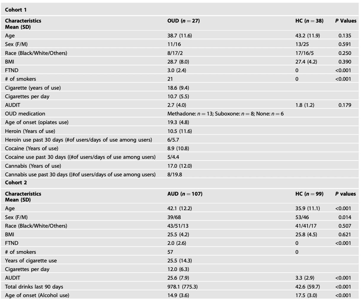
We used data from two cohorts to investigate brain states in OUD (Cohort 1) and AUD (Cohort 2), respectively. In Cohort 1, data from 27 OUD participants, who were recruited from treatment programs, or the community and 38 HC were included for analyses. OUD participants met the criteria for a DSM-5 OUD diagnosis in their lifetime and had a minimum 5-year history of opiate misuse. Twenty-one (out of 27) OUD participants were being treated with medications for OUD (buprenorphine or methadone). In Cohort 2, data from 107 AUD participants and 99 HC were included for analyses. Among AUD participants, 22 were non-treatment seeking and 85 were treatment seeking. Treatment-seeking participants were enrolled in a short-term inpatient detoxification program at the NIH clinical center and the average number of detoxification days prior to the scan day was 19.41 ± 7.52 days. A standardized clinical interview for DSM-IV or DSM-5 was used for AUD diagnosis. The Timeline Follow-back was used to assess daily alcohol consumption in the 90 days prior to the study. For the AUD cohort, we excluded participants with other SUDs apart from AUD or nicotine dependence, whereas for the OUD cohort, we did not exclude them if they had an additional SUD. For both cohorts, participants were given a breathalyzer and a urine drug screen (cocaine, tetrahydrocannabinol, opiates, amphetamine, methamphetamine, and oxycontin) on each day of testing. Participants who failed the drug screen were excluded, except for the presence of opiates in OUD. HC had no history of SUD or other psychiatric disorders nor current use of prescribed or over-the-counter psychoactive medications. The Alcohol Use Disorders Identification Test and the Fagerstrom Test for Nicotine Dependence (FTND) were used to identify harmful drinking behavior and nicotine dependence, respectively. All participants were asked not to smoke in the 2 h prior to their MRI scan and/or to remove their nicotine patch if they had one. There were no smokers among HC. Written informed consent approved by the Institutional Review Board at the NIH was obtained from all participants.

MRI acquisition

In Cohort 1, participants were scanned on a 3 T Magnetom Prisma scanner (Siemens Medical Solutions USA, Inc., Malvern, PA) with a 32-chaneel head coil. For rfMRI, data were collected for 8 min; a multi-echo, multiband echo-planar imaging (EPI) sequence was used (Multiband factor = 3, TR = 891 ms, TE = 16, 33 and 48 ms, FA = 57°, 45 slices with 2.9 × 2.9 × 3.0 mm voxels, 520 time points). Multi-echo images for each time point were combined using an echo-time weighted average of TEs. A fixation cross was presented on a black background and participants were instructed to keep their eyes open, using an LCD monitor. T1-weighted 3D MPRAGE (TR/TE = 2400/2.24 ms, FA = 8°) and T2-weighted variable flip angle turbo spin-echo (Siemens SPACE; TR/TE = 3200/564 ms) pulse sequences were used to acquire high-resolution anatomical brain images with 0.8 mm isotropic voxels FOV = 240 × 256 mm.

In Cohort 2, participants were scanned on a Siemens 3T Magnetom Skyra scanner with a 20-channel head coil. The rfMRI data were collected for 10 min with eyes open and an EPI sequence was used (TR = 2000 ms, TE = 30 ms, FA = 90°, 3.8 mm isotropic voxels, multi-slice mode: interleaved, 300 time points). High resolution structural images were collected using a T1-weighted MPRAGE sequence (TR/TE = 1900/3.09 ms, FA = 10°, FOV: 240 × 240 mm, 1 mm isotropic voxels).

Due to the differences in MRI acquisition, all analyses were conducted for Cohort 1 and Cohort 2 separately.

Replicate brain states with an independent dataset with a single-echo multiband EPI (Cohort 3)

An independent dataset with 55 healthy individuals (Age 42.0 ± 13.2; 30 Females) was used for validating identified brain states. A single-echo multiband EPI (multiband factor = 8, TR/TE = 720/37 ms, FA = 52° and 72 slices with 2 mm isotropic voxels, 1238 time points) were used to record resting-state BOLD responses for 15 min.

MRI preprocessing

The data were preprocessed using CONN toolbox 21a including rigid body realignment, spatial normalization to MNI space, smoothing (FWHM = 6 mm), band-pass filtering (0.01–0.08 Hz), linear detrending, head motion regression (three rotational, three translational and their derivatives), removal of signals within the CSF and the WM using aCompcor, a method for identifying principal components associated with segmented WM and CSF. Using custom MATLAB code, we further scrubbed volumes with a FD threshold of 0.25 mm and DVARS threshold of 150%. Participants who had a mean FD < 0.6 mm before scrubbing and the number of time frames greater than 180 after scrubbing were included in the analyses.

Analysis of brain states and dynamics

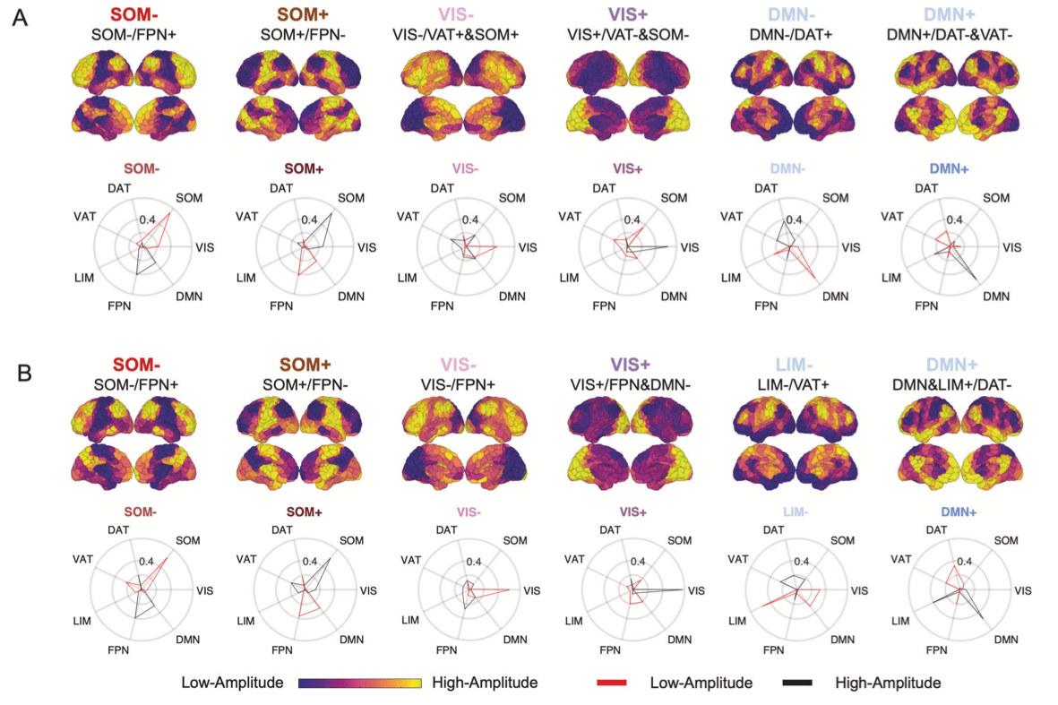
To identify brain states, i.e., brain co-activation patterns, denoised voxel-level data were first parcellated into 400-node Schaefer atlas [21] and demeaned. We then concatenated ROI timeseries from all participants combining HC and patients (Matrix row: NSubjects*Ntime points; column: NROIs) and applied k-means clustering. This approach allowed us to investigate brain dynamics with the maximal temporal resolution of 1 TR. We performed k-means clustering for k = 2–22 in Cohort 1 and k = 2–17 in Cohort 2, where k was the number of clusters (k2 must be less than the number of TRs to capture all transitions) using Pearson correlation as the distance metric and repeated 50 times with the random initializations before choosing the solution with the best data separation. The optimal number of clusters (k) was determined based on incremental variance explained by the lowest error solution at each value of k. In Cohort 1, k = 6 was chosen because the additional variance explained by increasing k beyond k = 6 was less than 1%. In Cohort 2, additional variance explained by increasing k beyond k = 5 was less than 1%. However, to keep consistency between the two cohorts, we chose k = 6 for the second cohort as well (Fig. S1). Another reason we chose k = 6 rather than k = 5 is that with k = 6 we were able to identify three anticorrelated pairs of brain states in both cohorts. To further ensure the reliability of our partitions, we independently repeated the process ten times and computed adjusted mutual information between each of the ten resulting partitions. We then selected the partition that shared the greatest adjusted mutual information with all other partitions for further analysis. Clusters were defined as brain states and labeled by assessing the cosine similarity of the positive and negative activations of their centroid with a binary presentation of seven a priori-defined brain functional networks. Positive values of cluster centroid reflect activations above mean (high amplitude), while negative values reflect activations below mean (low amplitude).

Following this, we then analyzed the dynamic characteristics of the six identified brain states. The fractional occupancy was defined as proportion of TRs assigned to each brain state. Dwell time was calculated by averaging the length of time (number of contiguous TRs *TR) spent in a brain state. Appearance rate was determined by the total number of times a state appeared per minute. Additionally, transition probability between states i and j was defined as the probability that state j occurs at the TR after state i, given that state i is occurring.

Analysis of static functional connectivity

Since instantaneous network co-activation and static functional connectivity provide complementary information on brain networks at rest, we additionally analyzed resting state functional connectivity between intrinsic networks. Denoised voxel-level data were first parcellated into seven networks using Yeo atlas. Pearson’s correlation coefficients between the network time courses were computed. Correlation coefficients were then converted to normally distributed Z-scores using the Fisher transformation for the second-level group analysis.

Statistics

For group comparisons (OUD/AUD vs HC), two sample t test were used. In OUD and AUD participants, we also assessed how co-morbid nicotine dependence affected brain state dynamics by calculating the correlations with the FTND scores. Additionally, we calculated the correlations between brain state dynamics with the AUDIT scores to examine the effect of co-morbid alcohol use in OUD and the effect of alcohol use severity in AUD. As the sample size in Cohort 2 was large enough, we also performed one-way ANOVA to investigate differences between AUD smokers (n = 57), AUD non-smokers (n = 50) and HC. Fisher’s LSD tests were used for ANOVA posthoc tests. SPSS 22 (IBM, Armonk, NY), MATLAB (R2022b) and RStudio were used for analyzes. Benjamini-Hochberg procedure (BH) was applied for corrections for multiple comparisons. Both uncorrected and BH-corrected p values as well as effect sizes were reported. Findings with puncorr < 0.05 were discussed.

Table 1. Demographic information and clinical characteristics.

Recurrent brain states

Clustering algorithm identified six co-activity patterns i.e., brain states in both cohorts (Fig. 1). The six brain states remained when identifying brain states by clustering groups (OUD/AUD vs HC) separately (Figs. S2 and S3). We further replicated our results with an independent dataset indicating the robustness of the identified brain states (Fig. S4). Based on cosine similarity to Yeo’s 7 networks, brain states were labeled as SOM+, SOM-, VIS+, VIS-, DMN+, DMN-/LIM-. In both cohorts, we also observed the hierarchical relationship among the six identified brain states, which could be grouped into three anti-correlated pairs (Fig. S5). Similar findings of anticorrelated sub-states have been previously reported by others using this method.

Fig. 1. Recurrent brain states.

Temporal dynamics of the recurrent brain states

To capture dynamic characteristics of the identified brain states, we calculated fractional occupancy (probability of occurrence), dwell time (duration of persistence) and appearance rates (frequency of appearance per minute). OUD participants showed lower fractional occupancy in DMN+ (t63 = −2.69, puncorr = 0.009, Cohen’s d = 0.68, pBH = 0.05 for six comparisons), shorter dwell time in DMN+ (t63 = −2.19, puncorr = 0.032, Cohen’s d = 0.55, pBH = 0.09) and DMN- (t63 = −2.28, puncorr = 0.026, Cohen’s d = 0.57, pBH = 0.09), and higher appearance rate in VIS+ (t63 = 2.22, puncorr = 0.030, Cohen’s d = 0.53, pBH = 0.10) (Fig. 2). There were no significant differences between AUD and HC when combining AUD smokers and non-smokers. Since age and sex differed between AUD and HC (Table 1), we further controlled them as covariates and did not find group differences.

Fig. 2. Brain dynamics in OUD vs HC.

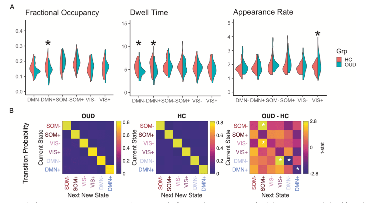
Transition probabilities between brain states were calculated for each participant. The probability of persistence in DMN+ and DMN- was 5% and 4% lower in OUD than HC, respectively (DMN + : t63 = −2.10, puncorr = 0.039, Cohen’s d = 0.51, pBH = 0.28 for 36 comparisons; DMN-: t63 = −2.80, puncorr = 0.007, Cohen’s d = 0.68, pBH = 0.08), consistent with shorter dwell time in DMN+ and DMN-. Compared to HC, higher probability of transitioning from states DMN- to VIS+ (2%, t63 = 2.80, puncorr = 0.007, Cohen’s d = 0.69, pBH = 0.08), from VIS- to SOM+ (2%, t63 = 2.24, puncorr = 0.029, Cohen’s d = 0.56, pBH = 0.26), and from SOM- to SOM+ (1%, t63 = 2.77, puncorr = 0.007, Cohen’s d = 0.65, pBH = 0.08) was observed in OUD (Fig. 2). Transition probabilities did not differ between AUD and HC when combining AUD smokers and non-smokers.

Static functional connectivity between networks

In Cohort 1, we found significant anticorrelations between SOM and FPN, and between DMN and DAT, which were consistent with their contra-activation in dynamic brain states i.e., SOM-/FPN+, SOM+/FPN-, DMN-/DAT+ and DMN+/DAT- (Figs. 1 and S6). Similarly, in Cohort 2, we found significant anticorrelations between SOM and FPN, and between DMN and DAT, as well as anticorrelations between VIS and all other networks that contributed to VIS+ and VIS- brain states. Strong functional connectivities between SOM and VAT and between LIM and DMN were also consistent with co-activation pattern in the SOM-, SOM+ and DMN+ brain states (Figs. 1 and S7). In sum, there were consistencies between resting functional connectivities and co-activation patterns of brain networks i.e., brain states.

OUD participants showed weaker anticorrelation between DMN and DAT (t63 = 2.33, puncorr = 0.023, Cohen’s d = 0.58) and greater functional connectivity between DMN and FPN than HC (t63 = 2.01, puncorr = 0.040, Cohen’s d = 0.51) (Fig. S6). Since findings from both brain states and resting-state functional connectivity indicated changes in DMN, we further explored the relationship between DMN functional connectivity and brain dynamics in OUD participants. Weaker anticorrelation between DMN and DAT but not DMN-FPN connectivity was correlated with shorter DMN- dwell time (r65 = −0.487, p = 0.010) and low probability of persisting in DMN- state (r65 = −0.416, p = 0.031). AUD participants had higher functional connectivity between SOM and VAT than HC (t63 = 2.69, puncorr = 0.008, Cohen’s d = 0.38) (Fig. S6). Controlling for age and sex did not change the results.

Effect of drug co-use

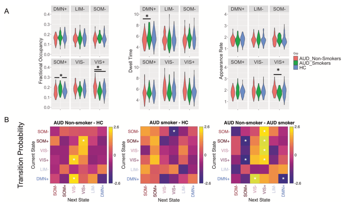
Among OUD participants, co-morbid nicotine dependence was associated with lowered fractional occupancy in VIS- (r27 = −0.487, puncorr = 0.010, pBH = 0.06 for six comparisons), dwell time in VIS+ (r27 = −0.396, puncorr = 0.041, pBH = 0.12), and VIS- (r27 = −0.453, puncorr = 0.018, pBH = 0.10), and reduced persistence in VIS- (r27 = −0.416, puncorr = 0.006, pBH = 0.22 for 36 comparisons) or VIS+ states (r27 = −0.427, puncorr = 0.004, pBH = 0.22). Also, nicotine dependence enhanced the transition probability from VIS- to DMN- (r27 = 0.567, puncorr = 0.002, pBH = 0.07), from VIS+/DMN- to DMN+ states (r27 = 0.418, puncorr = 0.030, pBH = 0.22/r27 = 0.533, puncorr = 0.004, pBH = 0.07) (Fig. 3A). Similarly in AUD participants greater nicotine dependence was associated with lower fractional occupancy (r107 = −0.268, puncorr = 0.005, pBH = 0.03) and appearance rate in VIS+ (r107 = −0.266, puncorr = 0.006, pBH = 0.03) and higher fractional occupancy (r107 = 0.232, puncorr = 0.016, pBH = 0.04) and longer dwell time in DMN+ state (r107 = 0.215, puncorr = 0.026, pBH = 0.16). In terms of transition probability, in AUD, higher FTND scores were associated with lower probabilities of transitioning from SOM-/VIS-/DMN+ to VIS+/VIS- state (all r107 < −0.190, puncorr < 0.050, all pBH > 0.05), and higher probabilities from VIS- to LIM-, from VIS- to SOM+, or of persisting in DMN+ state (all r107 > 0.200, all puncorr < 0.039, all pBH > 0.05 Fig. 3B). Consistently, we found different brain dynamics for AUD smokers and non-smokers. AUD smokers had longer dwell time in DMN+, higher fractional occupancy in SOM+, lower fractional occupancy and lower appearance rate in VIS+ than AUD non-smokers. Compared to HC, AUD non-smokers showed higher VIS+ fractional occupancy, while AUD smokers had higher SOM+ fractional occupancy (Fractional occupancy: VIS+: F(2,203) = 5.649, puncorr = 0.004, partial η2 = 0.053, pBH = 0.02; SOM+: F(2,203) = 4.519, puncorr = 0.012, partial η2 = 0.043, pBH = 0.03; Appearance rate: VIS+: F(2,203) = 3.277, puncorr = 0.040, partial η2 = 0.031, pBH = 0.24; Dwell time: DMN+: F(2,203) = 3.259, puncorr = 0.040, partial η2 = 0.031, pBH = 0.24; all post-hoc tests p < 0.05) (Fig. 4A). Regarding transition probabilities, compared to HC, there was higher transition to the VIS+ or VIS- states in AUD non-smokers (1–2%, all t147 > 2.08, puncorr < 0.039, Cohen’s d > 0.33, all pBH > 0.05), while lower transitions to VIS+ in AUD smokers (2%, t154 = 2.07, puncorr = 0.040, Cohen’s d = 0.36, pBH = 0.56). AUD smokers had higher transitions to SOM+ (2–3%), lower transition to VIS+ or VIS- brain states (1–3%) and higher persistence in DMN+ state (4%) than AUD non-smokers (all t105 > 2.15, puncorr < 0.034, Cohen’s d >0.42, all pBH > 0.05) (Fig. 4B). Differences in brain state dynamics were also reflected in static functional connectivity. Greater dwell time in DMN+ state and higher possibility of persistence in DMN+ state in AUD smokers was associated with stronger anticorrelation between DMN and VAT (Fig. S7) (all r206 < −0.349, all p < 0.001). In contrast, lower fractional occupancy and lower persistence in SOM+ in AUD non-smokers were associated with weaker anticorrelation between SOM-FPN (Fig. S7) (all r206 < −0.473, p < 0.001).

Fig. 3. The effect of nicotine dependence and alcohol use on brain state transition probabilities.Fig. 4. Brain dynamics in AUD smokers vs non-smokers.

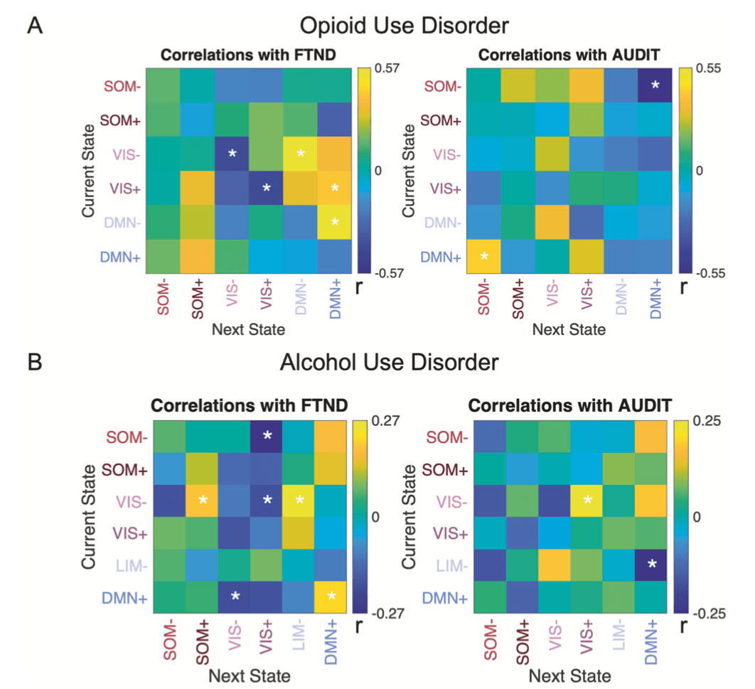
In OUD, co-morbid alcohol use was negatively associated with fractional occupancy (r27 = −0.501, puncorr = 0.008, pBH = 0.048 for six comparisons) and appearance rate in DMN+ (r27 = −0.500, puncorr = 0.008, pBH = 0.048). In line with this, severity of alcohol use problems decreased probability of transitioning from SOM- to DMN+ (r27 = −0.549, puncorr = 0.003, pBH = 0.11 for 36 comparisons), while it increased the transition probability from DMN+ to SOM- (r27 = 0.427, puncorr = 0.026, pBH = 0.47) in OUD participants (Fig. 3A). Higher AUDIT scores in AUD participants were associated with higher transition from VIS- to VIS+ (r99 = 0.235, p = 0.019, pBH = 0.34) and lower transition from LIM- to DMN+ (r99 = −0.246, puncorr = 0.014, pBH = 0.34) (Fig. 3B).

Effects of OUD medications and AUD detoxification

To assess if medications for OUD affected dynamic brain states and static functional connectivity in OUD participants, we compared those treated with Methadone, Buprenorphine, or without medications. Analyses showed no differences between OUD groups in brain state dynamics (one-way ANOVA, all F(2,24) < 3.405, all puncorr > 0.05) or static functional connectivity (one-way ANOVA, all F(2,24) < 1.188, all puncorr > 0.323).

In the AUD cohort who underwent inpatient detoxification, we investigated how days of detoxification affected brain state dynamics. Analyses showed that longer withdrawal days prior to the scan were associated with greater fractional occupancy in DMN+ (r61 = 0.27, puncorr = 0.038, pBH = 0.22), higher transition probabilities from VIS- to DMN+/DMN+ (all r61 > 0.28, all puncorr < 0.029, all pBH = 0.21), and lower transition probabilities from SOM+ to SOM- (r61 = −0.28, puncorr = 0.028, pBH = 0.21), from VIS- to VIS+ (r61 = −0.30, puncorr = 0.019, pBH = 0.21), from VIS+ to DMN- (r61 = −0.25, puncorr = 0.048, pBH = 0.27) and from DMN- to SOM+ (r61 = −0.35, puncorr = 0.006, pBH = 0.21).

Discussion

Aberrant brain functional connectivity has been reported in individuals suffering from various SUDs. Here, we revealed altered temporal dynamics in recurrent brain states in OUD and AUD participants. OUD and AUD non-smokers displayed similarities in brain dynamic changes including decreased fractional occupancy or dwell time in DMN-dominated brain states and increased fractional occupancy or appearance rate in VIS+ brain states, which were also reflected in transition probabilities of brain states. The observed alterations in brain state dynamics were greater in OUD participants who had more severe alcohol use and in the AUD participants with the more severe disorder. Interestingly, co-morbid nicotine dependence mitigated the disrupted brain states in individuals with OUD and AUD. Specifically, greater nicotine dependence was associated with higher DMN-dominated brain states and lower VIS-dominated brain states. Overall, the observed effect sizes of changes in brain state dynamics were greater in OUD than in AUD participants. Of note, OUD and AUD participants were from two different cohorts whose data were analyzed separately. Therefore, a direct comparison was not possible.

We identified six brain states comprised of combinations of active and inactive brain networks in both cohorts and validated the brain states in an independent sample scanned with different sample rate, scan lengths and EPI sequences (Figs. 1 and S4). The identified brain states were very robust and not affected by different scanning protocols. Compared to HC, OUD participants spent less time in brain states characterized by contraposition of DMN with DAT while they showed increased appearance rate in the brain state with high VIS activation. The findings of brain state dynamics and brain state transition probabilities were in agreement with each other. While DMN states were less persistent, the transition to states with high-amplitude activity in SOM and VIS was more likely in OUD than in HC. For AUD participants, we did not find significant differences from healthy controls when combining smokers and non-smokers, and differences only emerged when we compared HC with AUD who were non-smokers. Specifically, AUD non-smokers displayed very similar changes in brain dynamics and transitions as OUD participants i.e., decreased dwell time in DMN-dominated brain state, increased occurrence in VIS-dominated brain state accordant with higher probabilities of transitioning to a VIS-dominated state and lower probability of persisting in a DMN+ state. An additive effect on brain state differences in the participants with OUD and co-morbid alcohol misuse further supported the similar effects of chronic opioid and alcohol use on brain state dynamics. In both AUD and OUD participants, greater nicotine dependence was associated with lower occurrence and transition to a VIS-dominated state, whereas higher occurrence and transition to a DMN-dominated state. These results were also confirmed by an alternative analysis that compared AUD participants who were smokers versus non-smokers. As there were only six non-smokers among OUD participants, we were not able to compare OUD non-smokers with OUD smokers. We expect that changes in brain dynamics might be greater in OUD non-smokers than OUD smokers, but this will need to be confirmed in a larger sample size.

Our nicotine findings are consistent with previous reports of decreased dynamic functional connectivity density in the visual cortex and increased in the orbitofrontal cortex, anterior key node of the DMN, in chronic smokers compared to non-smokers. Similar findings on brain dynamics were reported in cocaine users who showed higher occurrence rate in the DMN state than HC. The counteracting effects of nicotine on the changes in brain state dynamics in AUD that we observed, are consistent with findings in two large datasets showing that participants with comorbid alcohol and nicotine misuse had smaller changes in static functional connectivity than participants who were only drinkers or smokers. Alcohol consumption increased ratings of desire to smoke and a similar effect was reported for opioid consumption and craving for cocaine. Combined use of drugs may serve to modulate the effects of one drug over the other e.g., stimulants to overcome mental state changes induced by sedatives and vice versa. The dynamics in DMN and VIS states might be important for shifting neural activity between internally and externally directed processes.

We interpret our findings as the result of neuroadaptations from chronic drug exposures. However, we cannot distinguish between temporal neuroadaptations that drive withdrawal (physical dependence) versus persistent neuroadaptations that sustain addiction. For AUD participants alcohol use was discontinued prior to the testing day. For the subgroup of AUD participants who were enrolled in an inpatient detoxification program, days in detoxification prior to the scan were associated with normalization of brain state dynamics as reflected by greater fractional occupancy and transition probability to the DMN+ state. For the OUD cohort, brain state dynamics did not significantly differ between participants based on medication status (Methadone vs Buprenorphine vs None). However, the lack of a significant effect of opioid medication could be attributed to the small sample size i.e., only six OUD participants were not under medication treatment (Table 1). Additionally, none of these six subjects used illicit opioids in the past 30 days indicating that these six OUD participants were in recovery even without OUD medication treatment. Thus, a larger cohort is needed to rule out the contribution of medication for OUD and the effects of long-term abstinence and recovery. As for nicotine, participants from both cohorts were asked to discontinue nicotine use 2 h before the scan. Based on the relatively low FTND scores and daily cigarette consumption of less than a pack per day (<20) in both AUD and OUD (Table 1), most participants were light smokers [11]. Thus, it is unlikely that they would have experienced severe nicotine withdrawal after 2 h of abstinence. However, since we did not record the time since last cigarette use nor did we assess withdrawal symptoms at the time of scanning, we could not control for these variables in the analyses. Therefore, our findings may reflect a mixture of short and long-term neuroadaptations associated with physical dependence and withdrawal and more persistent ones associated with addiction. Further, because we did not measure nicotine in plasma at the time of scanning, our findings could also be confounded by the presence of nicotine in plasma from the last cigarette, though the 2-hour nonsmoking period would have minimized this. Interestingly, a previous study that allowed chronic smokers (light and heavy smokers) to smoke before the scan to prevent withdrawal symptom, showed similar results of brain dynamics in chronic nicotine users indicating that effects cannot be attributed to nicotine withdrawal. In non-smokers, nicotine biases resting-state brain function away from the DMN+ and toward the salience network-dominated state, which is opposite to the effect observed in our study in participants with OUD or AUD who were also nicotine dependent. Therefore, the reported findings in the current study are more likely to relate to nicotine addiction than to withdrawal or to acute nicotine effects.

There are at least two kinds of neural underpinnings that might lead to altered brain state dynamics. First are structural changes. White matter damage has been associated with reduced DMN brain states occupancy in patients with cerebral small vessel disease. Damage of structural white matter connectivity has been reported in individuals with OUD and with AUD, which could impact brain states and transitions. Structural changes in gray matter such as cortical thinning in AUD and OUD could also affect brain state dynamics. OUD and AUD displayed common patterns of cortical thinning that were not present in patients with stimulant use disorder comorbid with AUD. Second, brain network dynamics are likely modulated by neurotransmitters including dopamine and serotonin. Since chronic opioid or alcohol use impacts dopaminergic and serotonergic signaling, altered neurotransmission in these systems might have contributed to the brain state dynamics imbalances seen in OUD and AUD participants. Moreover, alcohol stimulates the opioid system and opioid agonist medications (buprenorphine and methadone) as well as antagonists (naltrexone) reduce alcohol consumption in OUD suggesting that altered opioid signaling could also account for brain dynamics changes in OUD and AUD participants and might explain the greater effects observed in OUD than AUD. The counteracting effects of nicotine could be due to its interaction with opioid system. When used in combination, nicotine enhances opioid-induced antinociception. Furthermore, modulation of nicotinic acetylcholine receptors affects alcohol intake as well. Together, our finding suggests complex interactions among various neurotransmitter systems. Understanding the neural origins of altered brain state dynamics and transitions in SUD deserves further investigation.

The relationship between recurrent brain states and static functional connectivity is complex. Though static functional connectivity is associated with dynamic brain states, it cannot fully explain instantaneous coactivation of functional networks. In the current study, we found that anticorrelations between SOM and FPN and between DMN and DAT were reflected by brain states displaying contra-activations of these anti-correlated networks. Further, weaker/stronger DMN-DAT anticorrelation was associated with less/more time spent in DMN states. The neurocircuitry involved in addiction has been characterized by preclinical and clinical studies, however the dynamics underlying the stages of the addiction cycle (intoxication, withdrawal, and rumination/craving) and the transitions to remission and recovery are much less well understood. Our findings of a disruption in the dynamic mental states patterns in OUD and AUD participants, most of whom were studied during short-term detoxification (withdrawal) or while under OUD medication, suggests that addiction disrupts the stability of mental state functional networks towards externally dominated states over internal ones. Further work is needed to understand how brain state dynamics influence the risk for relapse including studies on how therapeutic interventions such as neuromodulation affect brain state dynamics and their relationship to symptom control.

The current study provides evidence for changes in brain state dynamics in OUD and AUD participants. We revealed similar effects of chronic opioid and alcohol use on brain dynamics and transitions at rest and a counteracting effect of nicotine dependence in these participants. Our findings highlight the importance of strategizing the treatment for drug co-use. For instance, one may consider nicotine replacement therapies as an adjunct intervention for treating AUD and OUD. Also, while discontinuing nicotine use in AUD and SUD, additional monitoring/intervention such as mindfulness training that prolongs the time spent in the DMN state, might be necessary to prevent worsening of symptoms. Future interventions that modulate brain network dynamics based on participants’ drug co-use might provide additional benefits to recovery.

Open Article as PDF

Abstract

Substance use disorder (SUD) is a chronic relapsing disorder with long-lasting changes in brain intrinsic networks. While most research to date has focused on static functional connectivity, less is known about the effect of chronic drug use on dynamics of brain networks. Here we investigated brain state dynamics in individuals with opioid use (OUD) and alcohol use disorder (AUD) and assessed how concomitant nicotine use, which is frequent among individuals with OUD and AUD, affects brain dynamics. Resting-state functional magnetic resonance imaging data of 27 OUD, 107 AUD, and 137 healthy participants were included in the analyses. To identify recurrent brain states and their dynamics, we applied a data-driven clustering approach that determines brain states at a single time frame. We found that OUD and AUD non-smokers displayed similar changes in brain state dynamics including decreased fractional occupancy or dwell time in default mode network (DMN)-dominated brain states and increased appearance rate in visual network (VIS)-dominated brain states, which were also reflected in transition probabilities of related brain states. Interestingly, co-use of nicotine affected brain states in an opposite manner by lowering VIS-dominated and enhancing DMN-dominated brain states in both OUD and AUD participants. Our finding revealed a similar pattern of brain state dynamics in OUD and AUD participants that differed from controls, with an opposite effect for nicotine use suggesting distinct effects of various drugs on brain state dynamics. Different strategies for treating SUD may need to be implemented based on patterns of co-morbid drug use.

Disrupted Brain State Dynamics in Opioid and Alcohol Use Disorder: Reduction with Nicotine Use

Introduction

Substance use disorder (SUD) is a long-term condition that causes a significant burden on global health. In a recent year, tens of thousands of deaths were linked to opioid overdoses in the United States, and over one hundred thousand deaths were caused by alcohol-related issues each year. Therefore, understanding the brain mechanisms involved in SUD is crucial for addressing its consequences.

Functional magnetic resonance imaging (fMRI) has advanced understanding of how large brain networks function when a person is at rest. Evidence suggests that long-term drug use can cause lasting changes in these brain networks, which can affect a person's thinking and emotions. These changes are important in how SUD develops and continues. Most studies on SUD have focused on measuring static functional connectivity, which assumes brain connections remain constant during a scan. Differences are often observed in networks like the default mode network (involved in self-thought), the salience network (directing attention), and the executive control network (cognitive control and goal-directed actions).

While static functional connectivity shows where networks are located and how strongly they are connected over time, dynamic functional connectivity provides insights into how these connections change over time. These changes may be key to understanding cognition, emotions, and behavior. Few studies have examined these brain dynamics in SUD. For example, in chronic smokers, changes were seen in the visual network, default mode network, and reward pathways. Importantly, dynamic analysis was more sensitive in detecting subtle differences than static analysis. This study examined brain states in individuals with opioid use disorder (OUD) and alcohol use disorder (AUD) and how using opioids, alcohol, and nicotine together affected brain state dynamics. It was hypothesized that both OUD and AUD participants would show unbalanced brain state dynamics compared to healthy controls. As opioids and alcohol are sedating, similar effects were expected, possibly worse with co-use, while nicotine, a stimulant, was predicted to have opposite effects. A data-driven clustering method was used to identify recurring patterns of brain network activity, known as brain states, and how they change over time.

Materials and methods

Participants

Data from two groups were used to study brain states in OUD and AUD. The first group included 27 OUD participants and 38 healthy controls. OUD participants had a diagnosis of opioid use disorder and at least five years of opioid misuse history. Many of them were receiving medication for OUD. The second group included 107 AUD participants and 99 healthy controls. Some AUD participants were seeking treatment through an inpatient detoxification program. A standard clinical interview was used for diagnosis, and alcohol consumption was assessed. Participants with other substance use disorders, except for nicotine dependence, were excluded in the AUD group. All participants were screened for drug use on testing days, and those who failed were excluded, except for OUD participants who tested positive for opiates. Healthy controls had no history of SUD or other mental health disorders. Participants were asked not to smoke for two hours before their brain scan. All participants gave their informed consent.

MRI acquisition

In the first group, brain scans were performed using a 3T MRI scanner for about 8 minutes while participants kept their eyes open and looked at a cross. Detailed anatomical brain images were also collected. In the second group, scans were done on a different 3T MRI scanner for 10 minutes, also with eyes open. High-resolution structural images were also collected. Because the MRI scanning methods were different for each group, all analyses were conducted separately for each group.

Replicate brain states with an independent dataset (Cohort 3)

An additional independent dataset of 55 healthy individuals was used to confirm the brain states identified in this study, ensuring their reliability.

MRI preprocessing

The raw brain imaging data underwent several cleaning and standardization steps. This included aligning images, placing them into a common brain space, smoothing, filtering, and removing motion-related signals and other unwanted noise from areas like cerebrospinal fluid and white matter. Data from participants with excessive head motion were excluded.

Analysis of brain states and dynamics

To identify brain activity patterns, or “brain states,” brain data from all participants were combined and analyzed using a clustering method. This method allowed for examining brain dynamics at the highest possible time resolution. The optimal number of brain states was determined based on how much additional variation they explained in the data. Clusters were then defined as brain states and labeled based on their similarity to known brain functional networks.

Following this, the dynamic characteristics of these brain states were analyzed. These characteristics included: "fractional occupancy," or how often a brain state was present; "dwell time," or how long a brain state persisted; and "appearance rate," or how frequently a state occurred per minute. Additionally, "transition probability" was calculated to determine the likelihood of switching from one brain state to another.

Analysis of static functional connectivity

To complement the dynamic analysis, the average connections between brain networks were also analyzed. This involved calculating the strength of connections between different brain regions and converting these values for group comparisons.

Statistics

Comparisons between participant groups were conducted to identify differences. For individuals with OUD and AUD, correlations were also calculated to assess how nicotine dependence affected brain state dynamics. Additionally, correlations were examined to understand the impact of alcohol use severity. Statistical adjustments were applied for multiple comparisons to ensure the reliability of the findings.

Recurrent brain states

The clustering analysis consistently identified six distinct patterns of brain activity, or "brain states," in both groups. These six brain states were also found when groups were analyzed separately. Furthermore, the findings were confirmed using an independent dataset, indicating the robustness of these identified brain states. Based on their similarity to established brain networks, these states were labeled, including those reflecting positive and negative activity in sensory-motor (SOM), visual (VIS), and default mode (DMN) networks. In both groups, these six brain states showed a hierarchical relationship, forming three pairs of opposite activity patterns.

Temporal dynamics of the recurrent brain states

To capture how brain states changed over time, measures like how often they occurred, how long they lasted, and how frequently they appeared were calculated. OUD participants showed a lower occurrence in a DMN-related brain state and spent less time in both DMN-related states. They also showed a higher frequency of appearance in a VIS-related state. The likelihood of staying in DMN-related states was lower for OUD participants compared to healthy controls. Additionally, OUD participants showed a higher likelihood of transitioning from a DMN-related state to a VIS-related state, and from one sensory-motor state to another. No significant differences were found between AUD and healthy controls when all AUD participants were combined.

Static functional connectivity between networks

In the OUD group, significant opposite connections were found between the sensory-motor network and the fronto-parietal network, and between the default mode network and the dorsal attention network. These findings aligned with the opposite activity patterns seen in the dynamic brain states. Similarly, in the AUD group, opposite connections were observed between the sensory-motor and fronto-parietal networks, and between the default mode and dorsal attention networks, as well as between the visual network and other networks. Overall, there was consistency between the average functional connections and the instantaneous co-activation patterns of brain networks.

OUD participants showed weaker opposite connections between the default mode network and the dorsal attention network, and stronger connections between the default mode network and the fronto-parietal network compared to healthy controls. Weaker opposite connections between the default mode and dorsal attention networks were linked to spending less time in the DMN-related state and a lower chance of staying in that state. AUD participants had stronger connections between the sensory-motor network and the ventral attention network than healthy controls.

Effect of drug co-use

Among OUD participants, greater nicotine dependence was associated with a lower occurrence and shorter duration in visual-related brain states, and reduced likelihood of staying in those states. Nicotine dependence also increased the likelihood of transitioning from visual-related or default mode network states to specific default mode states. Similarly, in AUD participants, greater nicotine dependence was linked to a lower occurrence and appearance rate in a VIS-related state, and a higher occurrence and longer duration in a DMN-related state. Consistent with these findings, AUD smokers showed different brain dynamics than AUD non-smokers. AUD smokers had longer duration in a DMN-related state, higher occurrence in a SOM-related state, and lower occurrence and appearance rate in a VIS-related state compared to AUD non-smokers. Regarding transitions, AUD non-smokers showed a higher likelihood of moving to VIS-related states compared to healthy controls, while AUD smokers showed lower likelihood of moving to VIS-related states. AUD smokers also had a higher likelihood of moving to SOM-related states, lower likelihood of moving to VIS-related states, and a higher chance of staying in a DMN-related state than AUD non-smokers. These differences were also reflected in the average brain connections.

In OUD participants, higher alcohol use was linked to a lower occurrence and appearance rate in a DMN-related state. Greater severity of alcohol use problems decreased the likelihood of transitioning into a DMN-related state, while increasing the likelihood of transitioning out of it. In AUD participants, higher alcohol use severity was associated with a higher likelihood of transitioning between visual-related states and a lower likelihood of transitioning to a DMN-related state.

Effects of OUD medications and AUD detoxification

To assess the impact of medication on brain dynamics in OUD participants, those on Methadone, Buprenorphine, or no medication were compared. No significant differences were found in brain state dynamics or static functional connectivity among these OUD groups.

In the AUD group who underwent inpatient detoxification, longer detoxification periods before the scan were associated with greater occurrence in a DMN-related state and higher likelihood of transitioning into DMN-related states. This also involved a lower likelihood of transitioning between sensory-motor states and visual states.

Discussion

Changes in brain functional connectivity have been observed in individuals with various substance use disorders. This study found altered temporal dynamics in recurring brain states in OUD and AUD participants. Both OUD and AUD non-smokers displayed similar changes in brain dynamics, including reduced time spent in default mode network-dominated states and increased occurrence in visual-dominated states. These changes were also reflected in brain state transitions. The observed alterations were more pronounced in OUD participants with more severe alcohol use and in AUD participants with a more severe disorder. Interestingly, nicotine dependence appeared to lessen the disrupted brain states in individuals with OUD and AUD. Specifically, greater nicotine dependence was associated with more time spent in DMN-dominated brain states and less time in visual-dominated brain states. Overall, the impact on brain state dynamics was greater in OUD than in AUD participants. Since OUD and AUD participants came from two different study groups and their data were analyzed separately, a direct comparison between the two disorders was not possible.

The study identified six robust brain states, consistent across both groups and an independent dataset, regardless of scanning protocols. Compared to healthy controls, OUD participants spent less time in brain states involving opposite activity between the default mode network and the dorsal attention network, but showed increased activity in the visual network. These findings regarding brain state dynamics and transitions were consistent. While default mode network states were less stable, transitions to states with high activity in the sensory-motor and visual networks were more likely in OUD than in healthy controls. For AUD participants, significant differences from healthy controls only emerged when comparing non-smokers. AUD non-smokers showed very similar changes in brain dynamics and transitions as OUD participants, including reduced time in DMN-dominated states and increased activity in VIS-dominated states. The combined effect of OUD and alcohol misuse further supported similar impacts of chronic opioid and alcohol use on brain state dynamics. In both AUD and OUD participants, greater nicotine dependence was linked to lower occurrence and transition to visual-dominated states, and higher occurrence and transition to default mode network-dominated states. These results were also supported by comparing AUD smokers versus non-smokers.

Findings regarding nicotine align with previous reports of changes in dynamic functional connectivity in chronic smokers. The counteracting effects of nicotine on brain state changes in AUD are consistent with studies showing that individuals with both alcohol and nicotine misuse had smaller changes in static functional connectivity than those using only one substance. Combining drugs may serve to alter the effects of one drug over another, for example, using stimulants to counteract mental state changes caused by sedatives. The dynamics within the default mode and visual states may be important for shifting brain activity between internal and external processes.

The observed brain changes are likely a result of long-term drug exposure. However, it is not possible to distinguish between temporary brain changes linked to withdrawal and more lasting changes that sustain addiction. For AUD participants, alcohol use had stopped before the testing day. In the subgroup of AUD participants in an inpatient detoxification program, longer detoxification periods before the scan were linked to a normalization of brain state dynamics, indicated by greater occurrence and transition to the DMN-related state. For the OUD group, medication status did not significantly impact brain state dynamics, possibly due to the small number of participants not on medication. While participants were asked to abstain from nicotine for two hours before the scan, and most were light smokers, it is unlikely they experienced severe nicotine withdrawal. However, these variables were not fully controlled, so findings might reflect a mix of short- and long-term changes. Interestingly, similar brain dynamics were observed in a previous study where chronic smokers were allowed to smoke before the scan, suggesting the effects are not solely due to withdrawal.

Altered brain state dynamics could stem from structural changes in the brain, such as white matter damage or cortical thinning, which have been observed in individuals with OUD and AUD. These structural changes could influence how brain states function and transition. Additionally, brain network dynamics are influenced by neurotransmitters like dopamine and serotonin, which are affected by chronic opioid or alcohol use. Altered signaling in these systems might contribute to the brain state imbalances seen in OUD and AUD participants. The interaction between alcohol and the opioid system, and nicotine's interaction with the opioid system, could also explain the observed counteracting effects of nicotine and the greater impact seen in OUD. Understanding the precise brain mechanisms behind altered brain state dynamics in SUD requires further research.

The relationship between recurring brain states and average functional connectivity is complex. While average connectivity is related to dynamic brain states, it cannot fully explain the instantaneous co-activation of functional networks. This study found that opposite connections between certain brain networks were reflected by brain states displaying opposite activity in these networks. Furthermore, weaker or stronger opposite connections were linked to less or more time spent in specific default mode network states. The brain circuits involved in addiction have been characterized by previous studies, but the dynamic changes underlying the stages of addiction (intoxication, withdrawal, craving) and transitions to recovery are less understood. The findings suggest that addiction disrupts the stability of brain functional networks, shifting them towards externally focused states rather than internally focused ones, particularly during withdrawal or under medication. Future research is needed to understand how brain state dynamics influence relapse risk and how treatments like neuromodulation can affect these dynamics and their link to symptom control.

This study provides evidence of changes in brain state dynamics in OUD and AUD participants. It revealed similar effects of chronic opioid and alcohol use on brain dynamics and transitions during rest, and a counteracting effect of nicotine dependence in these individuals. The findings highlight the importance of developing treatment strategies that consider co-occurring drug use. For example, nicotine replacement therapies might be considered as an additional intervention for treating AUD and OUD. Also, when individuals discontinue nicotine use during AUD and SUD treatment, additional support like mindfulness training, which can prolong time spent in certain default mode network states, might be necessary to prevent symptoms from worsening. Future interventions that adjust brain network dynamics based on an individual's co-occurring drug use could provide further benefits for recovery.

Open Article as PDF

Abstract

Substance use disorder (SUD) is a chronic relapsing disorder with long-lasting changes in brain intrinsic networks. While most research to date has focused on static functional connectivity, less is known about the effect of chronic drug use on dynamics of brain networks. Here we investigated brain state dynamics in individuals with opioid use (OUD) and alcohol use disorder (AUD) and assessed how concomitant nicotine use, which is frequent among individuals with OUD and AUD, affects brain dynamics. Resting-state functional magnetic resonance imaging data of 27 OUD, 107 AUD, and 137 healthy participants were included in the analyses. To identify recurrent brain states and their dynamics, we applied a data-driven clustering approach that determines brain states at a single time frame. We found that OUD and AUD non-smokers displayed similar changes in brain state dynamics including decreased fractional occupancy or dwell time in default mode network (DMN)-dominated brain states and increased appearance rate in visual network (VIS)-dominated brain states, which were also reflected in transition probabilities of related brain states. Interestingly, co-use of nicotine affected brain states in an opposite manner by lowering VIS-dominated and enhancing DMN-dominated brain states in both OUD and AUD participants. Our finding revealed a similar pattern of brain state dynamics in OUD and AUD participants that differed from controls, with an opposite effect for nicotine use suggesting distinct effects of various drugs on brain state dynamics. Different strategies for treating SUD may need to be implemented based on patterns of co-morbid drug use.

Introduction

Substance use disorder (SUD) is a long-lasting condition that contributes significantly to health problems worldwide. For example, in 2021, over 80,000 deaths in the United States were linked to opioid overdoses, and more than 140,000 deaths each year are related to alcohol use. Therefore, a deeper understanding of how these disorders affect the brain is essential for addressing their widespread consequences.

Functional magnetic resonance imaging (fMRI) performed while a person is at rest (resting-state fMRI or rfMRI) has improved understanding of how large-scale brain networks function when no specific task is being performed. Research indicates that ongoing drug use can lead to lasting changes in these brain networks, which can impair thinking and emotional functions important for developing and maintaining SUD. Most previous studies on SUD have focused on "static functional connectivity," which assumes that connections between brain regions remain constant during the scan. These studies often show differences in connectivity in networks like the default mode network (involved in self-reflection), the salience network (involved in directing attention), and the executive control network (involved in cognitive control).

While static connectivity shows how brain networks are spatially organized and the average strength of connections over time, "dynamic functional connectivity" offers additional insights into how these networks change over time. Although there is growing interest, few studies have explored brain dynamics in SUD. Earlier research on chronic smokers and cocaine users found altered dynamic connectivity, suggesting it might be more sensitive to subtle brain changes. This study investigated brain states in individuals with opioid use disorder (OUD) and alcohol use disorder (AUD), examining how co-use of opioids, alcohol, and nicotine affects these brain dynamics. The hypothesis was that OUD and AUD participants would show unbalanced brain state dynamics compared to healthy individuals. Since opioids and alcohol are sedatives, similar effects were expected, potentially worsening with co-use. Nicotine, being a stimulant, was predicted to have opposite effects. A data-driven clustering approach was used to identify recurring patterns of brain network co-activation, known as brain states, and their temporal changes.

Materials and Methods

This study used data from two groups to examine brain states in OUD (Cohort 1) and AUD (Cohort 2). Cohort 1 included 27 OUD participants with a history of opioid misuse and 38 healthy individuals. Most OUD participants were receiving medication for their disorder. Cohort 2 included 107 AUD participants and 99 healthy individuals. Some AUD participants were seeking treatment, while others were not. Participants were screened for drug use on testing days, with specific exclusions for other substance use disorders. Healthy individuals had no history of SUD or other psychiatric conditions. Standardized tests were used to assess alcohol and nicotine dependence. Participants were asked to avoid smoking for two hours before their MRI scan.

MRI scans were performed using different scanners for Cohort 1 and Cohort 2, which led to separate analyses for each cohort. Resting-state fMRI data were collected, with participants instructed to keep their eyes open. High-resolution anatomical brain images were also acquired. For validation, an independent dataset (Cohort 3) of healthy individuals was used to confirm the identified brain states, despite differences in scanning parameters.

The fMRI data underwent a series of preprocessing steps using specialized software. These steps included aligning images, standardizing brain size and shape, smoothing data, filtering out irrelevant frequencies, removing head motion, and cleaning up signals from non-brain tissues. Strict criteria were applied to ensure only high-quality data were included in the analyses, focusing on motion levels and the amount of usable data.

To identify distinct brain co-activation patterns, or "brain states," voxel-level data were divided into 400 regions. These time-series data from all participants were then grouped together, and a k-means clustering technique was applied. This method allowed the study to investigate brain dynamics at the highest possible temporal resolution, identifying brain states at each individual time point. The optimal number of clusters (brain states) was determined based on statistical variance. Six brain states were chosen for both cohorts to maintain consistency, and these states were labeled based on their similarity to known brain functional networks. After identifying the brain states, their dynamic characteristics were analyzed. These included "fractional occupancy" (the proportion of time spent in each state), "dwell time" (the average duration of remaining in a state), and "appearance rate" (how often a state occurred per minute). The probability of transitioning between different brain states was also calculated.

In addition to dynamic analyses, static functional connectivity between intrinsic brain networks was also examined. Denoised data were parcellated into seven networks, and Pearson's correlation coefficients were calculated between the time courses of these networks. These coefficients were then converted into Z-scores for group comparisons. Statistical analyses, including two-sample t-tests and one-way ANOVAs, were used for group comparisons (OUD/AUD vs. healthy controls) and to assess the effects of nicotine dependence and alcohol use severity. Correlations were also used to examine relationships between variables. The Benjamini-Hochberg procedure was applied to correct for multiple comparisons, and findings with uncorrected p-values less than 0.05 were discussed.

Recurrent Brain States

The clustering algorithm consistently identified six distinct co-activity patterns, or "brain states," in both cohorts. These six brain states remained consistent even when groups (OUD/AUD vs. healthy controls) were clustered separately. Validation with an independent dataset further confirmed the robustness of these identified brain states. Based on their similarity to established brain networks, the states were labeled, for instance, as SOM+ (sensorimotor active), SOM- (sensorimotor inactive), VIS+ (visual active), VIS- (visual inactive), DMN+ (default mode network active), and DMN-/LIM- (default mode network inactive/limbic inactive). Both cohorts also showed a hierarchical relationship among these six states, forming three anti-correlated pairs.

Temporal Dynamics of the Recurrent Brain States

To capture the dynamic features of the identified brain states, measures like fractional occupancy (how often a state occurs), dwell time (how long a state lasts), and appearance rates (how frequently a state appears) were calculated. Participants with OUD showed lower fractional occupancy and shorter dwell time in DMN+ brain states. They also had shorter dwell time in DMN- states and a higher appearance rate in VIS+ states. However, when combining AUD smokers and non-smokers, no significant differences were observed between AUD participants and healthy controls, even after accounting for age and sex.

Transition probabilities between brain states were also determined for each participant. In OUD participants, the probability of remaining in DMN+ and DMN- states was lower compared to healthy individuals, which aligned with the shorter dwell times observed in these states. OUD participants also showed a higher probability of transitioning from DMN- to VIS+, from VIS- to SOM+, and from SOM- to SOM+. Consistent with the other dynamic measures, transition probabilities did not differ between AUD participants (when combined) and healthy controls.

Static Functional Connectivity Between Networks

In Cohort 1, significant anti-correlations were found between the Sensorimotor (SOM) and Frontoparietal Network (FPN), and between the Default Mode Network (DMN) and Dorsal Attention Network (DAT). These findings were consistent with the contra-activation patterns seen in the dynamic brain states. Similarly, in Cohort 2, anti-correlations were observed between SOM and FPN, and between DMN and DAT, as well as between the Visual network (VIS) and other networks involved in VIS+ and VIS- brain states. Strong functional connections between SOM and Ventral Attention Network (VAT), and between Limbic (LIM) and DMN, also matched the co-activation patterns in some brain states. Overall, there was consistency between resting functional connectivity and the co-activation patterns of brain networks.

OUD participants exhibited weaker anti-correlation between DMN and DAT and stronger functional connectivity between DMN and FPN compared to healthy individuals. Weaker DMN-DAT anti-correlation, but not DMN-FPN connectivity, was linked to shorter DMN- dwell time and a lower probability of remaining in the DMN- state in OUD participants. AUD participants showed higher functional connectivity between SOM and VAT than healthy individuals, and these results remained consistent even when controlling for age and sex.

Effect of Substance Co-use

Among OUD participants, co-occurring nicotine dependence was associated with reduced fractional occupancy and dwell time in VIS- states, and reduced persistence in VIS+ or VIS- states. Nicotine dependence also increased the probability of transitioning from VIS- to DMN-, and from VIS+/DMN- to DMN+ states. Similarly, in AUD participants, greater nicotine dependence was linked to lower fractional occupancy and appearance rate in VIS+ states, and higher fractional occupancy and longer dwell time in DMN+ states. In terms of transition probabilities, higher nicotine dependence in AUD participants was associated with lower probabilities of transitioning from SOM-/VIS-/DMN+ to VIS+/VIS- states, and higher probabilities of transitioning from VIS- to LIM- or SOM+, or persisting in the DMN+ state.

Consistent with these findings, different brain dynamics were observed when comparing AUD smokers and non-smokers. AUD smokers had longer dwell time in DMN+, higher fractional occupancy in SOM+, and lower fractional occupancy and appearance rate in VIS+ compared to AUD non-smokers. When compared to healthy controls, AUD non-smokers showed higher VIS+ fractional occupancy, while AUD smokers had higher SOM+ fractional occupancy. Regarding transition probabilities, AUD non-smokers showed higher transitions to VIS+ or VIS- states compared to healthy controls. AUD smokers had higher transitions to SOM+, lower transitions to VIS+ or VIS- brain states, and greater persistence in the DMN+ state than AUD non-smokers. Differences in brain state dynamics were also reflected in static functional connectivity.

In OUD participants, co-occurring alcohol use was negatively linked to fractional occupancy and appearance rate in DMN+. Alcohol use severity also decreased the probability of transitioning from SOM- to DMN+ in OUD participants, while increasing the transition probability from DMN+ to SOM-. In AUD participants, higher alcohol use severity scores were associated with higher transition from VIS- to VIS+ and lower transition from LIM- to DMN+.

Effects of OUD Medications and AUD Detoxification

To determine if medications for OUD affected brain states, OUD participants treated with Methadone, Buprenorphine, or no medications were compared. Analyses showed no significant differences between these OUD groups in brain state dynamics or static functional connectivity. However, this lack of a significant effect might be due to the small sample size of OUD participants not receiving medication.

In the AUD cohort who participated in an inpatient detoxification program, the number of days in detoxification before the scan was investigated for its effect on brain state dynamics. Analyses indicated that longer withdrawal periods before the scan were associated with greater fractional occupancy in DMN+ states, higher transition probabilities to DMN+ from VIS- and DMN+, and lower transition probabilities between certain other states (e.g., from SOM+ to SOM-, from VIS- to VIS+).

Discussion

Changes in brain functional connectivity have been documented in individuals with various substance use disorders. This study revealed altered temporal dynamics in recurring brain states in OUD and AUD participants. OUD and AUD non-smokers exhibited similar changes in brain dynamics, including reduced time spent in DMN-dominated brain states and increased time or appearance rates in VIS+ brain states. These changes were also reflected in the probabilities of transitioning between brain states. The observed alterations in brain state dynamics were more pronounced in OUD participants with more severe alcohol use and in AUD participants with a more severe disorder. Interestingly, co-occurring nicotine dependence seemed to lessen the disrupted brain states in individuals with OUD and AUD. Specifically, greater nicotine dependence was linked to more time in DMN-dominated brain states and less time in VIS-dominated brain states. Overall, the observed effect sizes of changes in brain state dynamics were larger in OUD than in AUD participants. Since OUD and AUD participants came from two distinct cohorts, a direct comparison between them was not possible.

The study identified six robust brain states in both cohorts, which were consistent across different scanning protocols and validated in an independent sample. OUD participants spent less time in brain states characterized by the opposition of the Default Mode Network (DMN) and Dorsal Attention Network (DAT), while showing an increased rate of appearance in brain states with high Visual network (VIS) activation. These findings for brain state dynamics and transition probabilities were in agreement. DMN states were less stable, while transitions to states with high activity in the Sensorimotor (SOM) and VIS networks were more likely in OUD than in healthy controls. For AUD participants, no significant differences from healthy controls were found when smokers and non-smokers were combined. Differences only emerged when comparing healthy controls with AUD non-smokers. Specifically, AUD non-smokers displayed very similar changes in brain dynamics and transitions as OUD participants, including decreased dwell time in DMN-dominated states and increased occurrence in VIS-dominated states, along with higher probabilities of transitioning to VIS-dominated states and lower probability of persisting in DMN+ states. The additive effect on brain state differences in OUD participants with co-occurring alcohol misuse further supported the similar impacts of chronic opioid and alcohol use on brain state dynamics. In both AUD and OUD participants, greater nicotine dependence was associated with lower occurrence and transition to VIS-dominated states, and higher occurrence and transition to DMN-dominated states. These results were confirmed by an alternative analysis comparing AUD smokers versus non-smokers.

These findings regarding nicotine are consistent with previous reports of decreased dynamic functional connectivity in the visual cortex and increased connectivity in the orbitofrontal cortex (a key part of the DMN) in chronic smokers. Similar brain dynamics have been reported in cocaine users, who showed a higher occurrence rate in the DMN state compared to healthy controls. The observed counteracting effects of nicotine on brain state dynamics in AUD align with findings from large datasets showing that individuals with co-occurring alcohol and nicotine misuse had smaller changes in static functional connectivity than those who only drank or smoked. The combined use of drugs may help moderate the effects of one drug on the other; for example, stimulants might counteract mental state changes caused by sedatives. The dynamics within the DMN and VIS states could be important for shifting brain activity between internally and externally focused processes.

The observed findings are interpreted as the result of changes in the brain due to long-term drug exposure. However, the study cannot differentiate between temporary brain adaptations that drive withdrawal symptoms and persistent adaptations that maintain addiction. For AUD participants, alcohol use was stopped before testing. For a subgroup of AUD participants in a detoxification program, longer detoxification periods before the scan were associated with a normalization of brain state dynamics, indicated by greater fractional occupancy and transition probability to the DMN+ state. In the OUD cohort, brain state dynamics did not significantly differ based on medication status (Methadone vs. Buprenorphine vs. no medication). However, the small number of OUD participants not on medication limits the strength of this finding. Regarding nicotine, participants were asked to stop nicotine use two hours before the scan. Given their generally low nicotine dependence scores, severe withdrawal symptoms were unlikely. However, as the exact time since last use and withdrawal symptoms were not recorded, these factors could not be fully controlled. Therefore, the findings may reflect a mix of short-term and long-term brain adaptations related to physical dependence, withdrawal, and persistent addiction.

At least two types of neural mechanisms could contribute to altered brain state dynamics. First, structural changes, such as damage to white matter, have been linked to reduced DMN brain state occupancy in other conditions and are reported in individuals with OUD and AUD. Gray matter structural changes, like cortical thinning, also seen in AUD and OUD, could affect brain state dynamics. Second, brain network dynamics are likely influenced by neurotransmitters like dopamine and serotonin. Since chronic opioid or alcohol use impacts these systems, altered neurotransmission could contribute to the brain state imbalances seen in OUD and AUD. Furthermore, alcohol affects the opioid system, and opioid medications can reduce alcohol consumption, suggesting that altered opioid signaling might explain the greater effects observed in OUD than AUD. The counteracting effects of nicotine could be due to its interaction with the opioid system, as nicotine can enhance opioid effects and influence alcohol intake. Overall, the findings suggest complex interactions among various neurotransmitter systems.

The relationship between recurring brain states and static functional connectivity is complex. While static functional connectivity is related to dynamic brain states, it cannot fully explain instantaneous co-activation of functional networks. This study found that anti-correlations between SOM and FPN, and between DMN and DAT, were reflected by brain states showing opposing activity in these networks. Furthermore, weaker or stronger DMN-DAT anti-correlation was associated with less or more time spent in DMN states. The brain circuits involved in addiction are well-characterized, but the dynamic changes underlying the stages of the addiction cycle (intoxication, withdrawal, craving) and transitions to remission and recovery are less understood. The findings of disrupted dynamic mental state patterns in OUD and AUD participants, many of whom were studied during short-term detoxification or on OUD medication, suggest that addiction disrupts the stability of mental state functional networks, shifting them towards externally focused states over internal ones. Further research is needed to understand how brain state dynamics influence the risk for relapse and how interventions like neuromodulation affect these dynamics and symptom control.

This study provides evidence for changes in brain state dynamics in OUD and AUD participants. It revealed similar effects of chronic opioid and alcohol use on brain dynamics at rest and a counteracting effect of nicotine dependence in these individuals. These findings highlight the importance of developing treatment strategies that address the co-use of different substances. For example, nicotine replacement therapies might be considered as an additional intervention for treating AUD and OUD. Also, when individuals with AUD and SUD stop using nicotine, additional monitoring or interventions, such as mindfulness training that prolongs time spent in the DMN state, might be necessary to prevent symptoms from worsening. Future interventions that adjust brain network dynamics based on a participant's substance co-use could offer further benefits for recovery.

Open Article as PDF

Abstract

Substance use disorder (SUD) is a chronic relapsing disorder with long-lasting changes in brain intrinsic networks. While most research to date has focused on static functional connectivity, less is known about the effect of chronic drug use on dynamics of brain networks. Here we investigated brain state dynamics in individuals with opioid use (OUD) and alcohol use disorder (AUD) and assessed how concomitant nicotine use, which is frequent among individuals with OUD and AUD, affects brain dynamics. Resting-state functional magnetic resonance imaging data of 27 OUD, 107 AUD, and 137 healthy participants were included in the analyses. To identify recurrent brain states and their dynamics, we applied a data-driven clustering approach that determines brain states at a single time frame. We found that OUD and AUD non-smokers displayed similar changes in brain state dynamics including decreased fractional occupancy or dwell time in default mode network (DMN)-dominated brain states and increased appearance rate in visual network (VIS)-dominated brain states, which were also reflected in transition probabilities of related brain states. Interestingly, co-use of nicotine affected brain states in an opposite manner by lowering VIS-dominated and enhancing DMN-dominated brain states in both OUD and AUD participants. Our finding revealed a similar pattern of brain state dynamics in OUD and AUD participants that differed from controls, with an opposite effect for nicotine use suggesting distinct effects of various drugs on brain state dynamics. Different strategies for treating SUD may need to be implemented based on patterns of co-morbid drug use.

How Opioid and Alcohol Use Affect Brain States, and Nicotine's Impact

Introduction

Substance use disorders, which include opioid and alcohol addiction, are serious and widespread health problems. Researchers are working to better understand how these disorders affect the brain. One area of focus is "brain states," which are patterns of activity across different brain networks that change over time. This study looked at how opioid use disorder (OUD) and alcohol use disorder (AUD) change these dynamic brain states. Researchers also explored whether using nicotine at the same time affected these changes. It was expected that opioid and alcohol use would disrupt brain state balance, and that nicotine, being a stimulant, might have opposite effects.

Methods

To investigate brain states, the study included two groups of participants: those with OUD and those with AUD. Their brain activity was measured using functional magnetic resonance imaging (fMRI) while they rested. These measurements were compared to those from healthy individuals. Researchers used a special computer method to identify six common "brain states," or patterns of activity. They then measured how often each state appeared, how long the brain stayed in that state, and how frequently it switched between states. This allowed them to understand the "dynamics," or changes in brain activity over time.

Key Findings

The study found that individuals with OUD spent less time in brain states linked to the "default mode network" (DMN), which is involved in self-reflection and internal thoughts. They spent more time in states linked to the visual network, suggesting a shift towards external focus. Their brains were also less stable in DMN states and more likely to shift into visual processing states. For AUD participants, similar changes were observed, particularly among those who did not use nicotine. Interestingly, for both OUD and AUD participants, greater nicotine use was associated with more time spent in DMN-related brain states and less time in visual-related states. This suggests that nicotine might counteract some of the disruptions seen with opioid and alcohol use. The study also noted that more severe alcohol use in OUD participants led to greater DMN state disruptions. Furthermore, AUD participants who had been detoxing for longer periods showed some normalization of their brain states, moving closer to patterns seen in healthy individuals.

Conclusion and Future Directions

This research shows that long-term opioid and alcohol use can significantly alter the brain's dynamic activity patterns. Specifically, these disorders tend to reduce the brain's engagement with networks involved in internal thought and increase its focus on external processing. A key finding was the surprising role of nicotine, which appeared to lessen these disruptions. This suggests a complex interaction between different substances on brain function. The results highlight the importance of considering co-occurring substance use when planning treatments. For example, therapies that help stabilize brain states, like mindfulness, or strategies like nicotine replacement, might be beneficial. Further research is needed to fully understand how these dynamic brain changes relate to recovery and the risk of return to use.

Open Article as PDF

Abstract

Substance use disorder (SUD) is a chronic relapsing disorder with long-lasting changes in brain intrinsic networks. While most research to date has focused on static functional connectivity, less is known about the effect of chronic drug use on dynamics of brain networks. Here we investigated brain state dynamics in individuals with opioid use (OUD) and alcohol use disorder (AUD) and assessed how concomitant nicotine use, which is frequent among individuals with OUD and AUD, affects brain dynamics. Resting-state functional magnetic resonance imaging data of 27 OUD, 107 AUD, and 137 healthy participants were included in the analyses. To identify recurrent brain states and their dynamics, we applied a data-driven clustering approach that determines brain states at a single time frame. We found that OUD and AUD non-smokers displayed similar changes in brain state dynamics including decreased fractional occupancy or dwell time in default mode network (DMN)-dominated brain states and increased appearance rate in visual network (VIS)-dominated brain states, which were also reflected in transition probabilities of related brain states. Interestingly, co-use of nicotine affected brain states in an opposite manner by lowering VIS-dominated and enhancing DMN-dominated brain states in both OUD and AUD participants. Our finding revealed a similar pattern of brain state dynamics in OUD and AUD participants that differed from controls, with an opposite effect for nicotine use suggesting distinct effects of various drugs on brain state dynamics. Different strategies for treating SUD may need to be implemented based on patterns of co-morbid drug use.

How Opioid and Alcohol Use Change Brain States, and Nicotine's Role

Substance use disorder is a long-lasting health problem that leads to many deaths each year from opioids and alcohol. Because of this, it is important to understand how the brain changes in these conditions. Brain scans help scientists see how different major brain parts work together. Long-term substance use can change these brain connections, affecting how people think and feel. This study looked at how these brain changes happen over time in people with opioid use disorder (OUD) and alcohol use disorder (AUD), and how using nicotine might affect them. The researchers expected opioids and alcohol to cause similar brain changes, and nicotine to have an opposite effect.

Scientists studied two groups of people with substance use disorder, one with OUD and one with AUD, and compared them to healthy people. They used special brain scans to measure how different brain parts turned on and off together moment by moment. This showed how the brain moved between different 'states' or ways of working. They also looked at how brain parts were connected on average. The study then used math tools to find important differences between the groups.

The study found six main ways the brain works, called 'brain states.' People with OUD spent less time in brain states linked to 'self-thinking' or being focused inward. Instead, their brains were more often in states linked to seeing things or being focused outward. People with AUD who did not smoke showed similar changes. The more severe a person's alcohol usage, the bigger these brain changes were. Interestingly, for both OUD and AUD, using nicotine seemed to lessen these brain changes. It made the brain act more like a healthy brain, spending more time in the 'self-thinking' states and less time in the 'seeing' states. For those with AUD who stopped drinking, the longer they were off alcohol, the more their brain states seemed to return to normal.

These findings suggest that long-term opioid and alcohol use changes how the brain switches between different ways of working. This pushes it away from internal thoughts and more towards external focus. Nicotine seems to help balance these brain changes, bringing them closer to how a healthy brain works. This means that treatments for addiction might need to consider all the drugs a person uses. For example, therapies like nicotine replacement might help people with OUD or AUD. Also, methods such as mindfulness, which helps people focus inward, might be helpful during recovery, especially if a person stops using nicotine. More research is needed to fully understand why these brain changes happen and how different treatments can help.

Open Article as PDF

Footnotes and Citation

Cite

Zhang, R., Yan, W., Manza, P., Shokri-Kojori, E., Demiral, S. B., Schwandt, M., Vines, L., Sotelo, D., Tomasi, D., Giddens, N. T., Wang, G. J., Diazgranados, N., Momenan, R., & Volkow, N. D. (2024). Disrupted brain state dynamics in opioid and alcohol use disorder: attenuation by nicotine use. Neuropsychopharmacology : official publication of the American College of Neuropsychopharmacology, 49(5), 876–884. https://doi.org/10.1038/s41386-023-01750-w

    Highlights