Brain reward circuitry: The overlapping neurobiology of trauma and substance use disorders
Timothy I Michaels
Emily Stone
Sonali Singal
Vladan Novakovic
Robert L Barkin
SimpleOriginal

Summary

This review explores shared neural mechanisms linking trauma and substance use disorders, focusing on fear and reward circuitry. It proposes a unified hypothesis to inform treatment and research on high comorbidity and relapse rates.

2021

Brain reward circuitry: The overlapping neurobiology of trauma and substance use disorders

Keywords Posttraumatic stress disorder; substance use; Reward circuitry; Mesolimbic; Neurobiology

Abstract

Mental health symptoms secondary to trauma exposure and substance use disorders (SUDs) co-occur frequently in both clinical and community samples. The possibility of a shared aetiology remains an important question in translational neuroscience. Advancements in genetics, basic science, and neuroimaging have led to an improved understanding of the neural basis of these disorders, their frequent comorbidity and high rates of relapse remain a clinical challenge. This project aimed to conduct a review of the field’s current understanding regarding the neural circuitry underlying posttraumatic stress disorder and SUD. A comprehensive review was conducted of available published literature regarding the shared neurobiology of these disorders, and is summarized in detail, including evidence from both animal and clinical studies. Upon summarizing the relevant literature, this review puts forth a hypothesis related to their shared neurobiology within the context of fear processing and reward cues. It provides an overview of brain reward circuitry and its relation to the neurobiology, symptomology, and phenomenology of trauma and substance use. This review provides clinical insights and implications of the proposed theory, including the potential development of novel pharmacological and therapeutic treatments to address this shared neurobiology. Limitations and extensions of this theory are discussed to provide future directions and insights for this shared phenomena.

INTRODUCTION

Research focused on trauma and posttraumatic stress disorder (PTSD) dates back to 1889, when Pierre Janet, a prominent French psychiatrist, published L’Automatisme Psychologique, an early attempt to describe how the mind processes traumatic events[1]. Janet argued that patients suffering from dissociation and hysteria had unresolved traumatic memories and that this subconscious experience was routed in the physical effects of past negative experiences. When an individual experiences a traumatic event, they are overwhelmed with intensely negative emotions and are unable to accurately process and remember details surrounding the event[2]. The traumatic experience dissociates from conscious awareness. Janet believed that the individual would relive the memory of the trauma in fragmented pieces, such as emotional states, somatic conditions, visual images, or behavioral re-enactments. Janet was the first to identify dissociation as the crucial psychological technique involved in a variety of post-traumatic symptoms[1].

Decades of research on understanding the psychological and biological effects of trauma have provided support for many of Janet’s early observation[1]. Psychological distress following a traumatic event can manifest through a variety of symptoms including anxiety and exaggerated fear as well as anhedonia, dysphoria, anger, aggression, or dissociation[3]. The 5th edition of the Diagnostic and Statistical Manual of Mental Disorders (DSM-5) provides a description of seven different diagnoses which all relate to exposure to a prominent or stressful event, the most prevalent of these diagnoses are Acute Stress Disorder and PTSD. Criteria for PTSD include direct exposure to or indirect witnessing of a traumatic or stressful event, as well as the presence of intrusive symptoms, negative mood, dissociation, avoidance and/or arousal. These symptoms must cause clinically significant distress or impairment and must not be attributable to the effects of a substance, a medical condition, or a brief psychotic disorder[3]. Patients with PTSD must also display at least one or more symptom across each category (intrusive, avoidance, negative alterations in cognition and mood, alterations in arousal and reactivity). Interpersonal trauma is one of the most common criteria A events and is more likely to lead to poorer functional outcomes[3].

Given the diversity and complexity of individual responses to trauma, it has been difficult to fully understand the neurobiological substrates involved in PTSD. The majority of basic neuroscience and neuroimaging research on PTSD has focused primarily on two areas; the effect of stress on sympathetic nervous system functioning and the impact of trauma on frontal striatal brain circuitry[4]. Although such research has begun to shed light on the adverse neurobiological effects of repeated stress, many patients continue to suffer from the effects of trauma and are resistant to empirically validated treatments for PTSD[5]. An emerging area of research that may help to further elucidate the neurobiological mechanism of trauma relates to the important role of reward circuitry in translational models of PTSD. After providing an overview of animal and clinical research on the biological effects of PTSD, this article will review recent research that suggests an important role for the brain’s reward pathway in understanding the neurobiological effects of complex trauma.

THE PHYSIOLOGY AND NEUROBIOLOGY OF PTSD

The human stress response and PTSD

During stress, the sympathetic nervous system prepares an individual for action while the pituitary-adrenocortical system dampens initial physiological aspects of arousal[6]. The hypothalamic (HPA) axis releases corticotrophin releasing factor (CRF) which stimulates the release of cortisol from the adrenal cortex, and increases the release of catecholamine neurotransmitters within several regions of the brain[7]. Catecholamines have an integral role in the adaptive response to stress through the breakdown of glycogen, the suppression of the insulin release, and increased functioning of the cardiovascular system[6]. Increased levels of norepinephrine (NE) and epinephrine (EPI) during stress result in increased neuronal activity in limbic areas such as the amygdala, and hypothalamus, and decreased activation of cortical areas involved in higher-order cognitive functioning[8].

For individuals at risk for developing PTSD, traumatic experiences can alter the normal functioning of the sympathetic nervous system. Many of the core symptoms of PTSD reflect a state of hyperarousal including exaggerated startle response, initiating and maintaining sleep and poor concentration[9]. Patients with PTSD demonstrate exaggerated sympathetic nervous system responses including tachycardia and skin conductance during acute stress and increased sensitivity of the HPA axis[10]. Compared to healthy controls, patients with PTSD have reduced baseline cortisol levels, and increased levels of CRF[11]. Several studies have demonstrated that patients with PTSD have higher urinary secretion levels of NE, and EPI, compared to controls and that neurotransmitters levels correlate with the severity of self-reported PTSD symptoms[10,12]. Previous PTSD studies have reported related abnormalities in sensory processing including deficits in the P50 and P300 evoked potential component[13].

Abnormalities in HPA axis and neurotransmitter function can alter neural circuitry both structural and functional changes of, especially in brain circuits integral to affective and cognitive processing. While high levels of cortisol enhance the formation of emotional memories (mediated by the increased amygdala function) and facilitates fear conditioning, high levels of cortisol upon trauma exposure decreases hippocampus function, resulting in memory and learning deficits[14]. Structural changes occur as well; high levels of stress can result in dendritic hypertrophy of the prefrontal cortex (PFC), and dendritic remodeling of the amygdala[15].

Between stimulus and response

How does the brain process sensory information that may be perceived as a threat? LeDoux was among the first to demonstrate the underlying neural circuitry of fear (Figure 1) For example, imagine that a door slams shut in the middle of the night, waking you up from sleep. The initial sensory information about the threat is relayed to the thalamus and then quickly sent to the amygdala, activating the stress response and generates an immediate reaction. The body begins to sweat, a diaphoretic response that precipitates a rapid response. The hippocampus and PFC process contextual information about the stimulus by providing reasoning (perhaps it was a windy day) and episodic memory (a slamming door has never caused you any harm) that dampens the stress response and allow an individual to relax, returning to baseline. The signal from the thalamus to the amygdala is rapid, while signals from the hippocampus and PFC are transmitted with less velocity[16]. Recent translational research has demonstrated how structural, chemical and functional differences in each of these brain areas evolve in the neurobiology of PTSD.

Given the role of intrusive memories in PTSD, many have speculated that hippocampal dysfunction is a critical component of the underlying neurobiology. The hippocampus has a high concentration of corticosteroid receptors that are involved in the termination of the stress response through the negative feedback of the HPA axis[17]. Both animal and human studies have demonstrated that high levels of stress can damage the hippocampus, resulting in memory impairments[18]. During an acute stress response, high levels of cortisol can diminish dendritic branching in the hippocampus while augmenting neurogenesis in the amygdala, enhancing the emotional salience of the event, but impairing memory functioning[15,19]. When compared with healthy controls, patients with PTSD demonstrate impairments in short-term memory and some studies (but not all) have demonstrated reduced hippocampal volumes in patients with PTSD[20]. Patients with PTSD often have difficulty describing details related to traumatic events and some studies have correlated hippocampal regional cerebral blood flow with PTSD symptom severity[21,22].

The amygdala receives input from the thalamus as well as sensory processing regions of the neocortex, and transmits signals to autonomic brain structures, thereby playing a critical role in both the sympathetic and parasympathetic stress response[23,24]. In human studies, the amygdala has been demonstrated to have a critical role in processing emotional stimuli, and in the formation of emotionally salient memories[25]. It also has a role in fear conditioning, whereby a neutral conditioned stimulus is associated with an unconditioned stressful stimulus. Exposure to the conditioned stimulus initiates the stress response, activates the amygdala, and engages the autonomic nervous system[26]. Lesion of the amygdala interrupts fear learning and the conditioned response in animals[27,28]. Human neuroimaging studies have confirmed the involvement of this region in fear learning, conditioning and extinction[29,30]. Compared to healthy controls, patients with PTSD demonstrate increased activation of the amygdala when presented with trauma-related cues, as well as when presented with unrelated affective stimuli[31,32]. Amygdala response in patients with PTSD has been found to correlate with self-reported symptom severity[33,34].

Alterations in prefrontal cortical (PFC) activity may help to link the role of memory impairment in PTSD as well as increased amygdala activation during the stress response[20]. In patients with PTSD, repeated exposure to trauma damages these neural structures. The ability to extinguish emotional memories involves the ventromedial PFC as well as the amygdala while extinguishing conditioned fear involves the anterior cingulate cortex and the amygdala[24,35]. Activation of the medial PFC also occurs when inhibiting fearful responses or altering one’s perception of a negative emotional event and therefore decreased functioning of the PFC may explain why patients with PTSD exhibit difficulties in extinguishing fearful memories[36,37].

Fig 1

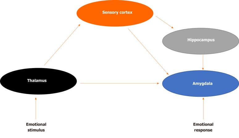
Figure 1. Fear brain circuitry.

COMORBIDITY OF PTSD AND SUBSTANCE USE DISORDERS

Clinical observations, case reports and epidemiological studies first suggested high rates of comorbidity between PTSD and substance use disorders (SUD), prompting translational research examining the possibility of overlapping neural mechanisms. Many studies have demonstrated a high comorbidity of PTSD with drug addiction in both clinical and community samples[38,39]. Approximately 36% to 50% of those that meet criteria for SUD also meet criteria for lifetime PTSD, and those with PTSD predictably have a history of drug abuse or dependence[40]. Comorbidity of these disorders is associated with negative treatment outcomes, increased risk for chronic diseases, and poorer functionality[41]. Co-twin studies have also demonstrated a link between childhood trauma and the later development of SUD[42].

Robinson and Berridge[43] proposed a model of addiction that demonstrates how repeat drug use disrupts normal reward processing. The Incentive-Sensitization Theory postulates that although increased pleasure is initially an important part of addiction behavior, regular substance use increases an individual’s sensitivity to drug cues, causing them to become hyper-responsive to drug cravings, even in the absence of pleasure[43]. This hyper-sensitization produces goal-directed behavior (“wanting”) not only in the absence of subjective pleasure, but also in the absence of consciously being aware of “wanting”. Recent research has supported this theory, demonstrated that substance use can alter brain reward circuitry[43].

BRAIN REWARD CIRCUITRY AND SUD

Data from several studies suggest that the reward circuit of the brain (the mesocortical dopamine pathway) provides a common molecular pathway with which to understand SUD. The mesolimbic pathway involves connections between the ventral tegmental area (VTA), the nucleus accumbens (NAc) in the ventral striatum, and the PFC. The mesolimbic dopamine reward circuit controls the reinforcing and rewarding effects related to food, sex, and social interaction[44]. Drug-induced adaptations in mesolimbic dopamine system (includes common adaptations to many different drugs) mediate changes in reward mechanisms that in part underlie addiction — including tolerance, dependence-withdrawal, sensitization, and relapse. Drug-induced adaptations include regulation of dopamine and opioid systems (mechanisms of tolerance and sensitization), regulation of glutamate systems (influences drug-related memories), upregulation of the cyclic adenosine monophosphate (cAMP) pathway, and transcription factor cAMP Response Element-Binding (CREB) protein, (mechanisms of drug tolerance, dependence, and withdrawal), structural changes in VTA neurons (influences drug tolerance), and structural changes in NAc neurons (influences drug sensitization)[45].

Many studies show that dopamine and accumbens neurons often become most active in anticipation of rewards, not during the reward phase, and also activated by the anticipation of aversive stimuli and events[46,47]. The role of the mesolimbic DA system is to increase the salience of stimuli and events associated with activation of the system. Stimuli are imbued with salience, making them “wanted” incentive stimuli. Alcaro et al[46] have theorized the role of the mesolimbic pathway as driving an organism toward “seeking” behaviors, searching to boost the salience of activities that are life-promoting while avoiding those that are harmful to survival. This proposed role is not only congruent with evidence of the importance of SUD, but also a mechanism that explains the connection of the VTA to the hippocampus, amygdala, and PFC, all of which have been implicated in the neurobiology of PTSD.

OVERLAPPING NEUROBIOLOGY OF PTSD AND SUD

Both animal models and clinical studies of PTSD have noted deficits in reward processing consistent with hypofunctionality of the mesolimbic pathway. Upon exposure to chronic stress, animal models demonstrate reduced striatal dopaminergic activity and decreased reward-seeking behavior that mimic symptoms of anhedonia experienced by PTSD patients[48]. Corral-Frias et al[49] utilized a novel animal model of PTSD to demonstrated that inactivation of the VTA can lead to long-term behavioral changes that mimic the clinical symptoms of PTSD. Inactivation of the VTA through either a dopamine antagonist or bilateral dissection can also lead to chronic changes in baseline VTA dopaminergic cell firing, demonstrating that trauma can lead to long-term alterations of the reward pathway. Evidence of deficits in the brain reward and reinforcement circuits in patients with PTSD also supports the involvement of the mesolimbic dopamine reward circuit[50,51]. In clinical studies, PTSD patients spend less time engaging in reward-seeking behavior compared to controls, report lower levels of reward expectation and are less satisfied with monetized rewards[52,53]. When compared to healthy controls, patients with PTSD demonstrate reduced bilateral striatal activation when responding to positive to reward gains and reported significantly higher levels of motivational and social deficits[50]. Collectively, these findings suggest a strong overlap in the brain regions involved in both fear processing and addiction. In particular, the VTA, through its connections to the amygdala, hippocampus and PFC, may serve as the common substrate of this overlapping circuitry, explaining the high co-morbidity in PTSD and SUD.

Several rodent models provide converging evidence for the overlapping circuitry of these two disorders, including the dorsal and ventral subdivisions of medial PFC and their respectively outputs to the amygdala and NAc (Figure 2). The prelimbic (PL) cortex projects to the basal (BA) nucleus of the amygdala, which excites the central (CE) nucleus of the amygdala, thereby promoting the expression of conditioned fear[54]. The BA also receives excitatory input from lateral amygdala, which also drives the expression of conditioned fear. The infralimbic (IL) cortex, in contrast, excites a class of GABAergic inhibitory neurons (the intercalated cell masses) which inhibit the CE, thereby promoting extinction of the conditioned fear[55]. PL and IL control drug seeking via their differential projections to the core and shell subdivisions of the NAc. The PL projects to the core, which promotes the expression of drug-seeking behavior. The IL projects to the shell, which also promotes the expression of extinction[56].

Functional magnetic resonance imaging studies provide consistent support for similar networks in human studies of fear and addiction (Figure 3). The dorsal portion of the anterior cingulate cortex is associated with fear expression during conditioning behavioral tasks, and overlaps with proximal regions that are activated when SUD patients respond regarding their craving levels after exposure to cocaine-related cues[57-59]. These findings are congruent with results from positron emission tomography mapping of cerebral blood flow using 15O-labeled water[60]. The ventral medial PFC (vmPFC) is activated during fear extinction recall and during recall of addiction cues in individuals with SUD disorders[57,61]. During states of cocaine craving, the vmPFC is deactivated, suggesting a failure to engage extinction[54]. Collectively, these studies suggest that the vmPFC is homologous to rodent IL, whereas the dorsal regions of anterior cingulate cortex are homologous to rodent PL.

Figure 2

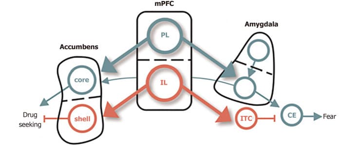
Figure 2. Conditioned fear in cocaine use. The dorsal and ventral subdivisions of medial prefrontal cortex are shown at the center, with their respective outputs to the amygdala controlling fear shown at right, and those to the nucleus accumbens, controlling cocaine, seeking shown at left. Green depicts pathways that activate fear and cocaine seeking. Red depicts pathways that inhibit fear and cocaine seeking[65]. Citation: Peters J, Kalivas PW, Quirk GJ. Extinction circuits for fear and addiction overlap in prefrontal cortex. Learn Mem 2009; 16(5): 279-288. Copyright ©Cold Spring Harbor Laboratory Press 2009. Published by Cold Spring Harbor Laboratory Press[65].

Fig 3

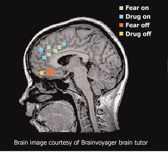
Figure 3. Functional magnetic resonance imaging and positron emission tomography studies of fear and addiction. Green dots represented human dorsal anterior cingulate cortex that correlated with fear expression functional magnetic resonance imaging[65]. Blue dots represent regions that correspond with drug cravings after exposure to cocaine-related cues. Red dots represent regions associated with fear extinction recall. Yellow dots represent regions activated during addiction-related cues. Citation: Peters J, Kalivas PW, Quirk GJ. Extinction circuits for fear and addiction overlap in prefrontal cortex. Learn Mem 2009; 16(5): 279-288. Copyright ©Brain Innovation 2009. Published by Cold Spring Harbor Laboratory Press[65].

CONCLUSION

This review examined evidence in support of a shared neurological origin between PTSD and SUD, in an effort to explain the high rates of comorbidity. It is clear that abnormalities in the PFC and VTA are central to the pathology of both disorders. The VTA is negatively affected during trauma and stress, and results in a decrease of dopaminergic activity and a subsequent alteration in the reward pathway. The PFC is involved in drug seeking behavior as well as the extinction of fear conditioning, playing a role in both addiction and PTSD. This review did not examine the genetic vulnerabilities nor neurodevelopmental pathways that may confer increased risk for either or both disorders, and it remains an important question whether the shared biology reviewed here is due to more distal risk factors, or are a result of one disorder conferring increased risk for the other.

It is important to gain a better understanding of the connection between PTSD and SUD in order to develop improved treatments that target both disorders. Despite shared neurobiology, there are few treatment options that target both, although notably some do exist (e.g., Seeking Safety). Yet many patients are not able to benefit from combined treatment interventions during the earlier stages of substance use recovery, and clinicians often struggle to determine the priority of treatment[62]. Many individuals diagnosed with comorbid PTSD-SUD believe that the outcomes of their disorders are interconnected, yet are not offered treatment for PTSD alongside SUD interventions[63].

Understanding comorbidity may also further prevention efforts, consistent with the “self-medication” hypothesis, as individuals with untreated trauma utilize substance as unhealthy coping mechanisms[64]. Earlier identification, access to care, and treatment of trauma across the lifespan is critical for intervening before the development of SUD or other maladaptive behaviours. Further research must leverage mechanisms between these two disorders, to ensure a more effective and efficient treatment option.

Open Article as PDF

Abstract

Mental health symptoms secondary to trauma exposure and substance use disorders (SUDs) co-occur frequently in both clinical and community samples. The possibility of a shared aetiology remains an important question in translational neuroscience. Advancements in genetics, basic science, and neuroimaging have led to an improved understanding of the neural basis of these disorders, their frequent comorbidity and high rates of relapse remain a clinical challenge. This project aimed to conduct a review of the field’s current understanding regarding the neural circuitry underlying posttraumatic stress disorder and SUD. A comprehensive review was conducted of available published literature regarding the shared neurobiology of these disorders, and is summarized in detail, including evidence from both animal and clinical studies. Upon summarizing the relevant literature, this review puts forth a hypothesis related to their shared neurobiology within the context of fear processing and reward cues. It provides an overview of brain reward circuitry and its relation to the neurobiology, symptomology, and phenomenology of trauma and substance use. This review provides clinical insights and implications of the proposed theory, including the potential development of novel pharmacological and therapeutic treatments to address this shared neurobiology. Limitations and extensions of this theory are discussed to provide future directions and insights for this shared phenomena.

INTRODUCTION

Research into trauma and posttraumatic stress disorder (PTSD) began as early as 1889 with Pierre Janet, a notable French psychiatrist. He described how the mind processes traumatic events. Janet proposed that individuals experiencing dissociation and hysteria had unresolved traumatic memories. He believed this subconscious experience was rooted in the physical effects of past negative events. When a person experiences trauma, they can be overwhelmed by strong negative emotions, making it difficult to accurately process and remember event details. The traumatic experience then separates from conscious awareness. Janet suggested that individuals would relive these memories in fragmented ways, such as emotional states, physical sensations, images, or behavioral repetitions. He was the first to identify dissociation as a key psychological process involved in various post-traumatic symptoms.

Decades of research aimed at understanding the psychological and biological effects of trauma have largely supported Janet's early observations. Psychological distress following a traumatic event can appear through various symptoms including anxiety, heightened fear, anhedonia (inability to feel pleasure), dysphoria (general unease), anger, aggression, or dissociation. The 5th edition of the Diagnostic and Statistical Manual of Mental Disorders (DSM-5) describes several diagnoses related to exposure to stressful events, with Acute Stress Disorder and PTSD being the most common. Criteria for PTSD include direct exposure to or indirect witnessing of a traumatic event, along with intrusive symptoms, negative mood, dissociation, avoidance, or heightened arousal. These symptoms must cause significant distress or impairment and not be due to substance use, a medical condition, or a brief psychotic disorder. Individuals with PTSD must also show at least one symptom across specific categories: intrusive symptoms, avoidance, negative changes in thinking and mood, and alterations in arousal and reactivity. Trauma experienced in interpersonal relationships is a frequent cause and often leads to poorer long-term outcomes.

Understanding the brain's complex responses to trauma has been challenging due to the wide range of individual reactions. Most basic neuroscience and brain imaging research on PTSD has focused primarily on two areas: how stress affects the sympathetic nervous system and how trauma impacts the brain's frontal striatal circuits. While this research has begun to clarify the negative brain effects of repeated stress, many individuals continue to suffer from trauma's impact and do not respond to established PTSD treatments. A new area of research that may help further explain the brain mechanisms of trauma involves the important role of the brain's reward circuits in models of PTSD. Following a review of animal and human research on the biological effects of PTSD, this discussion will explore recent findings that suggest the brain's reward pathway plays a significant role in understanding the neurobiological effects of complex trauma.

THE PHYSIOLOGY AND NEUROBIOLOGY OF PTSD

The human stress response and PTSD

During stress, the sympathetic nervous system prepares the body for action, while the pituitary-adrenocortical system helps to reduce the initial physical signs of arousal. The hypothalamic-pituitary-adrenal (HPA) axis releases corticotropin-releasing factor (CRF), which then stimulates the release of cortisol from the adrenal cortex. This also increases the release of catecholamine neurotransmitters in several brain regions. Catecholamines are vital for the body's adaptive response to stress by breaking down glycogen, suppressing insulin release, and increasing cardiovascular system activity. Higher levels of norepinephrine (NE) and epinephrine (EPI) during stress lead to increased brain cell activity in limbic areas like the amygdala and hypothalamus, while decreasing activity in cortical areas involved in higher-level thinking.

For individuals at risk of developing PTSD, traumatic experiences can alter the normal function of the sympathetic nervous system. Many core symptoms of PTSD, such as an exaggerated startle response, difficulty falling or staying asleep, and poor concentration, reflect a state of hyperarousal. Individuals with PTSD show exaggerated sympathetic nervous system responses, including rapid heart rate and changes in skin conductance during acute stress, and increased sensitivity of the HPA axis. Compared to healthy individuals, those with PTSD have lower baseline cortisol levels and increased levels of CRF. Several studies have shown that individuals with PTSD have higher urinary levels of NE and EPI compared to controls, and that these neurotransmitter levels relate to the severity of self-reported PTSD symptoms. Previous PTSD studies have also reported related abnormalities in sensory processing.

Abnormalities in HPA axis and neurotransmitter function can lead to both structural and functional changes in neural circuits, particularly in brain circuits essential for emotional and cognitive processing. While high cortisol levels enhance the formation of emotional memories (through increased amygdala function) and facilitate fear conditioning, high cortisol levels immediately after trauma can decrease hippocampus function, leading to memory and learning difficulties. Structural changes also occur; high levels of stress can cause an increase in dendrites in the prefrontal cortex (PFC) and changes in dendrites of the amygdala.

Between stimulus and response

The brain processes sensory information that might be perceived as a threat. LeDoux was among the first to illustrate the underlying brain circuits involved in fear. For instance, if a door slams shut in the middle of the night, startling an individual from sleep, the initial sensory information about the perceived threat is quickly sent to the thalamus and then to the amygdala, activating the stress response and causing an immediate reaction. The body may begin to sweat, a physical response that precedes a rapid reaction. Meanwhile, the hippocampus and prefrontal cortex (PFC) process contextual information about the stimulus, such as considering if it was a windy day or recalling that a slamming door has never caused harm before. This contextual processing helps to reduce the stress response, allowing the individual to relax and return to a normal state. The signal from the thalamus to the amygdala is very fast, whereas signals from the hippocampus and PFC are transmitted more slowly. Recent research has shown how structural, chemical, and functional differences in these brain areas contribute to the neurobiology of PTSD.

Given the role of intrusive memories in PTSD, many have suggested that problems with the hippocampus are a crucial part of the underlying neurobiology. The hippocampus has a high concentration of corticosteroid receptors, which are involved in ending the stress response through negative feedback to the HPA axis. Both animal and human studies have shown that high levels of stress can damage the hippocampus, leading to memory problems. During an acute stress response, high levels of cortisol can reduce dendritic branching in the hippocampus while increasing the growth of new brain cells in the amygdala. This enhances the emotional significance of the event but impairs memory function. Compared with healthy individuals, those with PTSD show impairments in short-term memory, and some studies (though not all) have found reduced hippocampal volumes in PTSD patients. Individuals with PTSD often have difficulty describing details related to traumatic events, and some studies have linked hippocampal regional blood flow to the severity of PTSD symptoms.

The amygdala receives input from the thalamus and sensory processing regions of the neocortex, and it sends signals to brain structures that control automatic bodily functions. This makes it critical for both sympathetic and parasympathetic stress responses. In human studies, the amygdala has been shown to play a vital role in processing emotional stimuli and forming emotionally significant memories. It also contributes to fear conditioning, where a neutral stimulus becomes associated with a stressful one. Exposure to the now-conditioned stimulus initiates the stress response, activating the amygdala and engaging the autonomic nervous system. Damage to the amygdala in animals interrupts fear learning and the conditioned response. Human brain imaging studies have confirmed the involvement of this region in fear learning, conditioning, and extinction. Compared to healthy individuals, those with PTSD show increased amygdala activation when presented with trauma-related cues, and also with unrelated emotional stimuli. Amygdala response in PTSD patients has been found to correlate with their self-reported symptom severity.

Changes in prefrontal cortical (PFC) activity may help explain both memory impairments in PTSD and increased amygdala activation during the stress response. In individuals with PTSD, repeated exposure to trauma can damage these neural structures. The ability to extinguish emotional memories involves the ventromedial PFC and the amygdala, while extinguishing conditioned fear involves the anterior cingulate cortex and the amygdala. Activation of the medial PFC also occurs when an individual inhibits fearful responses or changes their perception of a negative emotional event. Therefore, decreased PFC function may explain why individuals with PTSD struggle to extinguish fearful memories.

COMORBIDITY OF PTSD AND SUBSTANCE USE DISORDERS

Clinical observations, case reports, and epidemiological studies first indicated high rates of co-occurrence between PTSD and substance use disorders (SUD), leading to research exploring possible overlapping brain mechanisms. Many studies have shown a high rate of co-occurrence of PTSD with drug addiction in both clinical and community settings. Approximately 36% to 50% of individuals who meet criteria for SUD also meet criteria for lifetime PTSD, and those with PTSD commonly have a history of drug abuse or dependence. The co-occurrence of these disorders is linked to negative treatment outcomes, an increased risk for chronic diseases, and poorer overall functioning. Studies involving twins have also shown a connection between childhood trauma and the later development of SUD.

Robinson and Berridge proposed a model of addiction showing how repeated drug use disrupts normal reward processing. The Incentive-Sensitization Theory suggests that while increased pleasure is initially an important part of addictive behavior, regular substance use increases an individual's sensitivity to drug cues. This causes them to become overly responsive to drug cravings, even when there is no pleasure. This heightened sensitivity creates goal-directed behavior, or "wanting," not only without subjective pleasure but also without consciously realizing the "wanting." Recent research supports this theory, demonstrating that substance use can alter the brain's reward circuitry.

BRAIN REWARD CIRCUITRY AND SUD

Data from several studies suggest that the brain's reward circuit, specifically the mesocortical dopamine pathway, provides a common biological explanation for substance use disorders (SUD). The mesolimbic pathway involves connections between the ventral tegmental area (VTA), the nucleus accumbens (NAc) in the ventral striatum, and the prefrontal cortex (PFC). This mesolimbic dopamine reward circuit controls the reinforcing and rewarding effects related to natural pleasures like food, sex, and social interaction. Drug-induced changes in the mesolimbic dopamine system, common across many different drugs, mediate alterations in reward mechanisms that underlie addiction. These include tolerance, dependence-withdrawal, sensitization, and relapse. Drug-induced changes involve the regulation of dopamine and opioid systems (mechanisms of tolerance and sensitization), glutamate systems (influencing drug-related memories), an increase in the cyclic adenosine monophosphate (cAMP) pathway and the transcription factor cAMP Response Element-Binding (CREB) protein (mechanisms of drug tolerance, dependence, and withdrawal), and structural changes in VTA and NAc neurons (influencing drug tolerance and sensitization, respectively).

Many studies show that dopamine and accumbens neurons often become most active in anticipation of rewards, rather than during the reward itself. They are also activated by the anticipation of unpleasant stimuli and events. Researchers have theorized that the mesolimbic dopamine system's role is to increase the prominence or "salience" of stimuli and events associated with its activation. Stimuli become "imbued with salience," making them "wanted" incentive stimuli. This proposed role is consistent with evidence of the importance of SUD and also explains the VTA's connections to the hippocampus, amygdala, and PFC, all of which have been implicated in the neurobiology of PTSD.

OVERLAPPING NEUROBIOLOGY OF PTSD AND SUD

Both animal models and clinical studies of PTSD have identified deficits in reward processing, which are consistent with reduced function in the mesolimbic pathway. When exposed to chronic stress, animal models show reduced dopamine activity in the striatum and decreased reward-seeking behavior, mimicking symptoms of anhedonia experienced by individuals with PTSD. One animal model of PTSD demonstrated that inactivating the VTA can lead to long-term behavioral changes similar to clinical PTSD symptoms. Inactivating the VTA, either through a dopamine blocker or surgical disruption, can also cause chronic changes in baseline VTA dopamine cell firing, indicating that trauma can lead to long-term alterations in the reward pathway. Evidence of deficits in the brain's reward and reinforcement circuits in individuals with PTSD also supports the involvement of the mesolimbic dopamine reward circuit. In clinical studies, individuals with PTSD spend less time engaging in reward-seeking behavior compared to controls, report lower levels of reward expectation, and are less satisfied with monetary rewards. Compared to healthy individuals, those with PTSD show reduced activation in both sides of the striatum when responding to positive reward gains and report significantly higher levels of motivational and social deficits. Collectively, these findings suggest a strong overlap in the brain regions involved in both fear processing and addiction. The VTA, through its connections to the amygdala, hippocampus, and PFC, may serve as the common underlying structure explaining the high rate of co-occurrence between PTSD and SUD.

Several rodent models provide strong evidence for the overlapping circuits of these two disorders, including the dorsal and ventral parts of the medial prefrontal cortex (PFC) and their respective connections to the amygdala and nucleus accumbens (NAc). The prelimbic (PL) cortex connects to the basal (BA) nucleus of the amygdala, which stimulates the central (CE) nucleus of the amygdala, thus promoting the expression of conditioned fear. The BA also receives excitatory input from the lateral amygdala, which also drives the expression of conditioned fear. In contrast, the infralimbic (IL) cortex stimulates a type of inhibitory neurons, which then inhibit the CE, thereby promoting the extinction of conditioned fear. The PL and IL control drug seeking through their different connections to the core and shell subdivisions of the NAc. The PL projects to the core, which promotes drug-seeking behavior. The IL projects to the shell, which also promotes the expression of extinction.

Functional magnetic resonance imaging (fMRI) studies consistently support similar brain networks in human studies of fear and addiction. The dorsal part of the anterior cingulate cortex is associated with the expression of fear during conditioning tasks, and these regions overlap with nearby areas that are activated when individuals with substance use disorders report their craving levels after exposure to drug-related cues. These findings are consistent with results from positron emission tomography (PET) mapping of cerebral blood flow. The ventral medial PFC (vmPFC) is activated during the recall of fear extinction and during the recall of addiction cues in individuals with substance use disorders. During states of drug craving, the vmPFC is deactivated, suggesting a failure to engage in extinction. Collectively, these studies suggest that the vmPFC is similar in function to the rodent IL, while the dorsal regions of the anterior cingulate cortex are similar to the rodent PL.

CONCLUSION

This review examined evidence supporting a shared neurological basis between PTSD and substance use disorders (SUD), aiming to explain their high rates of co-occurrence. It is clear that abnormalities in the prefrontal cortex (PFC) and ventral tegmental area (VTA) are central to the underlying problems in both disorders. The VTA is negatively affected during trauma and stress, leading to decreased dopamine activity and a subsequent alteration in the reward pathway. The PFC is involved in both drug-seeking behavior and the extinction of fear conditioning, playing a role in both addiction and PTSD. This review did not explore genetic vulnerabilities or neurodevelopmental pathways that might increase the risk for either or both disorders. It remains an important question whether the shared biology discussed here is due to more distant risk factors or if one disorder increases the risk for the other.

A better understanding of the connection between PTSD and SUD is crucial for developing improved treatments that address both disorders. Despite shared neurobiology, there are few treatment options that specifically target both, although some notable examples do exist, such as Seeking Safety. Yet, many individuals cannot benefit from combined treatment interventions during the earlier stages of substance use recovery, and clinicians often struggle to determine the priority of treatment. Many individuals diagnosed with co-occurring PTSD and SUD believe that the outcomes of their disorders are interconnected, yet they are not always offered PTSD treatment alongside SUD interventions.

Understanding co-occurrence may also enhance prevention efforts, consistent with the "self-medication" hypothesis, which suggests that individuals with untreated trauma use substances as unhealthy coping mechanisms. Early identification, access to care, and treatment of trauma across the lifespan are critical for intervening before the development of SUD or other unhelpful behaviors. Further research must leverage the mechanisms shared between these two disorders to ensure more effective and efficient treatment options.

Open Article as PDF

Abstract

Mental health symptoms secondary to trauma exposure and substance use disorders (SUDs) co-occur frequently in both clinical and community samples. The possibility of a shared aetiology remains an important question in translational neuroscience. Advancements in genetics, basic science, and neuroimaging have led to an improved understanding of the neural basis of these disorders, their frequent comorbidity and high rates of relapse remain a clinical challenge. This project aimed to conduct a review of the field’s current understanding regarding the neural circuitry underlying posttraumatic stress disorder and SUD. A comprehensive review was conducted of available published literature regarding the shared neurobiology of these disorders, and is summarized in detail, including evidence from both animal and clinical studies. Upon summarizing the relevant literature, this review puts forth a hypothesis related to their shared neurobiology within the context of fear processing and reward cues. It provides an overview of brain reward circuitry and its relation to the neurobiology, symptomology, and phenomenology of trauma and substance use. This review provides clinical insights and implications of the proposed theory, including the potential development of novel pharmacological and therapeutic treatments to address this shared neurobiology. Limitations and extensions of this theory are discussed to provide future directions and insights for this shared phenomena.

INTRODUCTION

Research into trauma and posttraumatic stress disorder (PTSD) began as early as 1889 with Pierre Janet, a notable French psychiatrist. His work, L’Automatisme Psychologique, offered an early framework for understanding how the mind processes difficult experiences. Janet proposed that individuals experiencing dissociation—a disconnection from thoughts, memories, feelings, or identity—and hysteria often had unresolved traumatic memories. He believed these subconscious experiences were rooted in the physical effects of past negative events. When a person endures a traumatic event, intense negative emotions can overwhelm them, making it difficult to accurately process and recall details of the event. This traumatic experience may then become separated from conscious awareness. Janet suggested that individuals would later re-experience fragments of the trauma, such as emotional states, physical sensations, visual images, or repetitive behaviors. He was the first to identify dissociation as a key psychological process involved in various post-traumatic symptoms.

Decades of subsequent research into the psychological and biological impacts of trauma have largely supported Janet's initial observations. Psychological distress following a traumatic event can manifest through a range of symptoms, including anxiety, exaggerated fear, anhedonia (inability to experience pleasure), dysphoria (a state of unease), anger, aggression, or dissociation. The 5th edition of the Diagnostic and Statistical Manual of Mental Disorders (DSM-5) describes several diagnoses related to exposure to stressful events, with Acute Stress Disorder and PTSD being the most common. PTSD diagnostic criteria include direct exposure to, or indirect witnessing of, a traumatic event, along with intrusive symptoms, negative mood, dissociation, avoidance, and/or heightened arousal. These symptoms must cause significant distress or functional impairment and not be due to substance use, a medical condition, or a brief psychotic disorder. Individuals with PTSD must also exhibit at least one symptom from each category: intrusive symptoms, avoidance, negative alterations in thinking and mood, and changes in arousal and reactivity. Interpersonal trauma is frequently a causal event for PTSD and often leads to worse long-term outcomes.

Understanding the neurobiological basis of PTSD has been challenging due to the diverse and complex ways individuals respond to trauma. Most basic neuroscience and neuroimaging research on PTSD has primarily focused on two areas: the effects of stress on the sympathetic nervous system and the impact of trauma on frontal striatal brain circuits. While such research has provided insights into the negative neurobiological effects of repeated stress, many patients continue to suffer from trauma's effects and do not respond to existing evidence-based treatments for PTSD. A developing area of research that may further clarify trauma's neurobiological mechanisms involves the crucial role of the brain’s reward circuitry in translational models of PTSD. Following an overview of animal and clinical research on the biological effects of PTSD, this discussion will explore recent findings suggesting a significant role for the brain’s reward pathway in understanding the neurobiological effects of complex trauma.

THE PHYSIOLOGY AND NEUROBIOLOGY OF PTSD

During stressful situations, the sympathetic nervous system prepares the body for immediate action, while the pituitary-adrenocortical system helps to moderate the initial physiological arousal. The hypothalamic-pituitary-adrenal (HPA) axis releases corticotropin-releasing factor (CRF), which in turn stimulates the adrenal cortex to release cortisol. This process also increases the release of catecholamine neurotransmitters, such as norepinephrine (NE) and epinephrine (EPI), in several brain regions. Catecholamines are essential for the body's adaptive response to stress, facilitating energy release (through glycogen breakdown), suppressing insulin, and increasing cardiovascular system activity. Elevated levels of NE and EPI during stress enhance neuronal activity in limbic areas like the amygdala and hypothalamus, while decreasing activity in cortical areas responsible for higher-order cognitive functions. For individuals at risk of developing PTSD, traumatic experiences can disrupt the normal functioning of the sympathetic nervous system. Many core PTSD symptoms, such as an exaggerated startle response, difficulty sleeping, and poor concentration, reflect a state of hyperarousal. Patients with PTSD show heightened sympathetic nervous system responses, including a rapid heart rate and increased skin conductance during acute stress, along with increased sensitivity of the HPA axis. Compared to healthy individuals, those with PTSD often have reduced baseline cortisol levels but increased levels of CRF. Studies have also shown that PTSD patients have higher urinary levels of NE and EPI, and these neurotransmitter levels correlate with the severity of reported PTSD symptoms.

Abnormalities in HPA axis and neurotransmitter function can lead to both structural and functional changes in neural circuits, especially those vital for emotional and cognitive processing. While high cortisol levels typically enhance the formation of emotional memories (mediated by increased amygdala function) and facilitate fear conditioning, high cortisol levels during trauma exposure can impair hippocampus function, leading to memory and learning deficits. Structural changes also occur; high levels of stress can cause the prefrontal cortex (PFC) to experience dendritic hypertrophy (increased branching of nerve cell extensions) and the amygdala to undergo dendritic remodeling.

The brain processes sensory information that may be perceived as a threat through a specific circuitry. Pioneering work by LeDoux illustrated the neural pathways involved in fear. For instance, if a loud noise is heard, initial sensory information is rapidly transmitted to the thalamus and then quickly to the amygdala, triggering an immediate stress response and physical reactions like sweating. Simultaneously, the hippocampus and PFC process contextual details about the stimulus, providing reasoning (e.g., the sound was a door slamming due to wind) and episodic memory (e.g., a similar sound has not caused harm before). This contextual information helps to dampen the stress response, allowing the individual to relax and return to a baseline state. The signal from the thalamus to the amygdala is very fast, whereas signals from the hippocampus and PFC are transmitted more slowly.

Given the presence of intrusive memories in PTSD, many researchers hypothesize that hippocampal dysfunction is a crucial part of the underlying neurobiology. The hippocampus contains a high concentration of corticosteroid receptors that help terminate the stress response through negative feedback to the HPA axis. Both animal and human studies indicate that high levels of stress can damage the hippocampus, leading to memory impairments. During an acute stress response, elevated cortisol levels can reduce dendritic branching in the hippocampus while increasing neurogenesis (the formation of new neurons) in the amygdala. This enhances the emotional salience (importance) of the event but can impair memory functioning. Compared to healthy individuals, patients with PTSD often show impairments in short-term memory, and some studies have observed reduced hippocampal volumes in this population. Individuals with PTSD frequently struggle to describe details related to traumatic events, and some research has correlated hippocampal regional cerebral blood flow with the severity of PTSD symptoms.

The amygdala receives input from the thalamus and sensory processing regions of the neocortex, then sends signals to autonomic brain structures. This makes it critical for both the sympathetic and parasympathetic stress responses. In humans, the amygdala plays a vital role in processing emotional stimuli and forming emotionally significant memories. It is also involved in fear conditioning, where a neutral stimulus becomes associated with a stressful one. Exposure to the now-conditioned stimulus then triggers the stress response, activating the amygdala and engaging the autonomic nervous system. Damage to the amygdala in animals disrupts fear learning and the conditioned response. Human neuroimaging studies confirm the amygdala's involvement in fear learning, conditioning, and extinction (the reduction of a conditioned response). Compared to healthy individuals, patients with PTSD show increased amygdala activation when presented with trauma-related cues and even with unrelated emotional stimuli. Amygdala response in PTSD patients has been found to correlate with their self-reported symptom severity.

Changes in prefrontal cortical (PFC) activity may link memory impairment in PTSD with increased amygdala activation during stress. In patients with PTSD, repeated exposure to trauma can damage these neural structures. The ability to lessen or remove emotional memories involves the ventromedial PFC and the amygdala, while reducing conditioned fear involves the anterior cingulate cortex and the amygdala. Activation of the medial PFC also occurs when inhibiting fearful responses or altering one's perception of a negative emotional event. Therefore, decreased PFC functioning may explain why patients with PTSD have difficulty reducing fearful memories.

COMORBIDITY OF PTSD AND SUBSTANCE USE DISORDERS

Clinical observations, case reports, and widespread studies first indicated high rates of co-occurrence between PTSD and substance use disorders (SUD), leading to research exploring possible overlapping neural mechanisms. Numerous studies have demonstrated a significant co-occurrence of PTSD and drug addiction in both clinical settings and general populations. Approximately 36% to 50% of individuals who meet criteria for SUD also meet criteria for lifetime PTSD, and those with PTSD commonly have a history of drug abuse or dependence. The co-occurrence of these disorders is linked to less favorable treatment outcomes, an increased risk for chronic illnesses, and poorer overall functioning. Studies involving twins have also shown a connection between childhood trauma and the later development of SUD.

Robinson and Berridge proposed a model of addiction illustrating how repeated drug use disrupts normal reward processing. Their Incentive-Sensitization Theory suggests that while initial pleasure is important in addiction behavior, regular substance use increases an individual's sensitivity to drug cues. This heightened sensitivity causes a hyper-responsiveness to drug cravings, even in the absence of pleasure. This hyper-sensitization drives goal-directed behavior—a "wanting"—even without subjective pleasure or conscious awareness of that "wanting." Recent research supports this theory, showing that substance use can alter the brain's reward circuitry.

BRAIN REWARD CIRCUITRY AND SUD

Data from several studies indicate that the brain's reward circuit, specifically the mesocortical dopamine pathway, offers a common molecular framework for understanding SUD. The mesolimbic pathway involves connections between the ventral tegmental area (VTA), the nucleus accumbens (NAc) in the ventral striatum, and the prefrontal cortex (PFC). This mesolimbic dopamine reward circuit controls the reinforcing and pleasurable effects associated with natural rewards like food, sex, and social interaction. Drug-induced changes in the mesolimbic dopamine system, which are common across many different drugs, mediate alterations in reward mechanisms that contribute to addiction. These changes include the regulation of dopamine and opioid systems (related to tolerance and sensitization), glutamate systems (influencing drug-related memories), and the cyclic adenosine monophosphate (cAMP) pathway and its associated transcription factor CREB protein (involved in drug tolerance, dependence, and withdrawal). Structural changes in VTA and NAc neurons also occur, influencing drug tolerance and sensitization, respectively.

Many studies reveal that dopamine and nucleus accumbens neurons are often most active in anticipation of rewards, rather than during the reward experience itself. They are also activated by the anticipation of unpleasant stimuli. The mesolimbic dopamine system's role is to increase the salience—or importance—of stimuli and events associated with its activation, making them desired or "wanted" incentive stimuli. Researchers like Alcaro and colleagues have theorized that the mesolimbic pathway drives an organism toward "seeking" behaviors, motivating the pursuit of life-sustaining activities and the avoidance of harmful ones. This proposed role not only aligns with evidence of its importance in SUD but also offers a mechanism explaining the VTA's connections to the hippocampus, amygdala, and PFC—all implicated in the neurobiology of PTSD.

OVERLAPPING NEUROBIOLOGY OF PTSD AND SUD

Both animal models and clinical studies of PTSD have identified deficits in reward processing, consistent with reduced function in the mesolimbic pathway. When exposed to chronic stress, animal models show reduced dopaminergic activity in the striatum and decreased reward-seeking behavior, mirroring the anhedonia symptoms experienced by PTSD patients. Corral-Frias and colleagues used an animal model of PTSD to show that inactivating the VTA can lead to long-term behavioral changes resembling clinical PTSD symptoms. Inactivating the VTA, either through a dopamine blocker or surgical dissection, can also cause chronic changes in baseline VTA dopaminergic cell firing, indicating that trauma can result in lasting alterations to the reward pathway. Evidence of deficits in the brain's reward and reinforcement circuits in PTSD patients also supports the involvement of the mesolimbic dopamine reward circuit. In clinical studies, PTSD patients spend less time pursuing rewards compared to controls, report lower expectations of reward, and show less satisfaction with monetary rewards. When compared to healthy individuals, patients with PTSD demonstrate reduced activation in both sides of the striatum when responding to positive reward gains and report significantly higher levels of motivational and social deficits. Collectively, these findings suggest a significant overlap in the brain regions involved in both fear processing and addiction. Specifically, the VTA, through its connections to the amygdala, hippocampus, and PFC, may serve as the common neurological basis for this overlap, explaining the high co-occurrence of PTSD and SUD.

Several rodent models provide strong evidence for the overlapping circuitry of these two disorders, including the dorsal and ventral subdivisions of the medial PFC and their respective outputs to the amygdala and nucleus accumbens (NAc). The prelimbic (PL) cortex projects to the basal (BA) nucleus of the amygdala, which excites the central (CE) nucleus of the amygdala, thereby promoting the expression of conditioned fear. The BA also receives excitatory input from the lateral amygdala, which further drives the expression of conditioned fear. In contrast, the infralimbic (IL) cortex activates inhibitory neurons (intercalated cell masses) that suppress the CE, thus promoting the extinction of conditioned fear. Both PL and IL cortices regulate drug seeking through their distinct projections to the core and shell subdivisions of the NAc. The PL projects to the NAc core, which enhances drug-seeking behavior. The IL, however, projects to the NAc shell, which promotes extinction of this behavior.

Functional magnetic resonance imaging studies in humans consistently support the existence of similar neural networks in fear and addiction. The dorsal part of the anterior cingulate cortex is linked to fear expression during conditioning tasks and overlaps with nearby regions activated when SUD patients report craving levels after exposure to cocaine-related cues. These findings align with results from positron emission tomography mapping of cerebral blood flow. The ventral medial PFC (vmPFC) is activated during the recall of fear extinction and during the recall of addiction cues in individuals with SUDs. During states of cocaine craving, the vmPFC shows reduced activity, suggesting a failure to engage extinction. Collectively, these studies indicate that the human vmPFC is similar to the rodent IL, while the dorsal regions of the anterior cingulate cortex are similar to the rodent PL.

CONCLUSION

This discussion examined evidence supporting a shared neurological origin between PTSD and SUD, aiming to explain their high rates of co-occurrence. It is clear that abnormalities in the prefrontal cortex (PFC) and ventral tegmental area (VTA) are central to the pathology of both disorders. The VTA is negatively affected during trauma and stress, leading to decreased dopaminergic activity and subsequent alterations in the reward pathway. The PFC is involved in both drug-seeking behavior and the extinction of fear conditioning, playing a role in both addiction and PTSD. This review did not explore genetic vulnerabilities or neurodevelopmental pathways that might increase the risk for one or both disorders. It remains an important question whether the shared biology discussed here results from broader risk factors or if one disorder directly increases the risk for the other.

A better understanding of the connection between PTSD and SUD is crucial for developing improved treatments that address both conditions. Despite their shared neurobiology, there are few integrated treatment options, though some do exist (e.g., Seeking Safety). However, many patients cannot benefit from combined interventions during the early stages of substance use recovery, and clinicians often find it challenging to prioritize treatment. Many individuals diagnosed with co-occurring PTSD and SUD believe their disorders are interconnected, yet they are often not offered PTSD treatment alongside SUD interventions.

Understanding this co-occurrence can also enhance prevention efforts, aligning with the "self-medication" hypothesis, which suggests that individuals with untreated trauma use substances as unhealthy coping mechanisms. Early identification, access to care, and treatment of trauma across the lifespan are critical for intervening before the development of SUD or other maladaptive behaviors. Further research must explore the mechanisms linking these two disorders to ensure more effective and efficient treatment options.

Open Article as PDF

Abstract

Mental health symptoms secondary to trauma exposure and substance use disorders (SUDs) co-occur frequently in both clinical and community samples. The possibility of a shared aetiology remains an important question in translational neuroscience. Advancements in genetics, basic science, and neuroimaging have led to an improved understanding of the neural basis of these disorders, their frequent comorbidity and high rates of relapse remain a clinical challenge. This project aimed to conduct a review of the field’s current understanding regarding the neural circuitry underlying posttraumatic stress disorder and SUD. A comprehensive review was conducted of available published literature regarding the shared neurobiology of these disorders, and is summarized in detail, including evidence from both animal and clinical studies. Upon summarizing the relevant literature, this review puts forth a hypothesis related to their shared neurobiology within the context of fear processing and reward cues. It provides an overview of brain reward circuitry and its relation to the neurobiology, symptomology, and phenomenology of trauma and substance use. This review provides clinical insights and implications of the proposed theory, including the potential development of novel pharmacological and therapeutic treatments to address this shared neurobiology. Limitations and extensions of this theory are discussed to provide future directions and insights for this shared phenomena.

INTRODUCTION

Research into trauma and post-traumatic stress disorder (PTSD) began in 1889. At that time, Pierre Janet, a well-known French psychiatrist, wrote a book called L’Automatisme Psychologique. This book was one of the first attempts to explain how the mind deals with upsetting events. Janet believed that patients experiencing certain mental states, like dissociation (feeling disconnected from reality) and hysteria, had unresolved traumatic memories. He thought these hidden experiences were rooted in the physical effects of past negative events. When a person goes through a traumatic event, they can feel overwhelmed by strong negative emotions. This makes it hard for them to clearly process and remember the details of what happened. The traumatic experience then becomes disconnected from their conscious thoughts. Janet proposed that individuals would later relive parts of the trauma, such as strong emotions, physical feelings, mental images, or repeated behaviors. He was the first to recognize that dissociation was a key psychological process in many symptoms following trauma.

Decades of studies on the psychological and biological impacts of trauma have since supported many of Janet's early ideas. After a traumatic event, psychological distress can appear in various ways. These symptoms include anxiety, exaggerated fear, a loss of pleasure in activities (anhedonia), a general feeling of unhappiness (dysphoria), anger, aggression, or dissociation. The Diagnostic and Statistical Manual of Mental Disorders (DSM-5), which doctors use to diagnose mental health conditions, describes seven different diagnoses linked to experiencing a very stressful event. The most common of these are Acute Stress Disorder and PTSD. To be diagnosed with PTSD, a person must have directly experienced or indirectly witnessed a traumatic or stressful event. They must also show symptoms like intrusive thoughts, a negative mood, dissociation, avoidance behaviors, or heightened arousal. These symptoms must cause significant distress or interfere with daily life and cannot be due to substance use, a medical condition, or a short psychotic episode. People with PTSD also need to show at least one symptom from each of the main categories: intrusive, avoidance, negative changes in thinking and mood, and changes in arousal and reactivity. Trauma involving other people is a common type of traumatic event that often leads to worse long-term outcomes.

Because people react so differently and complexly to trauma, it has been challenging to fully understand the brain changes involved in PTSD. Most basic brain and brain imaging research on PTSD has mainly focused on two areas: how stress affects the sympathetic nervous system (which controls the "fight or flight" response) and how trauma impacts brain circuits involving the front part of the brain. While this research has started to explain the harmful brain effects of ongoing stress, many people still suffer from trauma's effects and do not respond to common, proven treatments for PTSD. A new area of research is exploring how the brain's reward system might help explain the brain mechanisms of complex trauma. This article will first provide a general overview of animal and human research on the biological effects of PTSD. It will then discuss newer research suggesting that the brain's reward pathway plays an important role in understanding the brain changes caused by complex trauma.

The Human Stress Response and PTSD

When under stress, the sympathetic nervous system prepares the body for action, while another system, the pituitary-adrenocortical system, helps calm down the body's initial physical reactions. The hypothalamic-pituitary-adrenal (HPA) axis releases a substance called corticotrophin-releasing factor (CRF). This stimulates the release of cortisol from the adrenal glands and increases levels of chemical messengers called catecholamines in several brain areas. Catecholamines are vital for the body's ability to adapt to stress. They do this by breaking down stored energy, reducing insulin release, and increasing how well the heart and blood vessels work. Higher levels of norepinephrine (NE) and epinephrine (EPI) during stress lead to more brain cell activity in emotional areas like the amygdala and hypothalamus. At the same time, activity decreases in parts of the brain involved in higher-level thinking.

For individuals who are at risk of developing PTSD, traumatic experiences can change how the sympathetic nervous system normally works. Many key symptoms of PTSD show a state of being overly alert, or "hyperarousal." These include an exaggerated startle response, difficulty falling or staying asleep, and poor concentration. People with PTSD show stronger sympathetic nervous system responses, such as a faster heart rate (tachycardia) and changes in skin sweat during acute stress. They also have an increased sensitivity in their HPA axis. Compared to healthy individuals, patients with PTSD often have lower baseline cortisol levels but higher levels of CRF. Several studies have also shown that people with PTSD have higher levels of NE and EPI in their urine compared to others, and that the levels of these chemical messengers are related to how severe their reported PTSD symptoms are. Past PTSD studies have also found related problems in how the senses process information.

Problems with the HPA axis and the function of chemical messengers can change brain circuits, both in their structure and how they work. This is especially true in brain circuits important for emotions and thinking. While high levels of cortisol can improve the formation of emotional memories (through increased amygdala function) and make fear conditioning easier, high levels of cortisol during trauma exposure reduce hippocampus function. This leads to problems with memory and learning. Physical changes also happen in the brain; high levels of stress can cause certain brain cells in the prefrontal cortex (PFC) and amygdala to change their shape and connections.

Between Stimulus and Response

How does the brain process sensory information that might be seen as a threat? Scientists like LeDoux were among the first to show the brain circuits involved in fear. For example, consider a scenario where a door slams shut in the middle of the night, waking a person from sleep. The initial sensory information about the potential threat is sent to the thalamus. From there, it quickly travels to the amygdala, which then activates the stress response and creates an immediate reaction. The body might start to sweat, a physical reaction that leads to a quick response. Meanwhile, the hippocampus and PFC process other information about the sound. They provide reasoning (maybe it was a windy day) and past memories (a slamming door has never caused a person any harm before). This information helps to calm the stress response, allowing the individual to relax and return to normal. The signal from the thalamus to the amygdala is very fast, while signals from the hippocampus and PFC travel more slowly. Recent research has shown how structural, chemical, and functional differences in these brain areas develop in the neurobiology of PTSD. (Note: Figures 1 and 2 from the original text showing fear brain circuitry are not included in this summary.)

Given that intrusive memories are a key part of PTSD, many experts believe that problems with the hippocampus are a crucial part of the disorder's underlying brain mechanisms. The hippocampus has many corticosteroid receptors, which are involved in stopping the stress response through a process that gives negative feedback to the HPA axis. Both animal and human studies have shown that high levels of stress can damage the hippocampus, leading to memory problems. During a strong stress response, high levels of cortisol can reduce the branching of nerve cells in the hippocampus while increasing the growth of new cells in the amygdala. This makes the emotional importance of the event stronger but harms memory function. Compared to healthy people, patients with PTSD show problems with short-term memory, and some studies (though not all) have found that people with PTSD have smaller hippocampal volumes. Individuals with PTSD often find it hard to describe details related to traumatic events, and some studies have linked the amount of blood flow to certain parts of the hippocampus to the severity of PTSD symptoms.

The amygdala receives information from the thalamus and sensory processing areas of the outer brain layer (neocortex). It then sends signals to brain structures that control automatic body functions, playing a critical role in both the "fight or flight" and "rest and digest" stress responses. Human studies have shown that the amygdala is essential for processing emotional information and for forming memories that have strong emotional meaning. It also plays a role in fear conditioning, where a neutral cue becomes linked with a stressful event. When a person is exposed to that neutral cue, it then triggers the stress response, activates the amygdala, and engages the body's automatic nervous system. Damage to the amygdala stops fear learning and conditioned responses in animals. Human brain imaging studies have confirmed that this area is involved in learning, conditioning, and overcoming fear. Compared to healthy individuals, patients with PTSD show increased amygdala activity when they are shown cues related to trauma, as well as when they are shown other emotional stimuli. Amygdala responses in PTSD patients have been found to correlate with how severe they report their symptoms to be.

Changes in the activity of the prefrontal cortex (PFC) might help explain both the memory problems in PTSD and the increased amygdala activity during the stress response. In patients with PTSD, repeated exposure to trauma damages these brain structures. The ability to stop emotional memories involves the ventromedial PFC and the amygdala, while stopping conditioned fear involves the anterior cingulate cortex and the amygdala. Activation of the medial PFC also occurs when a person tries to control fearful responses or change their view of a negative emotional event. Therefore, reduced function of the PFC might explain why patients with PTSD have difficulty overcoming fearful memories.

Comorbidity of PTSD and Substance Use Disorders

Early observations, case studies, and studies of large populations first suggested that PTSD and substance use disorders (SUD) often occur together. This led to research exploring whether they share similar brain mechanisms. Many studies have shown that PTSD and drug addiction frequently co-exist in both clinical settings and general communities. About 36% to 50% of individuals who meet the criteria for SUD also meet the criteria for PTSD at some point in their lives, and those with PTSD are often found to have a history of drug abuse or dependence. When these disorders occur together, it is linked to worse treatment outcomes, a higher risk for chronic diseases, and poorer daily functioning. Studies involving twins have also shown a connection between childhood trauma and the later development of SUD.

Robinson and Berridge proposed a model of addiction that explains how repeated drug use disrupts the brain's normal reward processing. Their Incentive-Sensitization Theory suggests that while increased pleasure is initially an important part of addictive behavior, regular substance use makes a person more sensitive to drug cues. This causes them to become overly responsive to drug cravings, even if they no longer feel pleasure from the drug. This heightened sensitivity leads to goal-directed behavior (a strong "wanting") not only without conscious pleasure but also without being consciously aware of this "wanting." Recent research has supported this theory, showing that substance use can change the brain's reward circuits.

Brain Reward Circuitry and SUD

Information from several studies suggests that the brain's reward circuit, known as the mesocortical dopamine pathway, provides a common pathway for understanding substance use disorders. The mesolimbic pathway involves connections between the ventral tegmental area (VTA), the nucleus accumbens (NAc) in the ventral striatum, and the PFC. This mesolimbic dopamine reward circuit controls the reinforcing and pleasurable effects linked to activities like eating, sex, and social interaction. Drug use causes changes in the mesolimbic dopamine system. These changes, which are common to many different drugs, alter how the reward system works and contribute to addiction. This includes developing tolerance (needing more of the drug for the same effect), dependence and withdrawal symptoms, sensitization (increased response to the drug or cues over time), and relapse. Such drug-induced changes involve how dopamine and opioid systems are regulated (affecting tolerance and sensitization), how glutamate systems are regulated (affecting drug-related memories), an increase in the cyclic adenosine monophosphate (cAMP) pathway and a specific protein (CREB) (affecting tolerance, dependence, and withdrawal), and changes in the structure of neurons in the VTA and NAc (affecting tolerance and sensitization).

Many studies show that dopamine and nucleus accumbens neurons often become most active when a reward is expected, not necessarily during the reward itself. They also become active when something bad is anticipated. The mesolimbic dopamine system's role is to make stimuli and events associated with its activation seem more important or "salient." This gives stimuli a special significance, making them "wanted." Alcaro and colleagues have suggested that the mesolimbic pathway drives an organism towards "seeking" behaviors, searching for activities that promote survival while avoiding those that are harmful. This idea not only fits with the importance of substance use disorders but also helps explain the VTA's connections to the hippocampus, amygdala, and PFC—all brain areas known to be involved in the neurobiology of PTSD.

Overlapping Neurobiology of PTSD and SUD

Both animal and human studies of PTSD have found problems with how rewards are processed, which is consistent with the mesolimbic pathway not functioning well enough. When exposed to ongoing stress, animal models show reduced dopamine activity in the striatum and less reward-seeking behavior. These changes resemble the symptoms of anhedonia (loss of pleasure) experienced by people with PTSD. Corral-Frias and others used a new animal model of PTSD to show that inactivating the VTA can lead to long-term behavioral changes that are similar to the clinical symptoms of PTSD. Inactivating the VTA, either by using a drug that blocks dopamine or by cutting certain connections, can also cause lasting changes in the baseline firing of VTA dopamine cells. This suggests that trauma can lead to long-term changes in the reward pathway. Evidence of problems in the brain's reward and reinforcement circuits in patients with PTSD also supports the involvement of the mesolimbic dopamine reward circuit. In human studies, PTSD patients spend less time engaging in reward-seeking behaviors compared to control groups, report lower expectations of reward, and are less satisfied with money-based rewards. Compared to healthy individuals, patients with PTSD show reduced activity in both sides of the striatum when responding to positive rewards and report significantly higher levels of problems with motivation and social interactions. Together, these findings suggest a strong overlap in the brain regions involved in both fear processing and addiction. Specifically, the VTA, through its connections to the amygdala, hippocampus, and PFC, may be the common link in this overlapping circuitry, which helps explain why PTSD and SUD so often occur together. (Note: Figure 2 from the original text showing conditioned fear in cocaine use is not included in this summary.)

Several studies using rodent models provide more evidence for the overlapping brain circuits of these two disorders. These include the upper (dorsal) and lower (ventral) parts of the medial PFC and their connections to the amygdala and nucleus accumbens (NAc). The prelimbic (PL) cortex connects to the basal (BA) nucleus of the amygdala, which then excites the central (CE) nucleus of the amygdala, encouraging the expression of conditioned fear. The BA also receives exciting input from the lateral amygdala, which also drives the expression of conditioned fear. The infralimbic (IL) cortex, however, excites a type of inhibitory brain cells that then stop the CE, thus helping to reduce or extinguish conditioned fear. The PL and IL control drug-seeking behavior through their different connections to the core and shell parts of the NAc. The PL connects to the core, which promotes drug-seeking behavior. The IL connects to the shell, which also helps reduce drug-seeking behaviors.

Functional magnetic resonance imaging (fMRI) studies provide strong support for similar brain networks in human studies of fear and addiction. The upper part of the anterior cingulate cortex is linked to showing fear during conditioning tasks. This area also overlaps with nearby regions that become active when individuals with SUD report their craving levels after seeing cues related to cocaine. These findings match results from another type of brain imaging called positron emission tomography (PET), which maps blood flow in the brain. The ventral medial PFC (vmPFC) becomes active when a person recalls the extinction of fear and when individuals with SUD recall cues related to addiction. However, during states of cocaine craving, the vmPFC is less active, suggesting a failure to stop these cravings. Together, these studies suggest that the human vmPFC is similar to the rodent IL, while the dorsal regions of the anterior cingulate cortex are similar to the rodent PL. (Note: Figure 3 from the original text showing fMRI and PET studies of fear and addiction is not included in this summary.)

CONCLUSION

This review examined evidence supporting a shared brain origin between PTSD and substance use disorders (SUD), aiming to explain why these two conditions often appear together. It is clear that problems in the prefrontal cortex (PFC) and ventral tegmental area (VTA) are central to both disorders. The VTA is negatively affected during trauma and stress, leading to less dopamine activity and a change in the reward pathway. The PFC is involved in both drug-seeking behavior and the process of overcoming conditioned fear, playing a role in both addiction and PTSD. This review did not explore genetic factors or early brain development pathways that might increase the risk for one or both disorders. It remains an important question whether the shared brain biology discussed here is due to more distant risk factors or whether one disorder increases the risk for the other.

It is important to understand the connection between PTSD and SUD better to develop improved treatments that address both conditions. Despite their shared brain mechanisms, there are few treatment options that target both, although some notable examples do exist (such as "Seeking Safety"). Yet, many patients cannot benefit from combined treatments during the early stages of substance use recovery, and healthcare providers often find it difficult to decide which disorder to treat first. Many individuals diagnosed with both PTSD and SUD believe that the outcomes of their disorders are linked, but they are often not offered PTSD treatment alongside SUD interventions.

Understanding why these conditions co-exist might also help improve prevention efforts. This aligns with the "self-medication" idea, which suggests that individuals with untreated trauma use substances as unhealthy ways to cope. Early identification, access to care, and treatment of trauma throughout a person's life are crucial for stepping in before SUD or other harmful behaviors develop. Further research must explore the connections between these two disorders to ensure more effective and efficient treatment options.

Open Article as PDF

Abstract

Mental health symptoms secondary to trauma exposure and substance use disorders (SUDs) co-occur frequently in both clinical and community samples. The possibility of a shared aetiology remains an important question in translational neuroscience. Advancements in genetics, basic science, and neuroimaging have led to an improved understanding of the neural basis of these disorders, their frequent comorbidity and high rates of relapse remain a clinical challenge. This project aimed to conduct a review of the field’s current understanding regarding the neural circuitry underlying posttraumatic stress disorder and SUD. A comprehensive review was conducted of available published literature regarding the shared neurobiology of these disorders, and is summarized in detail, including evidence from both animal and clinical studies. Upon summarizing the relevant literature, this review puts forth a hypothesis related to their shared neurobiology within the context of fear processing and reward cues. It provides an overview of brain reward circuitry and its relation to the neurobiology, symptomology, and phenomenology of trauma and substance use. This review provides clinical insights and implications of the proposed theory, including the potential development of novel pharmacological and therapeutic treatments to address this shared neurobiology. Limitations and extensions of this theory are discussed to provide future directions and insights for this shared phenomena.

Introduction

Doctors have studied how the mind deals with upsetting events for a long time. Early ideas suggested that when a person goes through something very bad, their mind might not fully process it. This can lead to parts of the memory coming back as strong feelings or body reactions. Over the years, research has shown that very stressful events can cause problems like feeling worried, angry, or scared. Post-Traumatic Stress Disorder, or PTSD, is one such problem. People with PTSD may have upsetting memories that keep returning, feel distant from others, or be easily startled. Scientists are still learning how trauma changes the brain, especially areas linked to feelings of reward and pleasure.

What is PTSD?

When a person feels stressed or threatened, their body has a special way of getting ready to act. Certain brain chemicals are released, and specific brain parts become more active. For example, the amygdala, a brain area linked to fear, becomes very active. The front part of the brain, called the prefrontal cortex, which helps with clear thinking, might work less. In people with PTSD, this stress response can change. They might be easily startled, have trouble sleeping, or their body’s stress system might be too active all the time. Trauma can also change how brain parts work. For instance, the hippocampus, which helps with memories, can be affected. The amygdala might become too active, making fear stronger, while the prefrontal cortex might not work as well to help calm down. These brain changes make it hard for people to deal with upsetting memories.

PTSD, Drugs, and Alcohol

It is common for people who have PTSD to also struggle with using drugs or alcohol. Many studies show that these two problems often happen together. Some believe people might use substances to try and cope with their upsetting memories or feelings. Experts have found that using drugs regularly can change how the brain's "reward system" works. This system usually helps people feel good about important things like food or social connections. But with drug use, the brain can start to "want" the substance very strongly, even if it does not bring pleasure anymore. This strong "wanting" can make it very hard to stop using drugs.

PTSD, Addiction, and the Brain

Studies show that people with PTSD often feel less joy or reward from everyday things, which means their brain’s reward system is not working as it should. The same brain areas that are affected by trauma in PTSD, such as those involved in fear or making choices, are also involved in drug use. For example, a part of the brain that helps people lessen their fear can also help them stop wanting drugs. If these brain areas are damaged or changed by trauma, it can make it harder for someone to overcome both PTSD and addiction. This close connection in the brain helps explain why these two problems often happen together.

Conclusion

This information shows how Post-Traumatic Stress Disorder and drug use problems are linked by changes in the brain. It is clear that certain brain areas are affected in both problems. Since the brain changes are similar, it means that new and better treatments are needed that can help with both PTSD and drug use at the same time. Right now, there are not many such treatments available. It is also important to help people deal with upsetting events early in life. This may stop them from developing drug problems or other unhealthy ways to cope later on. More research is needed to find the most effective ways to help people with both of these challenges.

Open Article as PDF

Footnotes and Citation

Cite

Michaels, T. I., Stone, E., Singal, S., Novakovic, V., Barkin, R. L., & Barkin, S. (2021). Brain reward circuitry: The overlapping neurobiology of trauma and substance use disorders. World journal of psychiatry, 11(6), 222.

    Highlights