Biological aging processes underlying cognitive decline and neurodegenerative disease
Mitzi M. Gonzales
Valentina R. Garbarino
Erin Pollet
Juan P. Palavicini
Dean L. Kellogg Jr
SimpleOriginal

Summary

Alzheimer’s disease and related dementias are major causes of disability and death in older adults, with aging as the main risk factor. Targeting aging processes like mitochondrial dysfunction and inflammation may offer new treatments.

2022

Biological aging processes underlying cognitive decline and neurodegenerative disease

Keywords Alzheimer’s disease; related dementias; cognitive decline; brain morphology; neuropathological protein accumulation; aging processes; autophagy; mitochondrial dysfunction; cellular senescence; epigenetics; cerebrovascular dysfunction

Abstract

Alzheimer’s disease and related dementias (ADRD) are among the top contributors to disability and mortality in later life. As with many chronic conditions, aging is the single most influential factor in the development of ADRD. Even among older adults who remain free of dementia throughout their lives, cognitive decline and neurodegenerative changes are appreciable with advancing age, suggesting shared pathophysiological mechanisms. In this Review, we provide an overview of changes in cognition, brain morphology, and neuropathological protein accumulation across the lifespan in humans, with complementary and mechanistic evidence from animal models. Next, we highlight selected aging processes that are differentially regulated in neurodegenerative disease, including aberrant autophagy, mitochondrial dysfunction, cellular senescence, epigenetic changes, cerebrovascular dysfunction, inflammation, and lipid dysregulation. We summarize research across clinical and translational studies to link biological aging processes to underlying ADRD pathogenesis. Targeting fundamental processes underlying biological aging may represent a yet relatively unexplored avenue to attenuate both age-related cognitive decline and ADRD. Collaboration across the fields of geroscience and neuroscience, coupled with the development of new translational animal models that more closely align with human disease processes, is necessary to advance novel therapeutic discovery in this realm.

Introduction

By 2030, an estimated one in five Americans will be 65 years of age or older. As a consequence, the prevention and treatment of chronic age-related diseases are of growing public health significance. Alzheimer’s disease and related dementias (ADRD), which induce progressive cognitive and functional impairment, are among the top contributors to disability and mortality. As with many chronic conditions, aging is the greatest risk factor for the development of ADRD. After the age of 65, the incidence of ADRD nearly doubles every 5 years, and by the ninth decade of life, approximately one of every three adults meets criteria for dementia. Even among older adults who remain free of dementia throughout their lives, cognitive decline and neurodegenerative changes are appreciable with advancing age, suggesting shared pathophysiological mechanisms. Here we provide a concise overview of brain structure and function changes across the human lifespan, and mechanistic insights from translational studies highlighting biological aging processes as propagators of cognitive decline and neurodegenerative disease.

Cognitive changes across the lifespan

As early as the third decade of life, core cognitive abilities, including processing speed, reasoning, episodic memory, and spatial visualization, begin to decline. Rather than a precipitous drop in old age, multivariate growth curve models have demonstrated small yet consistent diminishment in abilities across the lifespan. Individual cognitive domains vary with regard to their underlying neuroanatomical substrates and may decline at different rates within individuals. In aggregate, so-called “fluid skills” such as processing speed, memory, and reasoning, which rely on integration of new information, speeded response, and problem solving, tend to decrease more saliently. In contrast, “crystallized skills,” such as vocabulary and fund of knowledge, which are overlearned, practiced, and enhanced by experience, typically demonstrate greater stability throughout the lifespan. Despite variability across domains, longitudinal studies estimate that 30% to 60% of intraindividual cognitive change is attributable to a “domain-general effect”, which accounts for the global declines with advancing age. Similarly, experiments conducted in rodents across the lifespan have revealed age-associated deficits in late adulthood, including decrements in spatial and avoidance learning and memory. Mice, like humans, also experience age-related changes in sensory modalities, including hearing and vision loss, which have been linked to accelerated cognitive decline. A recent review summarized mechanisms driving age-associated cognitive decline with a focus on changes in synaptic plasticity and intracellular calcium homeostasis. Other identified mechanisms entail hall marks of aging including epigenetic changes, cellular senescence, autophagy, mitochondrial function, and inflammation, which are discussed in greater detail in later sections.

Lifespan changes in brain morphology and function

In the absence of disease or trauma, most neurons persist throughout the lifespan, with preclinical studies suggesting that they may even outlive their host if transplanted into a longer-lived animal. However, in humans, cerebral gray matter volumetry gradually declines, beginning in the second decade of life, with the most appreciable changes in the frontal and parietal lobes. Rodent models similarly indicate a reduction of gray matter volumetry in advanced age, along with increased ventricle cerebrospinal fluid (CSF) and cerebral microbleeds. A growing appreciation for age-associated changes in neuronal chemistry, metabolism, and morphology coincident with neuronal dysfunction and inflammation has emerged.

The ability to engage in new learning and memory formation, as well as other complex cognitive processes, requires coordinated action of neurons across interconnected networks. Neuronal firing patterns induce changes in synaptic plasticity that can selectively strengthen or weaken network nodes. In aging and neurodegenerative disease, subpopulations of neurons demonstrate reductions in intrinsic excitability, while others exude hyperexcitability, altering the signal-to-noise output. Aberrant hyperexcitability, in particular, has been associated with detrimental cognitive outcomes in both human and animal models. In Caenorhabditis elegans, advancing age is associated with higher neuronal excitability, while dampening these changes enhances longevity. Exceptionally long-lived humans demonstrate upregulation of the RE1 silencing transcription factor (REST), as well as downregulation of genes implicated in excitatory transmission. More pronounced changes in neuronal hyperexcitability occur in the context of neurodegenerative disease, increasing seizure likelihood and accelerating cognitive decline. Neuropathological protein accumulation in Alzheimer’s disease (AD) disrupts the balance of inhibitory and excitatory synaptic transmission, propagating neuronal dysfunction and DNA damage. Other changes that occur in aging and neurodegenerative disease, such as reduced mitochondrial efficiency and higher production of reactive oxygen species, have also been shown to alter glutaminergic signaling and induce hyperexcitability. In mouse models of AD, suppressing neuronal hyperexcitability with levetiracetam prevented synaptic loss and preserved cognitive functioning. A phase III clinical trial of AGB101, HOPE4MCI, is currently evaluating the efficacy of targeting hyperexcitability in adults with neurodegenerative disease (NCT03486938; ClinicalTrials.gov).

Changes in metabolites across the lifespan have further revealed new molecular targets that may provide insights into cognitive impairment, including those suggestive of altered myelination of the white matter tracts. Cerebral white matter is composed of lipid-rich myelin, which is essential for efficient neuronal transmission. In humans, age-related declines in white matter integrity are most pronounced in anterior brain regions and have been shown to contribute to poorer processing speed and executive function. In older rats, the myelin sheath increasingly splits and becomes untethered to the axon, which has been attributed to decline in structural proteins such as myelin basic protein and cyclic nucleotide phosphodiesterase. Furthermore, the myelin-generating cells, oligodendrocytes, decline in normal aging, resulting in loss of myelination and age-related reductions in white matter integrity. White matter hyperintensities also become increasingly prevalent in older age. Histopathological studies attribute white matter hyperintensities to demyelination, gliosis, myelin parlor, and tissue rarefaction, which may be propagated by varied mechanisms including cerebral ischemia, neuroinflammation, and blood-brain barrier dysregulation. In animal models, age-related reductions in white matter capillary density, coupled with atherosclerosis of the small perforating arteries, increase vulnerability to hypoperfusion and ischemia, further damaging the white matter.

AD neuropathological burden in aging and disease

The pathological hallmarks of AD, the accumulation of senile plaques composed of amyloid-β (Aβ) and neurofibrillary tangles derived from the aggregation of hyperphosphorylated tau, gradually accrue over decades in the context of both normal aging and neurodegenerative disease. With improvements in neuroimaging techniques, Aβ and transentorhinal tau have been detected in adults beginning in middle adulthood (ages 30–49; ref. 39). Evidence from AD mouse models suggests that pathological tau may spread across the brain, converting normal tau proteins into the pathological hyperphosphorylated form. In wild-type mice, brain extracts from humans or transgenic mice with tauopathies have been shown to induce neurofibrillary tangles that can spread from the injection site to interconnected brain regions. Aβ has also been shown to display seeding properties. Furthermore, Aβ and hyperphosphorylated tau, as well as broader neuropathological proteins such as α-synuclein, may interact to accelerate the overall neuropathological burden in the brain. In Aβ-expressing mice, the addition of human tau dampens the expression of genes involved in synaptic regulation, further inducing deleterious effects on the CNS.

While accumulation of Aβ and tau is linked to AD, neuropathology in old age is common even in the absence of cognitive impairment. A postmortem study of 161 cognitively unimpaired adults reported that 86% displayed at least one type of neuropathology, with approximately two-thirds displaying multiple pathologies. Moreover, a recent meta-analysis of 4477 adults reported that approximately one-third of individuals with intermediate to high AD neuropathology remained free of dementia throughout their lives. Histological evidence suggests that individuals with high neuropathological burden and normal cognition may demonstrate resistance to the synaptic degradation that typically occurs with neuropathological protein accumulation. Several research groups are actively exploring mechanisms mediating cognitive resiliency

Biological aging hallmarks of cognitive decline and ADRD

Population studies have demonstrated that aging is the single most influential risk factor for the development of sporadic ADRD. In addition, processes linked to neurodegenerative disease, including cognitive decline, cerebral atrophy, white matter degradation, and neuropathological protein accumulation, gradually manifest across the lifespan even among individuals who will remain free of dementia throughout their lives. Therefore, biological pathways underlying normal cognitive aging and ADRD are likely to overlap, existing along a continuum. Targeting fundamental processes underlying biological aging may represent a yet relatively unexplored avenue to attenuate both age-related cognitive decline and ADRD. The biologyof-aging field has made substantial gains in identifying the pathophysiological processes that contribute to biological aging and multisystem organ decline. In a seminal paper, LopezOtin et al. defined nine hallmarks of aging: genomic instability, telomere attrition, epigenetic alterations, loss of proteostasis, dysregulated nutrient sensing, mitochondrial dysfunction, stem cell exhaustion, altered intercellular communication, and cellular senescence. These aging hallmarks and others have been implicated as pathogenic factors underlying numerous chronic age-related diseases, including ADRD (Figure 1). In animal models, targeting biological aging processes has extended both lifespan and healthspan, suggesting the possibility that these approaches may have beneficial effects for cognitive health as well. The following sections highlight selected aging processes that are differentially regulated in ADRD and have been mechanistically linked to pathogenesis.

Figure 1. Interactions of biological aging processes with CNS changes.

Fig 1

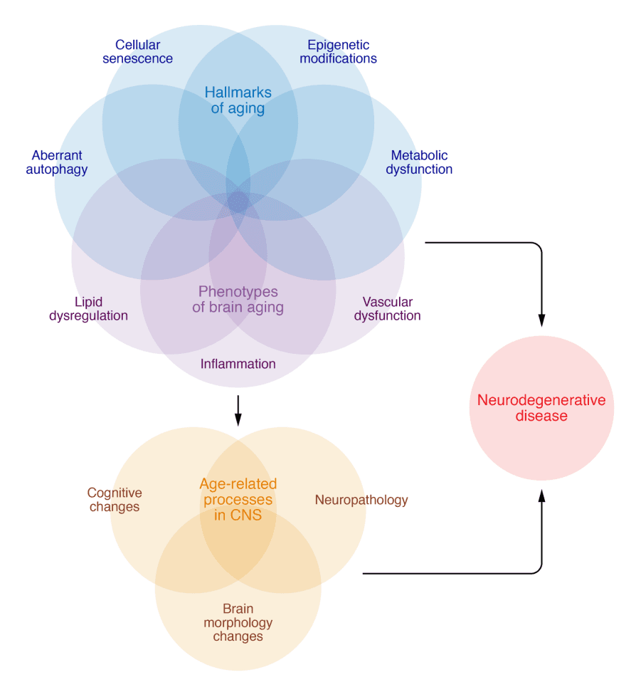
The hallmarks of aging, such as epigenetic modifications, cellular senescence, metabolic dysfunction, and aberrant autophagy, as well as other phenotypes of brain aging, including inflammation, vascular dysfunction and loss of blood brain barrier integrity, and lipid dysregulation, interact to contribute to age-related processes in the CNS, including cognitive decline, neuropathological protein accumulation, and brain morphology changes. These same factors are further dysregulated in neurodegenerative disease. Further investigations are necessary to determine the specific factors and sequences that force the transition between normative age-related changes and manifest neurodegenerative disease in some individuals while others remain cognitively resilient.

Aberrant autophagy. The inability of postmitotic cells, such as neurons, to dilute proteotoxic burden and cellular waste through cell division increases their vulnerability to proteotoxic insults. Autophagy, along with the ubiquitin-proteasome system, provides relief by catabolizing proteins. Autophagy subtypes (e.g., microautophagy, chaperone-mediated autophagy, and macroautophagy) result in lysosomal degradation of substrates, including pathogenic forms of aggregate-prone proteins (i.e., Aβ, tau, and α-synuclein), lipids, dysfunctional mitochondria, and other organelles. Healthy neurons maintain constitutively active, highly efficient autophagy. Neurons in aged brains display higher levels of polyubiquitinated proteins than those in young brains; the age-associated effect becomes further elevated in the context of neurodegenerative disease. The requirement of autophagy activation in memory formation further underscores the critical importance of its regulation for brain function. Postmortem examination of human brains with AD indicates aberrant autophagy; however, there have been conflicting reports about the directionality of dysfunction. Discrepancies may reflect methodological challenges associated with measuring and interpreting autophagic flux in tissue; differences in the brain regions, cell types, and species evaluated; the specific form of autophagy studied; the etiological factor(s) driving neurodegeneration; and differences in normalization controls.

Laser capture microdissection to evaluate autophagy in CA1 hippocampal neurons revealed elevated activation, but a progressive decline in lysosomal clearance across AD severity. Other studies indicate that Beclin-1, an autophagy-initiating protein, is reduced in AD compared with controls. Mechanistic studies in vitro and in vivo have demonstrated that a reduction in Beclin-1 can drive extracellular Aβ deposition, which protects neurons from toxic intracellular accumulation. Changes in Beclin-1 levels are important, as this protein negatively regulates transcription factor EB (TFEB), a master transcriptional regulator of lysosome biogenesis and autophagy. Levels of nuclear (i.e., active) TFEB have been shown to progressively decrease across advancing Braak stages. In rodent studies, increasing TFEB reduced pathogenic tau accumulation and neurodegeneration; exosomal exocytosis may have contributed to the clearance of intraneuronal tau. Chaperone-mediated autophagy (CMA) has emerged as a critical mediator of intraneuronal tau clearance. Wild-type tau is degraded primarily through CMA; however, tau acetylation blocks CMA and redirects it toward extracellular release, increasing pathogenic spread. These studies collectively highlight the role of autophagy in eliminating intracellular neurotoxic proteins by either degrading or secreting them, as well as the essential function of extracellular clearance mechanisms for preventing the subsequent propagation of neuropathological proteins.

Mitochondrial and metabolic dysfunction. Mitochondria utilize oxygen for cellular respiration, extracting, transferring, and producing energy from molecular substrates derived from glucose, fat, fatty acids, and amino acids. They also contribute to calcium and iron homeostasis, cell proliferation and cell death, cell signaling, and proteostasis, thereby broadly connecting mitochondrial function with cell viability and function, and other hallmarks of aging. The brain is a highly metabolically active organ that requires approximately 20% of the body’s basal oxygen to optimally function. Reactive oxygen species (ROS) are a by-product of oxidative phosphorylation that function as a critical signaling molecule; however, their accumulation (i.e., through dysfunctional mitochondria or poor antioxidant scavenging) can lead to oxidative stress, lipid peroxidation, and DNA damage. Mitochondrial changes have been proposed to drive aging (i.e., the free radical theory of aging) and AD (i.e., the “mitochondrial cascade” hypothesis of AD). The critical importance of balanced mitochondrial activity is evidenced by data demonstrating lifespan extension both by the increasing of cellular metabolism and antioxidant capacity in models and by interventions designed to decrease mitochondrial function or enhance ROS production. These longevity benefits may occur through a reduction in ROS production by which improving mitochondrial oxidative stress resistance increases lifespan, suggesting that a little mitochondrial stress may be beneficial.

Elegant studies designed to determine the role of mitochondrial dysfunction in driving aging and disease highlight its complexity. Levels of mitochondrial DNA (mtDNA) mutations increase with age; however, results from mtDNA mutator mice indicate that these mutations do not drive oxidative stress nor accelerated aging until at extreme levels far exceeding those found in aging humans. The level of total mtDNA decreases with age and is reduced more in AD than in cognitively normal age-matched controls. Single-cell analyses indicate an increase of mtD-NA deletions in AD neurons that is also observed in CSF and blood cells. Through elegant cybrid experiments (which involve transferring mtDNA from donor cells to those with identical nuclear DNA but lacking mtDNA), AD mtDNA was shown to be responsible for subtle differences in mitochondrial morphology, biogenesis, and membrane potential; oxidative stress; and calcium buffering capacity. The observed differences in mitochondrial phenotypes that co-occur in peripheral tissues of individuals with AD compared with controls suggest that systemic changes in mitochondrial status relevant to the brain may be identified and tracked in peripheral samples. Such data provide evidence that mitochondrial dysfunction may be upstream, and not a consequence of AD neuropathology. Nevertheless, pathogenic Aβ and tau negatively impact mitochondrial function, which may suggest that once mitochondrial dysfunction is initiated, a pathogenic feedback loop involving oxidative stress and pathogenic protein accumulation may ensue. Further studies are needed to determine whether disease conditions (like AD) represent exacerbated “normal” age-associated changes in mitochondrial function or unique divergent pathogenic processes.

Cellular senescence. Cellular senescence is a stress-induced cell state induced by macromolecular damage that culminates with cell cycle arrest and concomitant, often deleterious, secretory phenotype. Cells that become senescent evade cell death by upregulating antiapoptotic pathways and arresting the cell cycle. Senescent cells also secrete molecules including proinflammatory cytokines, chemokines, growth factors, extracellular remodeling proteins, and other signaling factors that alter the extracellular environment, collectively referred to as the senescence-associated secretory phenotype (SASP). In the absence of senescent cell clearance, the SASP causes tissue damage, cell death, or the transition of other cells to become senescent, thus propagating the phenotype. With advancing age, senescent cells increase in tissues throughout the body, including the brain.

Rodent studies have demonstrated senescent cell accumulation in the brain in response to accumulation of tau or Aβ protein; dysfunctional immune system; high-fat diet or obesity; insulin resistance; chronic unpredictable stress; environmental neurotoxins; and brain injury. Studies using postmortem human brain tissue have identified multiple senescent cell types in AD, including astrocytes, neurons, microglia, oligodendrocyte precursor cells, and endothelial cells. Unbiased single-cell transcriptomics on dorsolateral prefrontal cortex from human AD revealed excitatory neurons as a prominent senescent cell type driven by CDKN2D (encoding p19) that overlapped with neurons bearing neurofibrillary tangles (NFTs). In contrast, bioinformatics analyses of data derived from bulk tissue from healthy human tissue donors revealed that prominent senescent cell types in the brain included endothelial cells and microglia driven by CDKN1A. These studies, both conducted by our group, highlight potential differences in senescent cell types (a) in health versus disease; (b) possibly as a reflection of the starting material (i.e., single-cell, single-nucleus, or bulk tissue analyses); and (c) owing to differences in the predetermined criteria for senescence. Immunosenescence, described below, drives senescent cell accumulation in the brain. Microglia, the macrophage-like cells of the brain, clear NFT-bearing neurons that display phosphatidylserine on their surface. Given that microglia become senescent and dysfunctional after clearing these possibly senescent, NFT-bearing neurons, therapeutic strategies to help remove senescent cells from the brain may alleviate senescent cell burden, inflammation, and disease propagation. Clinical trials are currently under way to test this approach.

Epigenetic changes. Epigenetic processes allow cells to integrate external stimuli into their genome to impact gene expression without altering the DNA sequence. These dynamic, reversible modifications include DNA methylation, chromatin remodeling, histone modification, and noncoding RNA regulation (microRNAs). Neuronal epigenetic changes are crucial for synaptic plasticity and new memory formation. With age, DNA methylation in the brain trends toward global decreases, but there are sex-dependent dimorphisms. Given that DNA methylation inhibits gene transcription, these changes may result in elevated gene expression. Genes implicated in AD, including those coding for APP, MAPT, BDNF, ABCA7, ANK1, BIB1, SORL1, and SIRT1, show differential methylation between individuals with AD and controls. Breast cancer type 1 susceptibility protein (BRCA1), a DNA repair protein typically associated with breast cancer, is hypomethylated in AD. Elevated BRCA1 localizes to the cytosol, where it coaggregates with insoluble tau. In vitro studies suggest that this impacts neurite and dendritic spine morphology. Moreover, epigenetic age acceleration was found to be heritable in AD, where it was associated with neuropathological protein accumulation and cognitive decline. Collectively these data suggest that epigenetic changes may increase AD susceptibility.

The frequency and pattern of epigenetic changes, specifically DNA methylation at CpG sites, can be used to generate an algorithm for comparing chronological age with biological age, termed an epigenetic clock. There are currently more than seven different epigenetic clocks developed for human assessments and others for mouse models. These differ in numbers of methylated CpGs, tissue type, and study populations. The current clocks lack correlation among them. Nevertheless, understanding the relationships between DNA methylation, age, longevity, and age-related disease may hold promise to predict disease, including diseases relevant to the brain. While initial epigenetic clocks were based in blood, recent advances are moving to the brain to predict cortical age. The recently developed Cortical clock provides evidence supporting the use of the epigenome to inform regarding brain aging and pathologies. The Cortical clock was trained using postmortem cortical tissue from older adults, which tracked better with AD diagnosis and Aβ deposition than clocks trained using blood. While blood-based clocks correlated with chronological age at death when applied to cortical tissue, only the Cortical clock significantly associated with tau and Lewy body pathology, highlighting the importance of considering tissue-specific epigenetic changes in these predictions.

Chromatin remodeling and chromatin heterogeneity (or what has been termed epigenetic noise) also increase with age. Histone acetylation tends to decrease with aging, resulting in a more condensed chromatin structure and consequent transcriptional changes. A recent assessment of postmortem human brain tissue revealed an upregulation of two histone acetyltransferases, H3K27ac and H3K9ac, that were linked with Aβ pathology and neurodegeneration by human proteomics data and a transgenic fly model. Three AD mouse models and one nonhuman primate model displayed epigenetic changes that differed across models. This work again emphasizes the complexity of genetic and epigenetic influence on disease progression, as well the importance of matching model systems to the underlying pathogenic process in question.

Unlike the above-mentioned epigenetic alterations, microRNAs (miRNAs) influence gene expression post-transcriptionally by binding to mRNA. miRNAs play critical roles in AD pathology, including modulating Aβ and tau production/function, synaptic plasticity, neuronal growth, apoptosis, and inflammatory response. In AD, disruptions have been noted in several miRNAs, including miRNAs 9, 124, 125b, 132, 146a, and 155, which may have the potential to serve as both biomarkers and therapeutic agents.

Vascular dysfunction and diminished blood-brain barrier integrity. Epidemiological evidence supports an association between risk factors for cardiovascular disease, cerebrovascular dysfunction, and cognitive impairment. More than 50% of individuals with ADRD have concomitant vascular pathologies that increase with advancing age. Furthermore, growing evidence indicates that the molecular mechanisms associated with both vascular and ADRD pathologies act synergistically to compromise cognition. Vascular contributions to cognitive impairment and dementia (VCID) derive from age-related changes to the neurovascular unit (NVU), which is composed of nonfenestrated endothelial cells, pericytes, smooth muscle cells, astrocytes, microglia, oligodendroglia, and neurons. The NVU facilitates normal brain function by ensuring neurovascular coupling, the physiological mechanism whereby cerebral blood flow is matched to neuronal metabolic demands. With aging, and to a greater extent in neurodegenerative disease, there is a loss of pericytes, which has been associated with diminished cerebral blood flow delivery in both human and animal models. In mouse models of AD, pericyte loss has also been shown to reduce Aβ clearance, further propagating neuropathological protein accumulation. In addition, age-related changes in mitochondrial efficiency and the upregulation of ROS induce endothelial dysfunction, which diminishes the bioavailability of the vasodilator nitric oxide and further dampens neurovascular coupling.

The NVU is also important for the maintenance of the bloodbrain barrier (BBB), which controls transport of substances across the endothelium into the CNS through specific transporters on both the luminal and abluminal surfaces. BBB integrity declines in normal aging and even more dramatically in ADRD. Loss of BBB function induces capillary leakage, brain leukocyte infiltration, ingress of toxic substances, and upregulation of TGF-α signaling in astrocytes, resulting in disruption of the brain milieu and neuronal dysfunction. BBB leakage has been identified in the hippocampi of individuals with mild cognitive impairment, which correlates with CSF levels of PDGF-β, a marker of damaged pericytes. Loss of BBB integrity further drives neuroinflammation, which has been implicated in aging and ADRD.

Inflammaging. It has been well established that systemic inflammation increases with age, as evidenced by higher circulating levels of proinflammatory cytokines (i.e., IL-1β, IL-6, TNF-α) and immune dysregulation (loss of vaccine efficacy, increased morbidity upon infection, rises in cancer incidence, and enhanced autoimmunity). This “inflammaging,” a term originally coined by Claudio Franceschi, is thought to contribute to systemic pathologies that develop with age, including ADRD. Numerous studies have shown correlations between circulating proinflammatory mediators and progression of neurodegenerative diseases, suggesting that peripheral inflammation contributes to the development of chronic brain inflammation. In addition, recent studies using CSF to interrogate neuroinflammation directly in the CNS have shown mixed results. For example, in adults without measurable cognitive impairment, increased cytokine levels in the CSF were, surprisingly, associated with lower tau and Aβ levels. In addition, higher plasma levels of IL-12p70 and IFN-γ have been associated with protection against cognitive decline in cognitively unimpaired adults. Thus, it is possible that mild neuronal inflammation may provide some early protection. On the other hand, as disease etiology progresses, an association with neuroinflammatory markers, including C-reactive protein (CRP), triggering receptor expressed on myeloid cells 2 (TREM2), intercellular adhesion molecule 1 (ICAM1), IFNs, and the IL-1 family, is typically reported.

Aging elicits pleiotropic outcomes, reflecting many different factors that contribute to increased neuroinflammation; these have been extensively reviewed and will be only briefly mentioned here. For example, brain microglia, analogous to systemic macrophages, become activated by tissue damage or pathogens and release proinflammatory mediators (reviewed in ref. 164). Inflammation can also alter Aβ clearance through effects on the NLRP3 inflammasome. Age-associated changes in the cells of the adaptive immune system may contribute as well. For example, the proportion of CD4+ T cells that are phenotypically suppressive, designated Tregs (expressing FOXP3), increases with age. Tregs have been shown to play both protective and pathogenic roles in neurodegenerative diseases. Indeed, in a mouse model of AD, transient inactivation of Tregs showed improved cognition and decreased inflammation. T cells may also play a more direct role in neurodegenerative disease through recognition of their cognate antigen(s) through the cell surface T cell receptor (TCR) as is seen in multiple sclerosis, an autoimmune disorder in which pathogenic T cells recognizing myelin peptides damage the tissue. In pilot Aβ vaccination studies for AD, there was an induction of neuroinflammation, which in some cases led to a devastating meningoencephalitis due to proinflammatory CD4+ T cells. Even without immunization, autoimmune responses to neuronal peptides could develop, and in that case, one might expect to find a more restricted TCR repertoire due to selection of those antigen-specific T cells in the CNS. Indeed, this has recently been reported for CD4+ T cells in the CSF of individuals with AD. However, it is not clear whether the T cell clonotypes responding are “helper” T cells (CD4+ FOXP3– ) or “suppressive” Tregs (CD4+ FOXP3+ ), which could be either pathogenic or protective.

Lipid dysregulation. Genetic linkage, large-scale genome-wide association, and exome sequencing studies have also repeatedly linked lipid metabolism–related genes/loci and rare variants with AD, including apolipoprotein E (APOE), CLU, ABCA7, SORL1, TREM2, PICALM, INPP5D, and PLCG2 (reviewed in refs. 171–173). Several lipid-related gene variants, including APOE, have also been associated with human longevity. The first longevity-assurance gene (LAG1) discovered in yeast was found to code for a ceramide synthase. Ceramides comprise a class of lipids that play essential roles both as intermediates in the biosynthesis of more complex sphingolipids, and as signaling molecules that participate in a plethora of biological processes, including apoptosis, inflammation, insulin signaling, mitochondria function, cellular senescence, telomerase activity, and autophagy.

Alterations in brain lipid composition occur in both normal aging and neurodegenerative disease. The brain is the richest organ in terms of lipid content and diversity, largely owing to the abundance of lipid-rich myelin. Lipidomics, the large-scale study of pathways and networks of cellular lipids in biological systems, has revealed specific lipid profiles associated with AD and aging. For example, early accumulation of ceramide levels in the AD brain has been consistently reported by multiple groups. On the other hand, sulfatides, a class of sulfoglycolipids highly enriched in myelin, have been reported to be specifically and dramatically reduced at the earliest clinically recognizable stages of AD. Brain sulfatide levels in patients with AD and in animal models strongly correlate with the onset and severity of Aβ deposition. Mechanistic studies in animal models have revealed that sulfatide deficiency in AD occurs in an isoform-specific manner and that sulfatide losses are sufficient to induce AD-like neuroinflammation and cognitive decline. Moreover, levels of the phospholipid plasmalogen have been consistently shown to decline not only in the brains of individuals with AD, but also in circulation, with ethanolamine plasmalogen deficits closely associating with disease severity. Notably, human brain plasmalogen levels have also been reported to decline with normal aging, decreasing dramatically by around 70 years of age.

Conclusions

Chronological aging is accompanied by molecular, cellular, and systems-level processes with underlying biology that may modulate susceptibility to neurodegenerative disease. Applying current insights from the biology-of-aging field to age-associated neurodegenerative diseases offers an opportunity to explore and target new cellular and molecular processes. We have focused on a few selected hallmarks of aging for which interventions are moving to clinical trials in the context of mild cognitive impairment/ early AD. Though still an emerging field, geroscience-motivated approaches are appealing for the treatment of complex age-associated diseases, like AD. The synergistic interactions across biologyof-aging pathways raise optimism that effective targeting of one may exert broader beneficial influences. As highlighted above, the transition in these cellular and molecular processes over the course of the disease is complex and may be nonlinear. Early upregulation of specific processes, such as cellular respiration and senescence, may help mitigate neurodegenerative disease changes; however, these same processes may be detrimental over time by perpetuating oxidative stress and inflammation. Early trials exploring geroscience-motivated approaches for the treatment of AD will provide critical information on this strategy. For example, NCT04685590, led by our team, will focus on geroscience outcomes as well as AD biomarkers and cognitive changes. Other studies are targeting mitochondrial function with NAD+ precursors (NCT04078178, NCT04430517) and nutrient sensing and handling with rapamycin (NCT04200911, NCT04629495). As these early trials are under way, advances in the basic biology of aging are needed to continue shedding light on cell type specificity and interactions across biology-of-aging hallmarks, and to refine model systems through efforts including Model Organisms Development and Evaluation for Late-Onset Alzheimer’s Disease (MODEL-AD). Furthermore, cross-disciplinary training and collaboration across the fields of neuroscience and geroscience will be crucial for advancing treatments that target age-related dysfunction across systems in an effort to optimize both physical and cognitive functioning throughout the lifespan.

Open Article as PDF

Abstract

Alzheimer’s disease and related dementias (ADRD) are among the top contributors to disability and mortality in later life. As with many chronic conditions, aging is the single most influential factor in the development of ADRD. Even among older adults who remain free of dementia throughout their lives, cognitive decline and neurodegenerative changes are appreciable with advancing age, suggesting shared pathophysiological mechanisms. In this Review, we provide an overview of changes in cognition, brain morphology, and neuropathological protein accumulation across the lifespan in humans, with complementary and mechanistic evidence from animal models. Next, we highlight selected aging processes that are differentially regulated in neurodegenerative disease, including aberrant autophagy, mitochondrial dysfunction, cellular senescence, epigenetic changes, cerebrovascular dysfunction, inflammation, and lipid dysregulation. We summarize research across clinical and translational studies to link biological aging processes to underlying ADRD pathogenesis. Targeting fundamental processes underlying biological aging may represent a yet relatively unexplored avenue to attenuate both age-related cognitive decline and ADRD. Collaboration across the fields of geroscience and neuroscience, coupled with the development of new translational animal models that more closely align with human disease processes, is necessary to advance novel therapeutic discovery in this realm.

Introduction

The aging population is increasing, with older adults projected to represent a significant portion of Americans by 2030. This demographic shift highlights the growing importance of preventing and treating chronic age-related conditions, particularly Alzheimer’s disease and related dementias (ADRD). These conditions lead to a progressive decline in cognitive abilities and daily functioning, making them major contributors to disability and mortality. Aging itself is the primary risk factor for ADRD, with incidence rates increasing significantly after age 65. Even in individuals who avoid dementia, some cognitive decline and brain changes are common with age, suggesting shared underlying biological processes. This overview discusses changes in brain structure and function throughout life and explains how biological aging processes may contribute to cognitive decline and neurodegenerative diseases.

Cognitive Changes Across the Lifespan

Cognitive abilities, such as processing speed, reasoning, memory, and spatial visualization, begin to decline as early as the third decade of life. This decline is not sudden but a small, consistent reduction over many years. Different cognitive skills decline at varying rates. Skills that require new information processing, quick responses, and problem-solving, often called "fluid skills," tend to decrease more noticeably. In contrast, "crystallized skills," like vocabulary and general knowledge, which are well-learned and based on experience, usually remain more stable throughout life. Research suggests a significant portion of individual cognitive change is due to a general factor that accounts for overall declines with advancing age. Animal studies show similar age-related declines in learning and memory, and sensory losses (like hearing and vision) are also linked to faster cognitive decline. Mechanisms underlying age-associated cognitive decline include changes in how brain cells connect and communicate, as well as broader aging processes like altered gene expression, cellular aging, and inflammation.

Lifespan Changes in Brain Morphology and Function

Most brain cells (neurons) typically survive throughout life, even without disease or injury. However, the volume of gray matter in the brain gradually decreases starting in the second decade of life, with the most noticeable changes occurring in the front and top regions of the brain. Animal models show similar reductions in gray matter and increases in fluid-filled spaces. Research increasingly recognizes that aging brings changes in brain cell chemistry, metabolism, and structure, which are linked to cell dysfunction and inflammation.

Effective learning, memory, and complex thought require the coordinated activity of neurons across connected brain networks. Neuronal activity strengthens or weakens these connections. In aging and neurodegenerative diseases, some neurons become less excitable, while others become overly excitable, leading to disrupted brain signals. Excessive neuronal excitability, in particular, has been linked to negative cognitive outcomes in both human and animal studies. Reducing this hyperexcitability has shown benefits in animal models, preventing connection loss between brain cells and preserving cognitive function. Clinical trials are currently investigating therapies targeting this neuronal hyperexcitability in adults with neurodegenerative disease.

Changes in brain chemicals throughout life also reveal targets for understanding cognitive impairment, including those related to the protective covering of nerve fibers, called myelin. Myelin, rich in lipids, is crucial for efficient nerve signal transmission. With age, the integrity of white matter, where myelin is abundant, declines, particularly in the front of the brain, contributing to slower processing speed and reduced executive function. The cells that produce myelin also decline with age, leading to reduced myelination. Age-related white matter changes may also involve factors like reduced blood flow, inflammation, and problems with the brain's protective barrier.

AD Neuropathological Burden in Aging and Disease

The defining features of Alzheimer’s disease, such as the accumulation of amyloid-beta plaques and tau tangles, develop over decades in both normal aging and neurodegenerative disease. These pathological proteins can be detected in adults as early as middle age. Research suggests that pathological tau can spread through the brain, converting normal tau proteins into their disease-causing form. Amyloid-beta also shows similar "seeding" properties. Furthermore, these proteins, along with others like alpha-synuclein, can interact to worsen the overall burden of pathology in the brain.

Despite the link between amyloid-beta and tau accumulation and Alzheimer’s disease, these brain pathologies are common in older individuals who show no signs of cognitive impairment. Studies indicate that a large percentage of cognitively healthy older adults exhibit one or more types of brain pathology after death, with many showing multiple pathologies. This suggests that some individuals with significant brain pathology remain dementia-free due to mechanisms that provide resistance to the brain cell damage typically caused by protein accumulation. Researchers are actively investigating these mechanisms of "cognitive resilience."

Biological Aging Hallmarks of Cognitive Decline and ADRD

Aging is the most significant risk factor for developing typical forms of Alzheimer's disease and related dementias. Processes linked to neurodegenerative disease, such as cognitive decline, brain shrinkage, white matter degradation, and protein accumulation, gradually appear throughout life, even in individuals who remain dementia-free. This indicates a likely overlap and continuum between the biological pathways underlying normal cognitive aging and ADRD. Targeting fundamental biological aging processes may offer a new approach to reduce both age-related cognitive decline and ADRD.

The field of aging biology has made significant progress in identifying processes that contribute to biological aging and age-related decline in various body systems. These processes, known as hallmarks of aging, include instability of genetic material, shortening of protective caps on chromosomes (telomeres), changes in gene expression without altering DNA sequence, issues with protein cleanup, abnormal nutrient processing, mitochondrial dysfunction (energy production problems), stem cell exhaustion, altered cell communication, and cellular senescence (cells that stop dividing but remain active). These hallmarks are implicated in many chronic age-related diseases, including ADRD. Studies in animal models show that targeting these processes can extend lifespan and healthspan, suggesting potential benefits for cognitive health.

Specific aging processes are particularly relevant to ADRD:

  • Issues with cellular waste removal (Autophagy): Brain cells, especially neurons, are vulnerable to waste buildup because they do not divide to dilute harmful proteins. Autophagy is a process that cleans cells by breaking down waste proteins, dysfunctional mitochondria, and other cellular debris. In aged brains and particularly in neurodegenerative diseases, this process can become impaired, leading to accumulation of toxic proteins like amyloid-beta and tau.

  • Mitochondrial and metabolic dysfunction: Mitochondria are crucial for cellular energy production and overall cell health. The brain requires a large amount of energy. Dysfunction in mitochondria can lead to oxidative stress and DNA damage, which are implicated in both aging and Alzheimer's disease. While some studies suggest a little mitochondrial stress might be beneficial, overall, their proper function is critical for brain health.

  • Cellular senescence: This is a stress-induced state where cells stop dividing but remain metabolically active, secreting substances that can harm surrounding tissues and promote inflammation. Senescent cells increase in the brain with age and are found in neurodegenerative diseases. These cells can contribute to tissue damage and the spread of unhealthy cell states.

  • Epigenetic changes: These are modifications to DNA or its associated proteins that affect gene activity without changing the underlying genetic code. Such changes are vital for brain plasticity and memory formation. With age and in AD, distinct epigenetic changes occur, which may increase susceptibility to the disease by altering gene expression related to brain function and protein handling.

  • Vascular dysfunction and diminished blood-brain barrier integrity: Problems with blood vessels and the blood-brain barrier (BBB) are common in aging and significantly contribute to cognitive impairment and dementia. The BBB controls substance transport into the brain, and its decline with age or disease leads to leakage, inflammation, and neuronal dysfunction. Reduced blood flow and a compromised BBB can worsen protein accumulation and overall brain health.

  • Inflammaging: This term describes the chronic, low-grade inflammation that increases with age throughout the body, including the brain. Systemic inflammation is linked to neurodegenerative disease progression, though early inflammation in the brain might sometimes offer mild protection. As disease progresses, markers of neuroinflammation typically increase. Immune cells in the brain, like microglia, become activated and contribute to inflammation, which can affect the clearance of harmful proteins.

  • Lipid dysregulation: Genes related to lipid (fat) metabolism are repeatedly linked to Alzheimer’s disease and human longevity. The brain is rich in lipids, particularly in myelin. Changes in brain lipid composition occur with both normal aging and neurodegenerative disease. For example, specific lipids like ceramides accumulate in AD, while others, like sulfatides and plasmalogens, are significantly reduced, impacting brain function and contributing to neuroinflammation and cognitive decline.

Conclusions

Aging involves complex molecular, cellular, and systemic processes that influence susceptibility to neurodegenerative diseases. Applying insights from aging biology to age-associated neurodegenerative diseases provides new avenues for understanding and targeting these processes. Geroscience-motivated approaches, which aim to target fundamental aging processes, hold promise for treating complex age-related diseases like Alzheimer's. The interconnected nature of these biological aging pathways suggests that interventions targeting one pathway might have broader beneficial effects. However, the progression of these cellular and molecular changes during disease can be complex and non-linear. Early studies on geroscience-based treatments for Alzheimer's disease will provide crucial information on this strategy. Continued advances in basic aging biology and collaboration between neuroscience and geroscience are essential to developing treatments that optimize both physical and cognitive functioning across the lifespan.

Open Article as PDF

Abstract

Alzheimer’s disease and related dementias (ADRD) are among the top contributors to disability and mortality in later life. As with many chronic conditions, aging is the single most influential factor in the development of ADRD. Even among older adults who remain free of dementia throughout their lives, cognitive decline and neurodegenerative changes are appreciable with advancing age, suggesting shared pathophysiological mechanisms. In this Review, we provide an overview of changes in cognition, brain morphology, and neuropathological protein accumulation across the lifespan in humans, with complementary and mechanistic evidence from animal models. Next, we highlight selected aging processes that are differentially regulated in neurodegenerative disease, including aberrant autophagy, mitochondrial dysfunction, cellular senescence, epigenetic changes, cerebrovascular dysfunction, inflammation, and lipid dysregulation. We summarize research across clinical and translational studies to link biological aging processes to underlying ADRD pathogenesis. Targeting fundamental processes underlying biological aging may represent a yet relatively unexplored avenue to attenuate both age-related cognitive decline and ADRD. Collaboration across the fields of geroscience and neuroscience, coupled with the development of new translational animal models that more closely align with human disease processes, is necessary to advance novel therapeutic discovery in this realm.

Introduction

By 2030, a significant portion of the American population is expected to be 65 years of age or older. This demographic shift highlights the increasing importance of preventing and treating chronic diseases common in older age. Alzheimer’s disease and related dementias (ADRD) are conditions that lead to a gradual decline in thinking abilities and daily function, contributing significantly to disability and death. Advancing age is the primary risk factor for developing ADRD, with the chance of developing the disease nearly doubling every five years after age 65. Even in older adults who do not develop dementia, some cognitive decline and brain changes are common, suggesting shared underlying biological processes. This overview briefly describes how brain structure and function change throughout life and explores insights from research that show how biological aging processes contribute to cognitive decline and neurodegenerative diseases.

Cognitive Changes Across the Lifespan

Important cognitive abilities, such as processing speed, reasoning, memory for events, and spatial visualization, begin to decline as early as an individual's twenties. Rather than a sudden drop in old age, studies indicate a small but steady decrease in these abilities over a person's lifetime. Different cognitive skills rely on different brain areas and may decline at varying rates. Generally, "fluid skills," which involve new information, quick responses, and problem-solving (like processing speed, memory, and reasoning), tend to decrease more noticeably. In contrast, "crystallized skills," such as vocabulary and general knowledge, which are learned well, practiced, and enhanced by experience, usually remain more stable throughout life. Despite these differences, research suggests that a substantial portion of an individual's cognitive change can be attributed to a "general aging effect" that causes overall declines with age. Animal studies also show age-related deficits in learning and memory in older rodents, similar to human experiences. Researchers continue to identify mechanisms behind age-related cognitive decline, including changes in how brain cells communicate and how they manage certain chemicals. Other factors linked to aging, such as changes in genes, cell aging, waste removal, energy production, and inflammation, are also being explored.

Lifespan Changes in Brain Morphology and Function

In healthy individuals without disease or injury, most brain cells (neurons) last throughout a person's life. However, the volume of the brain's gray matter gradually decreases starting in the twenties, with the most significant changes seen in the front and top parts of the brain. Animal models show similar reductions in gray matter in old age, along with an increase in brain fluid and small brain bleeds. There is a growing understanding of how age affects the chemistry, metabolism, and structure of neurons, often alongside problems with neuron function and inflammation.

The ability to learn new things, form memories, and perform complex cognitive tasks requires neurons to work together in interconnected networks. The way neurons fire causes changes in connections (synaptic plasticity) that can strengthen or weaken specific parts of these networks. In aging and neurodegenerative diseases, some groups of neurons become less excitable, while others become overly excitable, affecting the brain's ability to process signals clearly. Excessive excitability has been linked to negative cognitive outcomes in both humans and animals. Studies on long-lived humans show changes in gene expression that reduce the excitability of brain cells. More pronounced changes in neuronal excitability are seen in neurodegenerative diseases, which can increase the risk of seizures and speed up cognitive decline. The buildup of abnormal proteins in Alzheimer's disease disrupts the balance of signals between brain cells, leading to neuron dysfunction and DNA damage. Other age-related changes, such as reduced energy efficiency in cells and increased harmful byproducts, also alter brain signaling and can cause over-excitability. In animal models of Alzheimer's, reducing this over-excitability helped prevent nerve cell loss and maintained cognitive function. Clinical trials are currently investigating treatments that target this over-excitability in adults with neurodegenerative disease.

Changes in brain chemicals over the lifespan have also pointed to new targets for understanding cognitive problems, including those related to changes in the white matter pathways. White matter in the brain is made of myelin, a fatty substance crucial for efficient nerve signal transmission. In humans, age-related declines in white matter health are most noticeable in the front brain regions and are linked to slower processing speed and reduced executive function. In older rats, the myelin sheath can split, detaching from the nerve axon, which is attributed to a decline in structural proteins. Furthermore, the cells that produce myelin, called oligodendrocytes, decrease with normal aging, leading to myelin loss and reduced white matter integrity. Bright spots on brain scans (white matter hyperintensities) also become more common in older age. These spots are associated with myelin loss, scarring, and thinning of tissue, potentially caused by reduced blood flow, inflammation, and problems with the blood-brain barrier. In animal models, age-related reductions in small blood vessel density in white matter, combined with hardening of small arteries, increase the risk of insufficient blood flow and damage to white matter.

AD Neuropathological Burden in Aging and Disease

The key markers of Alzheimer's disease (AD), which are clusters of amyloid-beta (Aβ) plaques and tau tangles, gradually build up over decades in both normal aging and in neurodegenerative disease. Advanced brain imaging has detected Aβ and tau in adults as early as middle age. Research using animal models of AD suggests that abnormal tau protein can spread throughout the brain, transforming normal tau into its disease-causing form. Aβ has also been shown to spread. Furthermore, Aβ and abnormal tau, as well as other abnormal proteins like alpha-synuclein, may interact to worsen the overall amount of disease pathology in the brain. In mice with Aβ, adding human tau reduces the expression of genes involved in synaptic regulation, leading to further harmful effects on the central nervous system.

While Aβ and tau accumulation are linked to AD, brain pathology is common in older age even in individuals without cognitive problems. A study of adults who remained cognitively healthy found that many showed at least one type of brain pathology, with a significant number having multiple types. Another large study reported that about one-third of individuals with moderate to high levels of AD brain pathology did not develop dementia during their lives. These findings suggest that individuals with significant brain pathology but normal cognition may be resistant to the nerve cell damage that typically occurs with abnormal protein buildup. Several research groups are actively investigating the mechanisms that allow some individuals to maintain cognitive health despite brain changes.

Biological Aging Hallmarks of Cognitive Decline and ADRD

Studies involving large populations have consistently shown that aging is the most significant risk factor for developing sporadic Alzheimer's disease and related dementias (ADRD). Additionally, processes linked to neurodegenerative disease, such as cognitive decline, brain shrinkage, white matter degradation, and the accumulation of abnormal proteins, gradually appear throughout life, even in individuals who remain dementia-free. This suggests that the biological pathways underlying normal cognitive aging and ADRD likely overlap and exist on a continuum. Targeting fundamental biological aging processes may offer a new approach to reduce both age-related cognitive decline and ADRD. The field of geroscience, which studies the biology of aging, has made significant progress in identifying processes that contribute to aging and the decline of multiple body systems. Key hallmarks of aging include issues with genetic stability, telomere shortening, changes in gene expression (epigenetics), problems with protein maintenance, dysregulated nutrient sensing, mitochondrial dysfunction, stem cell exhaustion, altered cell communication, and cellular senescence. These hallmarks, among others, are implicated in many chronic age-related diseases, including ADRD. In animal models, targeting these biological aging processes has extended both lifespan and healthspan, suggesting these approaches could also benefit cognitive health. The following sections highlight selected aging processes that are regulated differently in ADRD and have been linked to disease development.

Aberrant Autophagy

Brain cells, especially neurons which do not divide, are vulnerable to the buildup of toxic proteins and cellular waste. Autophagy, along with another system called the ubiquitin-proteasome system, helps clear this waste by breaking down proteins. Various types of autophagy lead to the degradation of harmful proteins (like Aβ, tau, and alpha-synuclein), fats, and malfunctioning mitochondria within cell structures called lysosomes. Healthy neurons maintain highly active and efficient autophagy. Neurons in older brains show higher levels of proteins marked for degradation than those in younger brains, and this age-related effect is even more pronounced in neurodegenerative disease. The importance of autophagy for brain function is also highlighted by its role in memory formation. Postmortem studies of human brains with Alzheimer's disease indicate abnormal autophagy, though reports on the exact nature of this dysfunction have varied. These differences may be due to challenges in measuring autophagy in tissue, variations in brain regions or cell types studied, and other factors.

Research examining autophagy in specific hippocampal neurons (CA1) showed increased activation but a progressive decline in waste clearance as Alzheimer's disease worsened. Other studies suggest that Beclin-1, a protein that starts autophagy, is reduced in AD compared to healthy controls. Studies in labs and animals have shown that a reduction in Beclin-1 can lead to the buildup of Aβ outside neurons, which may protect neurons from toxic accumulation inside. Changes in Beclin-1 levels are important because this protein controls TFEB, a master regulator of lysosome formation and autophagy. Levels of active TFEB have been observed to decrease progressively with advancing stages of AD. In rodent studies, increasing TFEB reduced harmful tau accumulation and neurodegeneration; this clearance may have involved the release of tau from neurons. Chaperone-mediated autophagy (CMA) is recognized as crucial for clearing tau from inside neurons. Normal tau is primarily broken down by CMA; however, certain chemical changes to tau can block CMA and redirect tau for release outside the cell, potentially increasing its spread. These studies collectively emphasize the role of autophagy in eliminating toxic proteins inside cells through degradation or secretion, and the importance of extracellular clearance mechanisms in preventing the spread of these abnormal proteins.

Mitochondrial and Metabolic Dysfunction

Mitochondria use oxygen to produce energy for cells from various nutrients, playing a central role in cell survival and function. The brain is highly active metabolically, requiring about 20% of the body's basic oxygen supply for optimal function. Reactive oxygen species (ROS) are byproducts of energy production that are important signaling molecules, but their excessive accumulation due to dysfunctional mitochondria or poor antioxidant defenses can lead to oxidative stress, fat damage, and DNA damage. Mitochondrial changes have been proposed as drivers of both aging and Alzheimer's disease. The importance of balanced mitochondrial activity is shown by data indicating that both increasing and decreasing cellular metabolism or antioxidant capacity can extend lifespan in models. This suggests that a small amount of mitochondrial stress might even be beneficial.

Studies investigating the role of mitochondrial dysfunction in aging and disease highlight its complexity. Levels of mitochondrial DNA (mtDNA) mutations increase with age; however, animal studies suggest that these mutations do not cause oxidative stress or accelerated aging until they reach extreme levels far beyond those seen in aging humans. The total amount of mtDNA decreases with age and is further reduced in Alzheimer's disease compared to healthy older adults. Single-cell analyses indicate an increase in mtDNA deletions in AD neurons, also observed in cerebrospinal fluid and blood cells. Experiments involving transferring mtDNA from donor cells to cells lacking their own mtDNA showed that AD mtDNA was responsible for subtle differences in mitochondrial structure, formation, and energy potential; oxidative stress; and calcium regulation. Observed differences in mitochondrial characteristics in peripheral tissues (e.g., blood) of individuals with AD suggest that systemic changes in mitochondrial status, relevant to the brain, might be identified and tracked using these samples. Such data indicate that mitochondrial dysfunction may occur early in the disease process, rather than being a consequence of AD pathology. Nevertheless, abnormal Aβ and tau proteins negatively affect mitochondrial function, suggesting that once mitochondrial dysfunction begins, a harmful cycle involving oxidative stress and abnormal protein accumulation may develop. Further studies are needed to determine if disease conditions like AD represent an exaggeration of "normal" age-associated changes in mitochondrial function or unique, separate disease processes.

Cellular Senescence

Cellular senescence is a stress-induced state where cells stop dividing and often develop a harmful secretory profile, often due to damage to large molecules. Senescent cells avoid cell death by activating anti-apoptotic pathways and halting their cell cycle. They also release molecules, including pro-inflammatory cytokines, chemokines, growth factors, and other signaling factors, which alter the surrounding environment. This collection of secreted molecules is known as the senescence-associated secretory phenotype (SASP). If senescent cells are not cleared, the SASP can cause tissue damage, cell death, or prompt other cells to become senescent, thus spreading the condition. With increasing age, senescent cells accumulate in tissues throughout the body, including the brain.

Animal studies have shown that senescent cells build up in the brain in response to the accumulation of tau or Aβ protein, a dysfunctional immune system, a high-fat diet or obesity, insulin resistance, chronic stress, environmental toxins, and brain injury. Studies using human postmortem brain tissue have identified various types of senescent cells in Alzheimer's disease, including astrocytes, neurons, microglia, oligodendrocyte precursor cells, and endothelial cells. Detailed single-cell analysis of brain tissue from human AD cases revealed that excitatory neurons were a prominent type of senescent cell, linked to specific gene activity and overlapping with neurons containing tau tangles. In contrast, analyses of bulk tissue from healthy human donors indicated that endothelial cells and microglia were prominent senescent cell types in the brain. These studies highlight potential differences in senescent cell types between healthy and diseased brains, possibly due to the type of tissue analyzed, or differences in the criteria used to define senescence. Immune system aging (immunosenescence) also contributes to senescent cell accumulation in the brain. Microglia, which are immune cells in the brain, clear neurons containing tau tangles that display certain markers on their surface. Since microglia themselves can become senescent and dysfunctional after clearing these cells, therapeutic strategies to remove senescent cells from the brain may reduce the burden of these cells, inflammation, and disease progression. Clinical trials are currently testing this approach.

Epigenetic Changes

Epigenetic processes allow cells to respond to external stimuli by affecting gene activity without changing the underlying DNA sequence. These dynamic and reversible modifications include DNA methylation, changes in chromatin structure, histone modifications, and regulation by noncoding RNAs (like microRNAs). In neurons, epigenetic changes are critical for brain cell plasticity and the formation of new memories. With age, DNA methylation in the brain generally decreases, though there are differences between sexes. Since DNA methylation often inhibits gene activity, these changes may lead to increased gene expression. Genes linked to Alzheimer's disease, including those for APP, MAPT, and others, show different methylation patterns between individuals with AD and healthy controls. Breast cancer type 1 susceptibility protein (BRCA1), a DNA repair protein usually associated with breast cancer, shows reduced methylation in AD. Elevated BRCA1 accumulates in the cell's fluid, where it aggregates with abnormal tau. Lab studies suggest this impacts the structure of nerve cell extensions. Furthermore, accelerated epigenetic aging has been found to be inherited in AD, correlating with abnormal protein accumulation and cognitive decline. Collectively, these data suggest that epigenetic changes may increase susceptibility to AD.

The frequency and pattern of epigenetic changes, particularly DNA methylation at specific sites, can be used to create an algorithm that compares chronological age with biological age, known as an epigenetic clock. Several different epigenetic clocks have been developed for humans and animal models, varying in the number of methylation sites measured, tissue type, and study populations. These clocks do not always correlate perfectly with each other. Nevertheless, understanding the relationships between DNA methylation, age, longevity, and age-related disease holds promise for predicting disease, including brain-related conditions. While initial epigenetic clocks were based on blood samples, recent advancements are focused on using brain tissue to predict cortical age. A recently developed "Cortical clock" provides evidence that the epigenome can inform about brain aging and diseases. This clock, trained using postmortem cortical tissue from older adults, showed a better correlation with AD diagnosis and Aβ deposition than clocks trained using blood. While blood-based clocks correlated with chronological age when applied to cortical tissue, only the Cortical clock significantly associated with tau and Lewy body pathology, emphasizing the importance of considering tissue-specific epigenetic changes in these predictions.

Changes in chromatin structure and its variability also increase with age. Histone acetylation generally decreases with aging, leading to a more condensed chromatin structure and subsequent changes in gene activity. A recent assessment of postmortem human brain tissue revealed an increase in two specific histone modifications (H3K27ac and H3K9ac) linked to Aβ pathology and neurodegeneration through human protein data and an animal model. Different animal models of AD and a non-human primate model showed varied epigenetic changes, underscoring the complex influence of genetics and epigenetics on disease progression, and the need to match model systems to the specific disease process being studied.

Unlike the epigenetic changes mentioned above, microRNAs (miRNAs) affect gene expression after messenger RNA (mRNA) is created by binding to it. miRNAs play critical roles in AD pathology, including influencing Aβ and tau production/function, brain cell plasticity, neuronal growth, cell death, and inflammatory responses. Disruptions in several miRNAs have been observed in AD, suggesting their potential as both disease markers and therapeutic targets.

Vascular Dysfunction and Diminished Blood-Brain Barrier Integrity

Epidemiological evidence supports a link between risk factors for heart disease, problems with brain blood vessels, and cognitive impairment. More than half of individuals with Alzheimer's disease and related dementias (ADRD) also have co-existing vascular problems, which increase with age. Growing evidence indicates that the molecular mechanisms associated with both vascular and ADRD pathologies work together to worsen cognitive function. Vascular contributions to cognitive impairment and dementia (VCID) arise from age-related changes to the neurovascular unit (NVU). The NVU is a complex structure made of various cell types that ensures proper blood flow to the brain, matching it to neuronal metabolic needs. With aging, and even more significantly in neurodegenerative disease, there is a loss of pericytes, which are cells around blood vessels. This loss has been linked to reduced brain blood flow in both humans and animals. In animal models of AD, pericyte loss has also been shown to reduce the clearance of Aβ, further promoting abnormal protein accumulation. Additionally, age-related declines in cellular energy efficiency and an increase in harmful reactive oxygen species cause dysfunction in endothelial cells lining blood vessels, which reduces the availability of nitric oxide (a substance that widens blood vessels) and further impairs the matching of blood flow to brain activity.

The neurovascular unit is also crucial for maintaining the blood-brain barrier (BBB), which controls the passage of substances from the bloodstream into the central nervous system through specific transporters. The integrity of the BBB declines in normal aging and even more dramatically in ADRD. Loss of BBB function leads to leaky capillaries, immune cell infiltration into the brain, entry of toxic substances, and changes in astrocyte signaling, resulting in disruption of the brain's internal environment and neuronal dysfunction. BBB leakage has been identified in the hippocampus of individuals with mild cognitive impairment, correlating with levels of a marker for damaged pericytes in cerebrospinal fluid. Loss of BBB integrity also fuels neuroinflammation, a factor implicated in both aging and ADRD.

Inflammaging

It is well established that systemic inflammation increases with age, evidenced by higher levels of pro-inflammatory substances in the blood and immune system dysregulation (e.g., reduced vaccine effectiveness, increased susceptibility to infection, higher cancer rates, and enhanced autoimmunity). This "inflammaging," a term coined by Claudio Franceschi, is believed to contribute to age-related body-wide pathologies, including ADRD. Numerous studies show correlations between circulating pro-inflammatory mediators and the progression of neurodegenerative diseases, suggesting that inflammation in the rest of the body contributes to chronic brain inflammation. However, recent studies directly examining neuroinflammation in the central nervous system (e.g., using cerebrospinal fluid) have yielded mixed results. For instance, in adults without measurable cognitive impairment, increased cytokine levels in the cerebrospinal fluid were, surprisingly, associated with lower tau and Aβ levels. Also, higher plasma levels of certain cytokines have been linked to protection against cognitive decline in cognitively unimpaired adults. This suggests that mild neuronal inflammation might provide some early protection. On the other hand, as disease etiology progresses, an association with neuroinflammatory markers is typically reported.

Aging leads to many factors that contribute to increased neuroinflammation. For example, brain microglia, which are similar to immune cells called macrophages, become activated by tissue damage or pathogens and release pro-inflammatory substances. Inflammation can also alter Aβ clearance through its effects on certain immune pathways. Age-related changes in the adaptive immune system cells may also contribute. For example, the proportion of specific T cells (CD4+ T cells that are suppressive, called Tregs) increases with age. Tregs have been shown to play both protective and harmful roles in neurodegenerative diseases. In an animal model of AD, temporary inactivation of Tregs improved cognition and reduced inflammation. T cells may also play a more direct role in neurodegenerative disease through recognizing specific targets, as seen in autoimmune disorders like multiple sclerosis. In early Aβ vaccination studies for AD, neuroinflammation was induced, in some cases leading to severe brain inflammation due to pro-inflammatory T cells. Even without immunization, autoimmune responses to neuronal peptides could develop, which might lead to a more restricted T cell profile in the central nervous system. This has recently been reported for CD4+ T cells in the cerebrospinal fluid of individuals with AD, though it is unclear if these T cells are protective or harmful.

Lipid Dysregulation

Genetic studies have consistently linked genes and genetic variations related to lipid (fat) metabolism with Alzheimer's disease (AD), including APOE, CLU, and others. Several lipid-related gene variants, including APOE, have also been associated with human longevity. The first gene found to promote longevity in yeast was found to code for a ceramide synthase, an enzyme involved in producing ceramides. Ceramides are a class of lipids that are essential for the production of more complex fats and act as signaling molecules involved in various biological processes, including cell death, inflammation, insulin signaling, mitochondrial function, cellular aging, and waste removal.

Alterations in brain lipid composition occur in both normal aging and neurodegenerative disease. The brain is the body's richest organ in terms of lipid content and diversity, largely due to the abundance of lipid-rich myelin. Large-scale studies of cellular lipids (lipidomics) have revealed specific lipid profiles associated with AD and aging. For example, the early accumulation of ceramide levels in the AD brain has been consistently reported. Conversely, sulfatides, a class of fats highly concentrated in myelin, have been reported to be significantly reduced at the earliest clinically recognizable stages of AD. Brain sulfatide levels in patients with AD and in animal models strongly correlate with the onset and severity of Aβ deposition. Studies in animal models have shown that sulfatide deficiency in AD occurs in a specific way and that sulfatide losses are sufficient to induce AD-like neuroinflammation and cognitive decline. Moreover, levels of the phospholipid plasmalogen have been consistently shown to decline not only in the brains of individuals with AD but also in circulation, with ethanolamine plasmalogen deficits closely associating with disease severity. Notably, human brain plasmalogen levels have also been reported to decline with normal aging, decreasing dramatically around 70 years of age.

Conclusions

Chronological aging is accompanied by molecular, cellular, and system-level processes with underlying biological mechanisms that may influence an individual's susceptibility to neurodegenerative disease. Applying current knowledge from the biology-of-aging field to age-associated neurodegenerative diseases provides an opportunity to explore and target new cellular and molecular processes. This document has focused on several selected hallmarks of aging for which interventions are progressing to clinical trials in the context of mild cognitive impairment and early Alzheimer's disease. While still a developing area, geroscience-driven approaches are appealing for treating complex age-related diseases like AD. The way that biological aging pathways interact synergistically suggests that effectively targeting one pathway might have broader beneficial effects. As highlighted, the changes in these cellular and molecular processes over the course of the disease are complex and may not be linear. Early increases in specific processes, such as cellular respiration and senescence, may help reduce neurodegenerative disease changes; however, these same processes may become harmful over time by perpetuating oxidative stress and inflammation. Early trials exploring geroscience-based approaches for AD treatment will provide crucial information on this strategy. For example, some trials are focusing on geroscience outcomes as well as AD biomarkers and cognitive changes, while others are targeting mitochondrial function and nutrient sensing. As these early trials are underway, continued advancements in the basic biology of aging are needed to shed more light on cell type specificity and interactions across biological aging hallmarks, and to refine model systems. Furthermore, interdisciplinary training and collaboration between neuroscience and geroscience fields will be vital for advancing treatments that target age-related dysfunction across body systems to optimize both physical and cognitive functioning throughout the lifespan.

Open Article as PDF

Abstract

Alzheimer’s disease and related dementias (ADRD) are among the top contributors to disability and mortality in later life. As with many chronic conditions, aging is the single most influential factor in the development of ADRD. Even among older adults who remain free of dementia throughout their lives, cognitive decline and neurodegenerative changes are appreciable with advancing age, suggesting shared pathophysiological mechanisms. In this Review, we provide an overview of changes in cognition, brain morphology, and neuropathological protein accumulation across the lifespan in humans, with complementary and mechanistic evidence from animal models. Next, we highlight selected aging processes that are differentially regulated in neurodegenerative disease, including aberrant autophagy, mitochondrial dysfunction, cellular senescence, epigenetic changes, cerebrovascular dysfunction, inflammation, and lipid dysregulation. We summarize research across clinical and translational studies to link biological aging processes to underlying ADRD pathogenesis. Targeting fundamental processes underlying biological aging may represent a yet relatively unexplored avenue to attenuate both age-related cognitive decline and ADRD. Collaboration across the fields of geroscience and neuroscience, coupled with the development of new translational animal models that more closely align with human disease processes, is necessary to advance novel therapeutic discovery in this realm.

Introduction

By 2030, about one in five Americans will be 65 years old or older. This means that preventing and treating chronic diseases linked to aging, like Alzheimer's disease and related dementias (ADRD), are becoming increasingly important for public health. ADRD causes a gradual loss of thinking abilities and daily function, and it is a major cause of disability and death.

Aging is the greatest risk factor for developing ADRD. After age 65, the chance of developing ADRD nearly doubles every five years. By age 90, about one in three adults meets the criteria for dementia. Even older adults who never develop dementia still experience noticeable changes in thinking and brain health as they age. This suggests that normal aging and ADRD may share some underlying causes. This document provides a brief overview of how the brain's structure and function change throughout life and highlights how biological aging processes contribute to cognitive decline and neurodegenerative diseases.

Cognitive changes across the lifespan

Core mental abilities, such as how quickly one processes information, reasoning skills, memory for events, and spatial awareness, begin to decline as early as the third decade of life. Rather than a sudden drop in old age, studies show a small but steady decrease in these abilities throughout life. Different thinking skills rely on different brain areas and may decline at varying rates for individuals.

Generally, "fluid skills"—which involve processing new information, responding quickly, and solving problems—tend to decline more noticeably. Examples include processing speed, memory, and reasoning. In contrast, "crystallized skills," like vocabulary and general knowledge, which are learned well and improved by experience, usually remain more stable throughout life. Despite these differences, research indicates that a large part of individual cognitive change with age is due to a general decline affecting multiple skills. Studies in animals also show age-related problems in learning and memory as they get older. Mechanisms driving age-associated cognitive decline include changes in how brain cells communicate, how calcium is managed inside cells, as well as broader aging signs such as changes in genes, cellular aging, waste removal processes, energy production, and inflammation.

Lifespan changes in brain morphology and function

Most brain cells (neurons) last throughout a person's life, even without disease or injury. However, the volume of gray matter in the human brain slowly decreases, starting in the second decade of life. The most significant changes occur in the front and top-middle parts of the brain. Animal studies also show a reduction in gray matter volume in old age, along with more cerebrospinal fluid in the brain and tiny brain bleeds. There is a growing understanding of how age-related changes in the chemistry, metabolism, and shape of neurons coincide with nerve cell dysfunction and inflammation.

The ability to learn new things, form memories, and perform other complex mental processes requires brain cells to work together in connected networks. When neurons fire, they change the strength of their connections, which can either strengthen or weaken network points. In aging and neurodegenerative diseases, some groups of neurons show reduced activity, while others become overly active. This excessive activity has been linked to harmful cognitive outcomes in both humans and animals. For example, in a type of worm, increased neuronal activity is seen with age, and reducing this activity can extend lifespan. People who live exceptionally long lives show changes in genes related to reducing overactive brain signals.

More significant changes in neuronal overactivity occur in neurodegenerative diseases, increasing the chance of seizures and speeding up cognitive decline. The buildup of abnormal proteins in Alzheimer's disease disrupts the balance of signals between brain cells, leading to neuron dysfunction and DNA damage. Other age-related changes, such as reduced energy efficiency in cells and increased harmful byproducts, also alter brain signaling and cause overactivity. In mouse models of Alzheimer's, stopping neuronal overactivity with a specific drug helped prevent the loss of cell connections and preserved thinking abilities. A clinical trial is currently testing a drug (AGB101) to target overactivity in adults with neurodegenerative disease.

Changes in brain chemicals across a person's life have also pointed to new targets for understanding cognitive problems, including those suggesting issues with the fatty insulation (myelin) around nerve fibers in white matter. White matter consists of myelin, which is crucial for efficient nerve signal transmission. In humans, age-related declines in white matter health are most noticeable in the front brain regions and are known to contribute to slower processing speed and problems with executive functions. In older rats, the myelin sheath increasingly splits and detaches from the nerve fiber. Furthermore, the cells that produce myelin, called oligodendrocytes, decline with normal aging, leading to myelin loss and reduced white matter health. "White matter hyperintensities," which are bright spots on brain scans, also become more common in older age. These are linked to myelin damage, scarring, and tissue thinning, caused by various factors like reduced blood flow, inflammation, and problems with the blood-brain barrier. In animal models, age-related reductions in blood vessel density in white matter, combined with hardening of small arteries, make the white matter more vulnerable to damage from lack of blood flow.

AD neuropathological burden in aging and disease

The key features of Alzheimer's disease—the buildup of amyloid-β (Aβ) plaques and tau tangles—gradually accumulate over decades in both normal aging and in people with neurodegenerative diseases. With better brain imaging techniques, Aβ and tau have been found in adults as early as middle age (30-49 years old). Evidence from Alzheimer's mouse models suggests that abnormal tau may spread throughout the brain, changing normal tau proteins into their harmful form. Aβ has also been shown to spread similarly. Furthermore, Aβ and harmful tau, as well as other abnormal proteins, may interact to speed up the overall protein burden in the brain.

While the buildup of Aβ and tau is linked to Alzheimer's, it is common for older adults to have these brain changes even without showing signs of cognitive impairment. One study of 161 adults with normal thinking abilities found that 86% had at least one type of brain pathology, and about two-thirds had multiple types. Additionally, a recent review of 4,477 adults reported that about one-third of individuals with moderate to high Alzheimer's pathology remained free of dementia throughout their lives. This suggests that people with significant brain pathology but normal cognition might resist the damage to brain cell connections that usually occurs with the accumulation of abnormal proteins. Several research groups are actively investigating the mechanisms that lead to this "cognitive resilience."

Biological aging hallmarks of cognitive decline and ADRD

Population studies have shown that aging is the single most important risk factor for developing Alzheimer's disease and related dementias (ADRD). Additionally, processes linked to neurodegenerative diseases, such as cognitive decline, brain shrinkage, white matter deterioration, and the buildup of harmful proteins, gradually appear across the lifespan, even in individuals who will never develop dementia. This suggests that the biological pathways underlying normal cognitive aging and ADRD likely overlap and exist along a continuous spectrum. Targeting fundamental processes of biological aging might offer a promising, relatively unexplored way to lessen both age-related cognitive decline and ADRD.

The field of aging biology has made significant progress in identifying the biological processes that contribute to overall aging and the decline of multiple organ systems. Experts have identified nine key "hallmarks of aging," including unstable genes, shortened telomeres (protective caps on chromosomes), changes in gene activity, loss of protein balance, problems with nutrient sensing, mitochondrial dysfunction, stem cell exhaustion, altered cell communication, and cellular senescence (cells that stop dividing but remain active). These aging hallmarks, among others, have been linked to many chronic age-related diseases, including ADRD. In animal models, targeting these biological aging processes has extended both lifespan and healthspan, suggesting that these approaches could also benefit brain health. The following sections highlight specific aging processes that are affected differently in ADRD and have been directly linked to the development of the disease.

Cells that do not divide, like neurons, have a harder time getting rid of harmful proteins and cellular waste, making them more vulnerable to damage. Autophagy, a process of "self-eating," helps by breaking down and clearing out proteins, fats, and other cellular waste. Healthy neurons maintain a very active and efficient autophagy system. Neurons in older brains show higher levels of unwanted proteins than those in younger brains, and this effect is even more pronounced in neurodegenerative diseases. The need for active autophagy in memory formation highlights how crucial its regulation is for brain function. Studies of human brains with Alzheimer's show abnormal autophagy, though the exact nature of the dysfunction is debated.

Mitochondria use oxygen to produce energy for cells from various nutrients. They also play roles in managing calcium and iron, cell growth and death, cell signaling, and protein balance, broadly connecting their function to cell health and viability. The brain is a highly active organ that requires about 20% of the body's basic oxygen to function optimally. Harmful byproducts of energy production, called reactive oxygen species (ROS), can accumulate and lead to cell stress and DNA damage. Mitochondrial changes have been proposed as a driving force behind both aging and Alzheimer's disease. The importance of balanced mitochondrial activity is shown by data indicating that both increasing and carefully decreasing mitochondrial function or ROS production can extend lifespan.

Cellular senescence is a stress-induced state where cells stop dividing but remain active and often release harmful substances that cause tissue damage and inflammation. With increasing age, senescent cells accumulate in tissues throughout the body, including the brain. Studies in rodents show senescent cell buildup in the brain in response to harmful proteins, immune system problems, unhealthy diets, stress, toxins, and brain injury. Postmortem human brain studies have identified multiple types of senescent cells in Alzheimer's disease. Since immune cells in the brain (microglia) can become senescent and dysfunctional after clearing damaged neurons, therapies to remove senescent cells from the brain might reduce cellular burden, inflammation, and disease progression. Clinical trials are currently testing this approach.

Epigenetic processes allow cells to adapt to external signals by changing gene activity without altering the DNA sequence itself. These changes are crucial for brain cell connections and memory formation. With age, overall DNA methylation in the brain tends to decrease, which can lead to increased gene activity. Genes linked to Alzheimer's disease show different methylation patterns between affected individuals and healthy controls. Epigenetic "age acceleration" has been found to be inherited in Alzheimer's, linking it to the buildup of harmful proteins and cognitive decline. These findings suggest that epigenetic changes may increase the risk of Alzheimer's. The patterns of these changes can even be used to create "epigenetic clocks" that estimate a person's biological age, which could help predict brain diseases.

Evidence supports a link between cardiovascular disease risk factors, blood vessel problems in the brain, and cognitive impairment. More than half of individuals with ADRD also have vascular problems, which increase with age. Growing evidence indicates that molecular processes associated with both vascular and ADRD pathologies work together to worsen cognitive function. Vascular contributions to cognitive problems come from age-related changes to the "neurovascular unit," a group of cells that ensures proper blood flow to the brain, matching it to neuron needs. With aging, and more so in neurodegenerative disease, there is a loss of cells that help regulate blood flow, which has been linked to reduced brain blood flow. This loss has also been shown to reduce the clearance of harmful proteins in Alzheimer's mouse models. Additionally, age-related changes in cellular energy efficiency and increased harmful byproducts lead to blood vessel dysfunction, which further reduces brain blood flow.

The neurovascular unit is also important for maintaining the blood-brain barrier (BBB), which controls the movement of substances into the brain. The integrity of the BBB declines with normal aging and even more dramatically in ADRD. Loss of BBB function leads to leaky capillaries, immune cell infiltration into the brain, entry of toxic substances, and inflammation, disrupting the brain's environment and causing neuron dysfunction. BBB leakage has been identified in individuals with mild cognitive impairment. This BBB breakdown further drives neuroinflammation, which is implicated in both aging and ADRD.

It is well-known that inflammation throughout the body increases with age, as shown by higher levels of inflammatory markers in the blood and immune system dysfunction. This "inflammaging" is thought to contribute to age-related health problems, including ADRD. Many studies show connections between circulating inflammatory markers and the progression of neurodegenerative diseases, suggesting that inflammation in the rest of the body contributes to chronic brain inflammation. However, some studies looking directly at brain inflammation using spinal fluid have had mixed results, with some suggesting that mild brain inflammation might even offer some early protection. As the disease progresses, an association with neuroinflammatory markers is typically reported. Aging causes many factors to contribute to increased neuroinflammation. For example, immune cells in the brain (microglia) become active from tissue damage or pathogens and release inflammatory chemicals. Inflammation can also affect the clearance of harmful proteins. Changes in the adaptive immune system, such as an increase in certain T cells, may also play a role.

Genetic studies have repeatedly linked genes and genetic variations related to fat metabolism with Alzheimer's disease. Several gene variants linked to lipids have also been associated with human longevity. The brain is the richest organ in terms of fat content and diversity, largely due to the abundance of myelin, which is rich in lipids. Studies of brain fats have revealed specific lipid profiles associated with Alzheimer's and aging. For example, the early buildup of certain lipid levels in the Alzheimer's brain has been consistently reported. On the other hand, certain fats called sulfatides, which are abundant in myelin, have been reported to be significantly reduced at the earliest stages of Alzheimer's. Brain sulfatide levels in Alzheimer's patients and in animal models strongly correlate with the start and severity of harmful protein buildup. Studies in animals have shown that a lack of sulfatides in Alzheimer's can induce Alzheimer-like brain inflammation and cognitive decline. Moreover, levels of another fat, plasmalogen, have been consistently shown to decline not only in the brains of individuals with Alzheimer's but also in their blood, with deficits closely linking to disease severity. Notably, human brain plasmalogen levels have also been reported to decline with normal aging, dropping sharply around 70 years of age.

Conclusions

Aging naturally involves molecular, cellular, and system-wide changes, and the biology behind these changes may influence a person's susceptibility to neurodegenerative disease. Applying current knowledge from the field of aging biology to age-related neurodegenerative diseases provides an opportunity to explore and target new cellular and molecular processes. Researchers have focused on several selected hallmarks of aging for which interventions are progressing into clinical trials for mild cognitive impairment or early Alzheimer's disease.

Although still a developing area, approaches motivated by geroscience (the science of aging) are appealing for treating complex age-related diseases like Alzheimer's. The way biological aging pathways interact suggests that effectively targeting one pathway might have broader beneficial effects. As highlighted, the changes in these cellular and molecular processes over the course of disease are complex and may not be linear. Initially, the increased activity of some processes, like cellular respiration and senescence, might help to lessen neurodegenerative disease changes. However, these same processes could become harmful over time by promoting oxidative stress and inflammation. Early trials exploring geroscience-based approaches for treating Alzheimer's will provide crucial information on this strategy. For example, some studies are targeting mitochondrial function and nutrient processing. As these early trials continue, further advances in the basic biology of aging are needed to better understand cell-type specificities and the interactions among aging hallmarks. Additionally, collaboration between neuroscience and geroscience will be vital for developing treatments that address age-related dysfunction across body systems to optimize both physical and cognitive functioning throughout life.

Open Article as PDF

Abstract

Alzheimer’s disease and related dementias (ADRD) are among the top contributors to disability and mortality in later life. As with many chronic conditions, aging is the single most influential factor in the development of ADRD. Even among older adults who remain free of dementia throughout their lives, cognitive decline and neurodegenerative changes are appreciable with advancing age, suggesting shared pathophysiological mechanisms. In this Review, we provide an overview of changes in cognition, brain morphology, and neuropathological protein accumulation across the lifespan in humans, with complementary and mechanistic evidence from animal models. Next, we highlight selected aging processes that are differentially regulated in neurodegenerative disease, including aberrant autophagy, mitochondrial dysfunction, cellular senescence, epigenetic changes, cerebrovascular dysfunction, inflammation, and lipid dysregulation. We summarize research across clinical and translational studies to link biological aging processes to underlying ADRD pathogenesis. Targeting fundamental processes underlying biological aging may represent a yet relatively unexplored avenue to attenuate both age-related cognitive decline and ADRD. Collaboration across the fields of geroscience and neuroscience, coupled with the development of new translational animal models that more closely align with human disease processes, is necessary to advance novel therapeutic discovery in this realm.

Introduction

Many more adults are getting older. By 2030, about one in five Americans will be 65 or older. Because of this, stopping and treating diseases that come with age is very important. Alzheimer's disease and other types of dementia cause people to lose their memory and thinking skills over time. These diseases are a main cause of disability and death. Getting older is the biggest reason people get Alzheimer's and dementia. Even older adults who do not get dementia still show changes in their thinking and brain over time. This document explains how the brain changes as people age and how aging can lead to thinking problems and brain diseases.

Cognitive changes across the lifespan

Thinking skills start to change as early as a person's 20s. These changes happen slowly over a lifetime, not just all at once when a person gets old. Some skills, like how fast a person thinks or solves problems, tend to get worse more quickly. These are called "fluid skills." Other skills, like knowing many words or general knowledge, stay strong for much longer. These are called "crystallized skills." Studies show that a big part of how thinking changes with age is due to a general slowing down of the brain. Animals also show signs of memory and learning problems as they get older, like humans. Problems with hearing and vision that come with age may also lead to faster thinking decline.

Lifespan changes in brain morphology and function

Most brain cells (neurons) live for a person's whole life. But the brain's size and shape change with age. The amount of "gray matter," which helps with thinking, slowly shrinks starting in a person's teens. The way brain cells talk to each other also changes. Sometimes, brain cells become too active, which can harm thinking. This is seen in both older adults and those with brain diseases.

The brain also has "white matter," which helps brain cells send messages quickly. This white matter can wear down with age, making thinking slower. Small problems in the brain's blood vessels can also damage white matter as people get older.

AD neuropathological burden in aging and disease

Alzheimer's disease is linked to sticky clumps called amyloid-beta (Aβ) plaques and twisted fibers called tau tangles in the brain. These changes slowly build up over many years, even in people who do not have dementia. Imaging tests can sometimes find these changes in adults as young as 30 to 49. These bad proteins can spread in the brain and make problems worse.

However, many older adults can have these amyloid and tau changes in their brain but still think clearly. Studies after death show that many older adults have these brain changes, even if they never showed signs of dementia. This means some people's brains can fight off the damage from these proteins better than others. Scientists are studying why some people stay sharp even with these brain changes.

Biological aging hallmarks of cognitive decline and ADRD

Getting older is the biggest reason people get Alzheimer's and other dementias. The normal ways the body ages also cause brain changes, even in people who don't get dementia. This means the problems seen in normal aging and in brain diseases might be connected. Scientists are looking into ways to slow down or stop these aging processes to help the brain.

Some important aging processes affect the brain. For example, brain cells need to clean up waste, but this process (called autophagy) can go wrong with age. Brain cells also need a lot of energy, which comes from tiny parts called mitochondria. When mitochondria do not work well, it can harm the brain. Old cells that stop dividing (called senescent cells) can build up and cause damage and swelling (inflammation). Problems with blood vessels and how the brain uses fats also play a role in aging and brain diseases. Understanding these changes can help find new ways to treat or prevent brain problems as people get older.

Conclusions

As people get older, changes happen in their body and brain at a tiny level. These changes can make a person more likely to get brain diseases. Using what is known about how people age can help find new ways to treat diseases like Alzheimer's. Scientists are now testing medicines in people that aim to fix these aging processes in the body.

It is still a new area of study, but these ways of fighting aging in the body look promising for treating complex diseases like Alzheimer's. Helping one aging process might help many others. Learning more about how aging affects different parts of the brain will help create better ways to keep brains healthy as people live longer.

Open Article as PDF

Footnotes and Citation

Cite

Gonzales, M. M., Garbarino, V. R., Pollet, E., Palavicini, J. P., Jr, D. L. K., Kraig, E., & Orr, M. E. (2022). Biological aging processes underlying cognitive decline and neurodegenerative disease. The Journal of Clinical Investigation, 132(10), e158453, https://doi.org/10.1172/JCI158453

    Highlights