Acute War Stress in Ukrainian Refugees: Neurobiological and Psychological Impact
Ivan Rektor
Monika Fňašková
Sofia Berezka
Martin Gajdoš
Pavel Říha
SimpleOriginal

Summary

Ukrainian refugee women exposed to ongoing war stress show elevated PTSD, depression, anxiety, and thalamic brain changes, highlighting neurobiological and psychological impacts.

2024

Acute War Stress in Ukrainian Refugees: Neurobiological and Psychological Impact

Keywords Ukrainian refugees; war stress; PTSD; depression; anxiety; neurobiology

Abstract

Purpose: We raised the question whether the neurobiological and psychological impact of war stress could be observed in Ukrainian refugees during an ongoing war. Ukrainian refugee women (UG, n=43) from the war-torn regions were investigated and were compared to Czech women control group without stress (CG, n=21). Men were not allowed to leave Ukraine due to the war conditions. Method: Psychological testing was performed using tests available in Ukrainian language: Posttraumatic stress disorder Check List; Zung Self- Rating Depression Scale; The State-Trait Anxiety Inventory; Test of Intellectual Potential and Digit Span - WAIS III. Magnetic Resonance Imaging included voxel-based morphometry, functional MR, seed-based connectivity. Finding: UG scored significantly higher than CG in stress (p<0.001), depression (p=0.019) and anxiety (p=0.001) and lower than CG in an abstract reasoning task (p<0.030). The groups did not differ significantly in memory and intellectual tasks. MRI showed an enlargement of the posterior and central parts of the thalamus that were related to the frontal orbital gyrus and insula indicating a connectivity with limbic system regions. Conclusion: The data show the impact of acute and ongoing war-related stress on psychological features as well as on the thalamus and connectivity of stress-related cortical areas. Acute and ongoing war stress had a significant neurobiological impact on refugees from war-torn Ukraine.

The conflict between Russia and Ukraine started with the annexation of Donbas, Luhansk, and Crimea in 2014; the actual war started in February 2022. Fighting has occurred mostly in the eastern and southern part of the country, but bombing has been widespread in most Ukrainian regions. There have been increases in civilian fatalities and injuries since the Russian invasion of Ukraine on February 24, 2022, and a long-term impact of stress on the health of generations of Ukrainians is expected. Many Ukrainian citizens have left the country. Based on 43,571 interviews with refugees from Ukraine conducted between May and November 2022, most of these refugees are women with higher levels of education (46% with university or higher degrees) and with diverse professional experience (UNHCR). Between March 28 and April 4, 2022, the international research agency 4Service, a collective member of the Ukrainian Association of Contact Centers (UACC), conducted a pan-European sociological survey of the mood of Ukrainians who were forced to leave their homes because of the war. The study covered 3,355 respondents in 36 European countries, including Poland, Germany, Czech Republic, Slovakia, Hungary, Romania, Austria, Italy, Spain, and Moldova. According to the survey, most refugees are women (92%). Moreover, 60.4% of the refugees left to save their children, and 19% left with their parents. In 97% of respondents, relatives and friends remained in Ukraine. More than 7.8 million refugees from Ukraine were recorded across Europe; 472,000 Ukrainian refugees were registered in the Czech Republic. The refugees received help from the Czech government and population; however, they remained under stress due to the continuation of the war and in most cases because family members stayed in Ukraine, and the husbands of the refugees often enlisted in the military.

We focused on the neurobiological and psychological impact of war-related stress in Ukrainian women. We investigated only women as most men were not allowed to leave the country due to the war conditions. Our study explored the impact of acute war stress on cerebral structure and connectivity imaging (Magnetic Resonance Imaging - MRI) and mental health during the ongoing conflict. We analyzed the relationship between war exposure, psychological symptoms, brain morphometry and connectivity in Ukrainian refugees. In the interview we explored refugees’ experience with the war, including exposure to traumatic events. The study is based on our previous research on the lifelong impact of extreme stress of Holocaust survivors. Psychological testing and MRI showed a lifelong neurobiological effect of extreme stress on brain structures and connectivity in three generations of Holocaust survivors 70–75 years after the war.

In this study we questioned whether the neurobiological (MRI) and psychological impact of acute war-related stress could be observed in refugees during an ongoing war. We investigated Ukrainian women who had left Ukraine during wartime and who were living in the Czech Republic as war refugees.

Methods

Participants and procedure The study was conducted at the Central European Institute of Technology (CEITEC), Centre for Neuroscience at Masaryk University in Brno. Ukrainian participants were recruited in cooperation with Masaryk University, and with the help of Ukrainian psychologists working with our team, and through social networks.

We compared two groups: Ukrainian refugees (UG) and Czech controls (CG). All participants were women. All but one respondent in the UG was born in Ukraine, and they all had Ukrainian citizenship. The participants of the Ukrainian group were civilians who had lived in different regions of Ukraine with an impact of war (Donetsk, Dnipro, Zaporizhzhia, Kyiv, Poltava, Sumy, Kharkiv, and Kherson regions) and who experienced psychological trauma from the war in Ukraine. They were hidden in basements during air raids and had lived in occupied cities or under systematic bombing, without water, electricity, or communication. In the interview, all UG respondents reported having had a personal experience of the war in Ukraine. The Ukrainian refugees were under war event between several weeks and 2 months. After that they left Ukraine and arrived at Czech Republic or other countries. In the UG group, 19% had experienced bombing, 26% had experienced shooting, 79% had seen people dead or wounded, and 88% had direct experience of fighting. Based on the exposure to the described traumatic events (bombing, shooting, fighting, etc), we presumed that they could be probably psychologically and biologically affected. Based on interview with all participants it is evident that all UG were stressed.

The median age of the Ukrainian refugee group (UG) (n=43) was 38 years (age range: 18 to 60). Participants who did not meet our quality standards were excluded from the study. Czech control group (CG) participants (n=21) were gender-matched Czech civilian volunteers with a median age of 35 years (age range: 21 to 58). The inviting of CG was published in press and in Masaryk University. None of the CG had participated in any military action and they were not under direct life-threatening danger. The age difference between UG and CG was not significant.

The inclusion criteria for the UG were female gender, residency in Ukraine during the war (February 2022 onwards), being a refugee, obtaining refugee status in the Czech Republic, and willingness to participate in the study. Only healthy Ukrainian participants were accepted in this study. They were interviewed by Ukrainian psychologists before the testing and their health status was considered by neurologist. The UG had no psychiatric disease and did not report any acute health problems.

The exclusion criterion for the UG and CG was any kind of severe brain impairment (brain injury, tumors, neurodegenerative diseases, cognitive decline, psychiatric drug treatment or alcoholism). Contraindications for MRI were metal implants, pacemakers, and claustrophobia.

The research was approved by the ethics committee at Masaryk University and informed written consent was obtained from all participants. All methods were performed in accordance with the relevant guidelines and regulations.

We explored refugees’ experience in the war with exposure to traumatic events. We analysed the relationship between war exposure, psychological symptoms, brain morphometry and connectivity in Ukrainian refugees compared to Czech controls.

Psychological testing The interview and psychological testing were performed by two Ukrainian psychologists (Sofia Berezka and Tetyana Evmenova – co-authors) who were also war refugees. They were integrated into Czech society or Masaryk University and coordinated with an experienced Czech psychologist (Marek Preiss) who had participated in our previous studies and published papers based on our study on the impact of the Holocaust on survivors and two generations of descendants. A Ukrainian-language version of the test was used.

  • Posttraumatic stress disorder (PTSD) Check List – Civilian Version (PCL-5) The PTSD Check List – Civilian Version (PCL-5) is a 17-item measure of the DSM-5 symptoms of PTSD derived from the PCL-Military Version (PCL-M). The PCL-5 is a self-administered screening instrument that asks respondents to consider a “list of problems and complaints that people sometimes have in response to stressful experiences” and to indicate how much they “have been bothered by each problem in the past month” on a scale of 1 (not at all) to 5 (extremely). A higher score is associated with a greater level of PTSD symptoms. The lower bound of internal consistency as measured by Cronbach’s alpha reached .874 for PCL-5. A cut-off raw score is 38. A Ukrainian-language version of the instrument was used.

  • Zung Self-Rating Depression Scale (SDS) The Zung Self-Rating Depression Scale is a short, self-administered survey to quantify the depressed status of a patient in the previous weeks. There are 20 items on the scale that rate the four common characteristics of depression: the pervasive effect, the physiological equivalents, other disturbances, and psychomotor activities. Each question is scored on a scale of 1 to 4 from “none or a little of the time,” “some of the time,” “a good part of the time,” and “most or all of the time.” A higher score is associated with a greater level of depressive symptoms. The total score ranges from 25–100. An alpha coefficient of 0.82 was reported by DeJonghe & Baneke and by Leung et al.

  • The State-Trait Anxiety Inventory (STAI) The STAI is a commonly used measure of trait and state anxiety. Trait anxiety describes anxiety as a part of the personality (stable and persistent) and/or way of seeing the world. State anxiety describes anxiety that occurs in response to stressful situations. The STAI consists of 40 self-report items on a 4-point Likert scale. A higher score is associated with a greater level of anxiety symptoms. The total score ranges from 20 to 80. Internal consistency coefficients for the scale have ranged from .86 to .95; test-retest reliability coefficients have ranged from .65 to .75 over a 2-month interval. A Ukrainian-language version of the inventory was used.

  • Abstract reasoning task – Test of intellectual potential (TIP) One of four subtests, the Test of Intellectual Potential contains 29 picture puzzles with six possible solutions of which only one is correct. The test provides a relatively language-independent measure of abstract reasoning. A higher score is associated with a higher level of abstract reasoning. The total score is calculated as the sum of the correct solutions in 12 minutes. The total raw score was used.

  • Verbal memory task – Digit Span from WAIS-III A digit-span task is used to measure verbal short-term memory and working memory’s number storage capacity and the capacity of attention/concentration. The task requires subjects to repeat series of digits of increasing length. Digit span forward is a measure of simple attention, and most healthy individuals perform within the “seven plus or minus two” span of apprehension. Digit Span Forward and Digit Span Backward tasks were used, and the overall score was calculated.

Interview Participants were asked about demographic data, information about close people, events preceding the war, their experience with the war in Ukraine, the effect of the war, psychological difficulties, the most stressful traumatic events, the experience of emigrating to the Czech Republic, their health and medical conditions, and the overall assessment of their own life in the professional and personal sphere. The interviews lasted about 60 to 90 minutes. The Ukrainian women experienced terrible psycho-traumatic events, such as being under fire for several days or weeks or living in occupied cities (these cities were later liberated by the Ukrainian army). They were often cut off from communication, without access to food and water, and had seen their homes burn or lost a loved one (parent, spouse, close friend). During the interviews, 74% of the interviewed women said that the war had changed them, making them more emotionally unstable and anxious. The refugees were still under stress because many of them have relatives in Ukraine, including their parents and their husbands, who are at risk or who are actively involved in the military operations. The Ukrainian refugee group in our study experienced constant anxiety and fear that a loved one could die at any moment.

Statistics Group differences in psychological parameters PCL-5, abstract reasoning task, verbal memory task, STAI – state anxiety, STAI – trait anxiety, and SDS were assessed using a two-sample t-test; the effect of age and education was corrected using general linear model.

Neuroimaging MR examinations were performed on a Siemens Prisma 3T scanner using a 64-channel head coil.

  • Morphometry The MRI protocol for voxel-based morphometry included 3D T1-weighted magnetization prepared rapid gradient echo (MPRAGE) sequence with TR = 2.3 s, TE = 2.33 ms, TI = 0.9 s, flip angle = 8, isometric voxel size 1 mm in FOV 224 × 224 mm and 240 slices. Anatomical MRI data were analysed using SPM12 and CAT12 toolbox running in Matlab R2020a. Individual data were adjusted for spatial inhomogeneity with an intensity normalization filter and then denoised with the Non-Local Means (SANLM) denoising filter. High resolution data were then segmented into gray matter (GM) using the SPM Tissue Probability Map (TPM) and registered into common MNI space using shooting template IXI555_MNI152_GS. Finally, spatially normalized, and modulated GM maps were smoothed with a 6 mm FWHM isotropic Gaussian kernel. Group statistics for stress effects were calculated with a second-level model using SPM12. The modulated GM images were multiplicatively corrected with total intracranial volume and then analysed. A two-sample t-test comparison of gray matter volume files between UG and the CG groups was performed; age was included as a nuisance variable. Group-level results were evaluated with cluster-level inference for non-stationary conditions with family-wise error (FWE) correction at p (FWE) < 0.05 with initial cut-off threshold of p (uncorrected) = 0.005.

  • Functional MRI data The functional protocol used a multi-echo multiband T2 echo-planar imaging sequence with TR = 0.85 s, TEs = (15, 36, and 58) ms, flip angle = 25, isometric voxel size 2.5 mm in FOV 200 × 200 mm, 60 slices, and 700 scans. Data were processed in SPM12, accompanied with in-house Matlab scripts. Preprocessing consisted of realign (motion correction), CNR-weighted combination of individual echoes, RETROICOR procedure to suppress physiological artifacts (resting state only), co-registration of anatomical scans to BOLD data, spatial normalization using MNI template, and spatial smoothing using a Gaussian kernel with FWHM of 6 mm. Subsequently, very low frequencies were removed from BOLD data using a high-pass filter with a cut-off of 128 s; other nuisance effects were regressed out of BOLD data, specifically white matter and CSF signals, and 24 movement parameters obtained from the realign procedure.

  • Seed-based connectivity In the resting-state imaging, seed-based connectivity maps from the left and right thalamus (based on morphological data showing the impact on the thalamus) were analysed. Individual connectivity was calculated based on correlations between the ROI signal (first principal component) and all other voxels in the brain. Group statistics for stress effects were calculated with a second-level model using SPM12. A two-sample t-test comparison of the individual connectivity maps of the UG and CG was performed; age was included as a nuisance variable. Group-level results were evaluated with cluster-level inference for non-stationary conditions with family-wise error (FWE) correction at p < 0.05 with initial cut-off threshold of p (uncorrected) = 0.005.

  • Data quality Data from all participants were manually checked for artifacts and pathology. Participants who did not meet our quality standards were excluded from the study. Furthermore, we automatically checked data for spatial abnormalities (such as dropouts, failures in normalization) with the Mask Explorer tool and for the presence of excessive movement using framewise displacement (FD) with the criterion of FD<1.5mm in any scan. Scans with FD>1.5mm were excluded from the analysis.

Results

Psychological testing Psychological testing was performed in Ukrainian language in two Ukrainian psychologists working in our team. UG scored significantly higher than CG in PCL-5 (t=3.907, df=61, p<0.001), in STAI – trait anxiety (t=3.466, df=61, p=0.001), and in depression (t=2.402, df=61, p=0.019). UG scored significantly lower than CG in an abstract reasoning task (t=-2.216, df=61, p=0.030). The groups did not differ significantly in a verbal memory task (t=-1.888, df=61, p=0.064), or in STAI – state anxiety (t=-0.928, df=61, p=0.357).
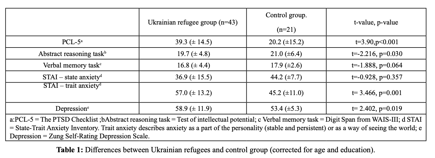
Table 1

Neuroimaging – Morphometry We compared gray matter volume in the UG with that of the CG. Both the right and left thalamus were significantly larger in the UG than in the CG. The largest changes were in the posterior and central part of the thalamus ([18–25 12] mm, 12,385 mm³, p(corrected) = 0.001); by contrast, the anterior part was not affected. Seed-based connectivity. We found stronger significant connectivity between the left thalamus and right frontal orbital gyrus and right insula ([38 23 -15] mm, 1 531 mm3) in the UG as compared to the CG (Figure 2). Seed connectivity from the right thalamus was not statistically significant.
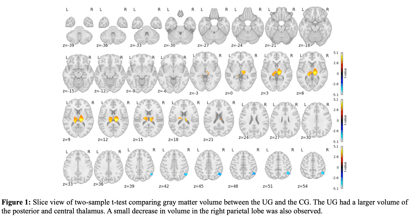
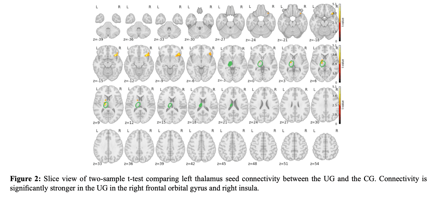
Figure 1Figure 2

Discussion

The psychological and neurobiological impact of acute and ongoing war stress was the target of this study. We questioned whether acute war stress impaired the psychological impact and the brain in healthy Ukrainian people who survived the war as compared to a Czech control group (CG) without stress. The study analysed the impact of recent acute and ongoing war stress on Ukrainian women who left Ukraine during the invasion that started in February 2022. Based on interview all of them experienced severe psychological trauma (bombing, hostility raids, occupied cities, etc.); leaving the country was also dangerous, with serious risks. The difference between acute and chronic trauma stress in Ukrainian refugees is difficult to distinguish. Acute trauma was certain during their presence in Ukraine during the war; however, even after they came to the Czech Republic, the war in their home country continues. The refugees received support from the Czech government and population; however, they all still experience stress as their family members and friends remained in the war-torn country. Some of their family members or friends were killed. In these stressed groups, there are people who lost a loved one (parent, spouse, close friend), who were in the war, who saw their homes burn, or who were under bombardment in the Ukrainian cities. Based on the interviews, it was evident that all Ukrainian refugees were stressed by the ongoing war. During the interviews, 74% of the UG said that the war had changed them by making them more emotionally unstable. The psychological tests showed that Ukrainian women experienced significantly more traumatic stress symptoms, depression, and anxiety than the control group. Higher levels of depression might accompany traumatic stress. The psychological data of the Ukrainian refugees were compared with data from a Czech control group who lived without war or extreme stress. The stress, anxiety and depression difference between Ukrainian refugees and Czech control group was significant. The first paper on Ukrainian stress was already published.

In the Magnetic Resonance Imaging (MRI) study, we performed structural imaging of the brain using voxel-based morphometry, functional data processing and seed-based connectivity. Our data show that acute and ongoing stress leads to the modification of the thalamus and of its connectivity to stress-related cortical areas. An enlargement of the bilateral posterior and central part of the thalamus was observed. A recent study noted abnormalities in the thalamus in patients with post-traumatic stress disorder (PTSD). The thalamic dysfunction in patients with PTSD may be related to the disorder’s psychopathology. The thalamus modulates activity in the limbic circuitry. The right thalamus connectivity was present but non-significant with limbic system; however, the left thalamus was a seed with significant connectivity with the right frontal orbital gyrus and insula. Both structures are clearly involved in stress. This connectivity confirms the involvement of the thalamus in stress-related circuitry. Connectivity with limbic circuitry was visible also in our Holocaust survivor research, 70 years after the extreme stress.

The mechanisms affecting the human brain through traumatic stress are not fully known. Posttraumatic stress disorder is not a single homogeneous disorder. It is based on various subcategories that may lead to different presentations and pathophysiological mechanisms. The stress may lead to autonomic nervous system dysfunction, altered concentrations of stress hormones, or noradrenergic hyper-reactivity linked with altered function of the hypothalamic-pituitary adrenal axis. The hypersensitivity of glucocorticoid receptors is modified by levels of cortisol. Evidence from previous studies suggest that traumatic stress affects the brain by altering the connections between neurons. Acute stress could promote the formation of new synapses or increase the existing synapses; this might explain the acute enlargement of the thalamus in our study. Psychological modifications have been reported in several other stress-related disorders. The involvement of the thalamus in anxiety has also been observed, in association with changes in the thalamic-limbic circuit. Several factors may activate this circuitry; however, it appears that the fear and stress that was present in the Ukrainian refugees had the inputs to the thalamus and limbic circuitry. Processing of the thalamic-limbic circuitry could contribute to multiple aspects of posttraumatic psychopathology.

In a previous study, we aimed to assess the lifelong impact of extreme stress on people who survived the Holocaust. We researched Holocaust survivors 70 to 75 years after the extreme stress of murder and war and their two generation descendants. We were able to show a neurobiological impact on the brain structures in survivors and MRI connectivity differences compared with control groups in the second and third generations. Areas with reduced gray matter correspond to the map of the impact of stress on the brain structure: bilateral insula, anterior cingulate, the sub-genu cingulate/orbitofrontal cortex, temporal pole, prefrontal cortex, and angular gyrus, however. The thalamus was not significantly involved. It is possible that in the future, the changes in the brains of Ukrainian refugees could be similar; however, the conditions are different as the Ukrainian refugees are being tested while suffering acute stress.

Acute war stress was studied also in Dutch soldiers before and after deployment to a combat zone in Afghanistan. It was found that combat stress increased amygdala and insula reactivity to biologically salient stimuli across the group of combat exposed individuals. PTSD was not present in this group. The amygdala increase due to a dangerous environment was normalized in a relatively safe setting within 1.5 years. Volume decreases of the amygdala and hippocampus were present in a Holocaust prenatal stress group, (children born by the end of the war), 70 to 75 years after the war. The volume modification present in the Dutch soldiers was not present in the Ukrainian refugees in our study, but the stress impact was probably different between soldiers and Ukrainian war victims. It is probable that different types of stress (Holocaust survivors, Holocaust prenatal stress, Dutch soldiers, Ukrainian refugees) may have variable impacts on brain and psychology.

Conclusion

Acute and ongoing war stress has a significant neurobiological impact on refugees from war-torn Ukraine. This study reveals significant impact on psychology, cerebral structures (thalamus) and connectivity as compared with Czech control group. The psychological tests in Ukrainian refugees showed significant stress, depression, and anxiety. The influence of ongoing stress is present in the posterior and central part of thalamus, connected with parts of the limbic circuitry. The lesion in the thalamus compared to Czech control group is significant. The modification of the limbic system observed in long-term stress is also involved in acute and ongoing Ukrainian stress. The data we obtained in Ukrainian refugees show the impact of war-related stress on psychological features as well as on cerebral structures in the thalamus and connectivity with stress-related cortical areas. The structural and connectivity data in this study reveal the characteristics of acute and ongoing stress in Ukrainian refugees.

Limitations

The sample of respondents included representatives of different regions of Ukraine and different social status and marital status; however, the entire UG was stressed in comparison to the control group and stress-related changes in MRI were present. Most men were not allowed to travel outside Ukraine at the time of our study; therefore, the study does not consider the gender aspect of stress. The interview and psychological testing were performed by two Ukrainian psychologists (Sofia Berezka and Tetyana Evmenova – co-authors) who were also war refugees. The psychological investigation was limited as we could use only psychological tests that were available in Ukrainian language. We performed approx. 45 to 60 minutes of MRI, up to 90–120 minutes of testing the psychological features, and 60 to 90 minutes of interviewing. The refugees provided subjective information about their psycho-emotional and physical state, life experiences, and experience of the war. This approach was subjective; however, all participants provided information about living through the stress of war. Sleep was not investigated; however, major sleep disturbances were not reported by the refugees.

Abstract

Purpose: We raised the question whether the neurobiological and psychological impact of war stress could be observed in Ukrainian refugees during an ongoing war. Ukrainian refugee women (UG, n=43) from the war-torn regions were investigated and were compared to Czech women control group without stress (CG, n=21). Men were not allowed to leave Ukraine due to the war conditions. Method: Psychological testing was performed using tests available in Ukrainian language: Posttraumatic stress disorder Check List; Zung Self- Rating Depression Scale; The State-Trait Anxiety Inventory; Test of Intellectual Potential and Digit Span - WAIS III. Magnetic Resonance Imaging included voxel-based morphometry, functional MR, seed-based connectivity. Finding: UG scored significantly higher than CG in stress (p<0.001), depression (p=0.019) and anxiety (p=0.001) and lower than CG in an abstract reasoning task (p<0.030). The groups did not differ significantly in memory and intellectual tasks. MRI showed an enlargement of the posterior and central parts of the thalamus that were related to the frontal orbital gyrus and insula indicating a connectivity with limbic system regions. Conclusion: The data show the impact of acute and ongoing war-related stress on psychological features as well as on the thalamus and connectivity of stress-related cortical areas. Acute and ongoing war stress had a significant neurobiological impact on refugees from war-torn Ukraine.

Summary

The conflict between Russia and Ukraine began with the annexation of Donbas, Luhansk, and Crimea in 2014, with full-scale war starting in February 2022. The fighting has been primarily in eastern and southern Ukraine, but bombings have occurred across most regions. This has led to increased civilian deaths and injuries, and a long-term impact of stress on the health of generations of Ukrainians is anticipated. Many Ukrainian citizens have become refugees. Interviews with over 43,000 refugees between May and November 2022 indicated that most are women, with high levels of education (46% holding university degrees or higher) and diverse professional backgrounds. A separate survey of 3,355 Ukrainian refugees in 36 European countries between March and April 2022 found that 92% were women. Of these, 60.4% left to protect their children, and 19% left with their parents. The survey also showed that 97% of respondents still had relatives and friends in Ukraine. Over 7.8 million Ukrainian refugees have been recorded across Europe, with 472,000 registered in the Czech Republic. While the Czech government and population provided support, refugees continued to experience stress due to the ongoing war and the fact that family members, often husbands who had joined the military, remained in Ukraine.

This study focused on the neurobiological and psychological effects of war-related stress in Ukrainian women. Men were generally not permitted to leave Ukraine due to wartime conditions. The research examined how acute war stress affects brain structure and connectivity, as observed through Magnetic Resonance Imaging (MRI), and mental health during the ongoing conflict. Researchers analyzed the connection between war exposure, psychological symptoms, and changes in brain structure and connectivity in Ukrainian refugees. Interviews explored the refugees' war experiences, including exposure to traumatic events. The study builds on previous research regarding the lifelong impact of extreme stress on Holocaust survivors, which revealed lasting neurobiological effects on brain structures and connectivity across three generations, 70–75 years after the war.

The primary question of this study was whether the neurobiological (MRI) and psychological effects of acute war-related stress could be observed in refugees during an ongoing conflict. The study investigated Ukrainian women who had left Ukraine during the war and were living as refugees in the Czech Republic.

Methods

Participants and procedure The study was conducted at the Central European Institute of Technology (CEITEC) Centre for Neuroscience at Masaryk University in Brno. Ukrainian participants were recruited through collaborations with Masaryk University, the assistance of Ukrainian psychologists working with the research team, and social networks.

Two groups were compared: Ukrainian refugees (UG) and Czech controls (CG). All participants were women. All but one participant in the UG were born in Ukraine and held Ukrainian citizenship. The Ukrainian participants were civilians who had lived in various war-affected regions of Ukraine (including Donetsk, Dnipro, Zaporizhzhia, Kyiv, Poltava, Sumy, Kharkiv, and Kherson regions) and had experienced psychological trauma from the war. They had sought shelter in basements during air raids, lived in occupied cities, or endured systematic bombing without access to water, electricity, or communication. All UG respondents reported personal war experiences during their interviews. Ukrainian refugees were exposed to war events for several weeks to two months before leaving Ukraine for the Czech Republic or other countries. In the UG group, 19% had experienced bombing, 26% shooting, 79% had seen dead or wounded individuals, and 88% had direct experience with fighting. Based on exposure to these traumatic events, it was presumed they might be psychologically and biologically affected. Interviews with all participants confirmed that all UG individuals were stressed.

The median age for the Ukrainian refugee group (UG) (n=43) was 38 years, with an age range of 18 to 60. Participants who did not meet quality standards were excluded. The Czech control group (CG) (n=21) consisted of gender-matched Czech civilian volunteers with a median age of 35 years and an age range of 21 to 58. Recruitment for the CG was advertised in the press and at Masaryk University. None of the CG participants had been involved in military action or faced direct life-threatening danger. The age difference between the UG and CG was not statistically significant.

Inclusion criteria for the UG included being female, residing in Ukraine during the war (from February 2022 onward), holding refugee status in the Czech Republic, and willingness to participate. Only healthy Ukrainian participants were included. They underwent interviews with Ukrainian psychologists before testing, and their health status was reviewed by a neurologist. The UG participants had no history of psychiatric illness and reported no acute health problems.

Exclusion criteria for both UG and CG included any severe brain impairment (e.g., brain injury, tumors, neurodegenerative diseases, cognitive decline, psychiatric drug treatment, or alcoholism). Contraindications for MRI included metal implants, pacemakers, and claustrophobia.

The research received approval from the ethics committee at Masaryk University, and all participants provided informed written consent. All methods followed relevant guidelines and regulations.

Researchers explored refugees' war experiences, including exposure to traumatic events. The study analyzed the relationship between war exposure, psychological symptoms, and brain structure and connectivity in Ukrainian refugees compared to Czech controls.

Psychological testing Interviews and psychological testing were conducted by two Ukrainian psychologists, Sofia Berezka and Tetyana Evmenova (co-authors), who were also war refugees. They had integrated into Czech society or Masaryk University and collaborated with an experienced Czech psychologist, Marek Preiss, who had contributed to previous studies on the impact of the Holocaust. A Ukrainian-language version of the tests was used.

  • Posttraumatic stress disorder (PTSD) Check List – Civilian Version (PCL-5) The PCL-5 is a 17-item self-report measure assessing DSM-5 symptoms of PTSD. It asks individuals to rate how much they have been bothered by problems related to stressful experiences in the past month, using a scale from 1 (not at all) to 5 (extremely). Higher scores indicate more severe PTSD symptoms. The internal consistency (Cronbach’s alpha) was 0.874. A raw score of 38 or higher indicates a potential PTSD diagnosis. A Ukrainian-language version was utilized.

  • Zung Self-Rating Depression Scale (SDS) The SDS is a short, self-administered questionnaire that quantifies depressive symptoms experienced in recent weeks. It consists of 20 items assessing four common characteristics of depression: mood, physical symptoms, other disturbances, and psychomotor activity. Each question is scored from 1 (none or a little of the time) to 4 (most or all of the time). Higher scores indicate greater depressive symptoms, with total scores ranging from 25–100.

  • The State-Trait Anxiety Inventory (STAI) The STAI measures both state anxiety (anxiety in response to specific situations) and trait anxiety (a stable, personality-based predisposition to anxiety). It contains 40 self-report items on a 4-point Likert scale. Higher scores indicate more severe anxiety symptoms. Total scores range from 20 to 80. The internal consistency coefficients for the scale ranged from 0.86 to 0.95, and test-retest reliability over a two-month period ranged from 0.65 to 0.75. A Ukrainian-language version of the inventory was used.

  • Abstract reasoning task – Test of intellectual potential (TIP) One of four subtests, the TIP comprises 29 picture puzzles, each with six possible solutions, only one of which is correct. This test provides a measure of abstract reasoning that is largely independent of language skills. Higher scores indicate higher levels of abstract reasoning. The total score is the sum of correct solutions completed within 12 minutes. The raw score was used.

  • Verbal memory task – Digit Span from WAIS-III The digit-span task assesses verbal short-term memory, working memory's capacity for number storage, and attention/concentration. Participants are asked to repeat sequences of digits of increasing length. Digit Span Forward measures simple attention, with most healthy individuals typically recalling seven plus or minus two digits. Both Digit Span Forward and Digit Span Backward tasks were used, and an overall score was calculated.

Interview Participants were asked about demographic information, close relationships, events leading up to the war, their war experiences in Ukraine, the war's impact, psychological difficulties, the most stressful traumatic events, their experiences immigrating to the Czech Republic, their health and medical conditions, and their overall assessment of their professional and personal lives. Interviews lasted approximately 60 to 90 minutes. The Ukrainian women interviewed had endured severe psycho-traumatic events, such as being under fire for extended periods or living in occupied cities that were later liberated. They often experienced communication blackouts, lacked food and water, witnessed their homes burn, or lost loved ones (parents, spouses, close friends). During the interviews, 74% of the women reported that the war had changed them, making them more emotionally unstable and anxious. Refugees continued to experience stress because many had relatives in Ukraine, including parents and husbands, who faced risks or were actively involved in military operations. The Ukrainian refugee group in this study experienced constant anxiety and fear regarding the potential death of a loved one at any moment.

Statistics Differences between groups in psychological parameters (PCL-5, abstract reasoning task, verbal memory task, STAI – state anxiety, STAI – trait anxiety, and SDS) were evaluated using a two-sample t-test. The influence of age and education was adjusted using a general linear model.

Neuroimaging MRI examinations were conducted using a Siemens Prisma 3T scanner with a 64-channel head coil.

  • Morphometry The MRI protocol for voxel-based morphometry included a 3D T1-weighted magnetization prepared rapid gradient echo (MPRAGE) sequence with specific parameters: TR = 2.3 s, TE = 2.33 ms, TI = 0.9 s, flip angle = 8, isometric voxel size 1 mm, FOV 224 × 224 mm, and 240 slices. Anatomical MRI data were analyzed using SPM12 and CAT12 toolbox within Matlab R2020a. Individual data were corrected for spatial inhomogeneity and denoised using the Non-Local Means (SANLM) filter. High-resolution data were then segmented into gray matter (GM) using the SPM Tissue Probability Map (TPM) and registered into common MNI space. Finally, spatially normalized and modulated GM maps were smoothed with a 6 mm FWHM isotropic Gaussian kernel. Group statistics for stress effects were calculated with a second-level model using SPM12. Modulated GM images were multiplicatively corrected for total intracranial volume and then analyzed. A two-sample t-test compared gray matter volume files between the UG and CG groups, with age included as a nuisance variable. Group-level results were evaluated with cluster-level inference for non-stationary conditions using family-wise error (FWE) correction at p (FWE) < 0.05, with an initial uncorrected threshold of p = 0.005.

  • Functional MRI data The functional protocol utilized a multi-echo multiband T2 echo-planar imaging sequence with TR = 0.85 s, TEs = (15, 36, and 58) ms, flip angle = 25, isometric voxel size 2.5 mm, FOV 200 × 200 mm, 60 slices, and 700 scans. Data processing involved SPM12 and custom Matlab scripts. Preprocessing steps included realignment (motion correction), CNR-weighted combination of individual echoes, RETROICOR procedure for physiological artifact suppression (resting state only), co-registration of anatomical scans to BOLD data, spatial normalization using an MNI template, and spatial smoothing with a 6 mm FWHM Gaussian kernel. Subsequently, very low frequencies were removed from BOLD data using a high-pass filter with a 128 s cut-off. Other nuisance effects, specifically white matter and CSF signals, and 24 movement parameters from the realign procedure, were regressed out of the BOLD data.

  • Seed-based connectivity In resting-state imaging, seed-based connectivity maps from the left and right thalamus (chosen due to morphological data indicating thalamic involvement) were analyzed. Individual connectivity was calculated based on correlations between the ROI signal (first principal component) and all other brain voxels. Group statistics for stress effects were calculated with a second-level model using SPM12. A two-sample t-test compared the individual connectivity maps of the UG and CG, with age included as a nuisance variable. Group-level results were evaluated with cluster-level inference for non-stationary conditions using family-wise error (FWE) correction at p < 0.05, with an initial uncorrected threshold of p = 0.005.

  • Data quality Data from all participants underwent manual checks for artifacts and pathology. Participants who did not meet quality standards were excluded. Additionally, data were automatically checked for spatial abnormalities (e.g., dropouts, normalization failures) using the Mask Explorer tool and for excessive movement using framewise displacement (FD), with a criterion of FD < 1.5mm in any scan. Scans exceeding FD > 1.5mm were excluded from the analysis.

Results

Psychological testing Psychological testing, conducted in Ukrainian by two Ukrainian psychologists on the research team, showed that the Ukrainian refugee group (UG) scored significantly higher than the Czech control group (CG) on the PCL-5 (t=3.907, df=61, p<0.001), STAI – trait anxiety (t=3.466, df=61, p=0.001), and depression (t=2.402, df=61, p=0.019). The UG scored significantly lower than the CG on an abstract reasoning task (t=-2.216, df=61, p=0.030). No significant differences were found between the groups for verbal memory tasks (t=-1.888, df=61, p=0.064) or STAI – state anxiety (t=-0.928, df=61, p=0.357).

Neuroimaging – Morphometry Comparison of gray matter volume revealed that both the right and left thalamus were significantly larger in the Ukrainian refugee group (UG) compared to the Czech control group (CG). The most pronounced changes were observed in the posterior and central parts of the thalamus ([18–25 12] mm, 12,385 mm³, p(corrected) = 0.001), while the anterior part remained unaffected. Seed-based connectivity analysis showed stronger significant connectivity between the left thalamus and the right frontal orbital gyrus and right insula ([38 23 -15] mm, 1,531 mm3) in the UG compared to the CG (Figure 2). Connectivity from the right thalamus did not show statistically significant differences.

Discussion

This study aimed to understand the psychological and neurobiological effects of acute and ongoing war stress. It investigated whether recent and current war stress impacted the mental health and brains of healthy Ukrainian women who survived the war, comparing them to a non-stressed Czech control group. All Ukrainian participants reported severe psychological trauma, including bombings, hostile raids, and living in occupied cities. The act of leaving the country was also risky. It is challenging to differentiate between acute and chronic traumatic stress in Ukrainian refugees. Acute trauma was certainly experienced during their time in Ukraine, but even after reaching the Czech Republic, the war in their home country continues. While refugees received support in the Czech Republic, they remained stressed because family members and friends were still in the war-affected country, with some having been killed. In these stressed groups, individuals had lost loved ones, experienced combat, witnessed their homes burn, or endured bombardment. Interviews clearly indicated that all Ukrainian refugees were stressed by the ongoing war, with 74% reporting that the war made them more emotionally unstable. Psychological tests confirmed that Ukrainian women experienced significantly more symptoms of traumatic stress, depression, and anxiety compared to the control group. Higher levels of depression often accompany traumatic stress. The significant differences in stress, anxiety, and depression between Ukrainian refugees and the Czech control group were evident. Prior research on Ukrainian stress has also been published.

The Magnetic Resonance Imaging (MRI) study involved structural imaging using voxel-based morphometry, functional data processing, and seed-based connectivity. The findings indicate that acute and ongoing stress leads to changes in the thalamus and its connections to stress-related areas of the brain's outer layer. An enlargement of the posterior and central parts of both the left and right thalamus was observed. Recent studies have also noted thalamic abnormalities in individuals with post-traumatic stress disorder (PTSD), suggesting that thalamic dysfunction may contribute to the disorder's symptoms. The thalamus plays a role in modulating activity within the limbic system, a network involved in emotion and memory. While right thalamus connectivity with the limbic system was observed but not statistically significant, the left thalamus showed significant connectivity with the right frontal orbital gyrus and insula. Both of these brain structures are clearly implicated in stress responses. This connectivity confirms the thalamus's involvement in stress-related brain networks. Similar connectivity within the limbic system was observed in our research on Holocaust survivors, even 70 years after their extreme stress.

The precise mechanisms by which traumatic stress affects the human brain are not fully understood. Post-traumatic stress disorder is not a uniform condition; it encompasses various subcategories that can lead to different symptoms and underlying biological processes. Stress can cause dysfunction in the autonomic nervous system, alter stress hormone levels, or lead to overactivity in the noradrenergic system, linked to changes in the hypothalamic-pituitary-adrenal axis. The sensitivity of glucocorticoid receptors is influenced by cortisol levels. Previous research suggests that traumatic stress alters the connections between brain cells. Acute stress might promote the formation of new connections or strengthen existing ones, which could explain the observed acute enlargement of the thalamus in this study. Psychological changes have also been documented in several other stress-related disorders. Thalamic involvement in anxiety has been noted, often in conjunction with changes in the thalamic-limbic circuit. Various factors can activate this circuit, and it appears that the fear and stress experienced by Ukrainian refugees provided input to the thalamus and limbic circuitry. The processing within the thalamic-limbic circuit may contribute to multiple aspects of post-traumatic psychological symptoms.

A previous study assessed the lifelong impact of extreme stress on Holocaust survivors. This research examined survivors 70 to 75 years after their extreme experiences of murder and war, as well as their second- and third-generation descendants. It demonstrated a neurobiological impact on brain structures in survivors and MRI connectivity differences compared to control groups in the second and third generations. Areas with reduced gray matter corresponded to the known effects of stress on brain structure: bilateral insula, anterior cingulate, sub-genu cingulate/orbitofrontal cortex, temporal pole, prefrontal cortex, and angular gyrus. However, the thalamus was not significantly affected in that study. It is possible that the brain changes in Ukrainian refugees could eventually become similar; however, the current conditions differ as Ukrainian refugees are being studied while experiencing acute stress.

Acute war stress has also been studied in Dutch soldiers before and after deployment to a combat zone in Afghanistan. This research found that combat stress increased the reactivity of the amygdala and insula to biologically relevant stimuli across the group of combat-exposed individuals. PTSD was not present in this group. The increased amygdala activity, which resulted from a dangerous environment, returned to normal within 1.5 years in a relatively safe setting. In contrast, volume reductions in the amygdala and hippocampus were observed in a group exposed to prenatal stress during the Holocaust (children born at the end of the war), 70 to 75 years later. The specific volume modifications seen in the Dutch soldiers were not observed in the Ukrainian refugees in this study. However, the impact of stress likely differs between soldiers and Ukrainian war victims. It is probable that different types of stress (e.g., experienced by Holocaust survivors, those with prenatal Holocaust stress, Dutch soldiers, and Ukrainian refugees) may have varying effects on the brain and psychology.

Conclusion

Acute and ongoing war stress significantly impacts refugees from war-torn Ukraine both psychologically and neurobiologically. This study reveals substantial effects on mental health, brain structures (specifically the thalamus), and connectivity when compared with a Czech control group. Psychological tests indicated significant levels of stress, depression, and anxiety in Ukrainian refugees. The influence of ongoing stress is evident in the posterior and central parts of the thalamus, which are connected to components of the limbic system. The observed changes in the thalamus, compared to the Czech control group, are significant. Modifications to the limbic system, typically seen in long-term stress, are also involved in the acute and ongoing stress experienced by Ukrainians. The data collected from Ukrainian refugees demonstrate the impact of war-related stress on psychological well-being and on cerebral structures within the thalamus, as well as its connections with stress-related cortical areas. The structural and connectivity data in this study characterize the nature of acute and ongoing stress in Ukrainian refugees.

Limitations

The study's sample included respondents from various regions of Ukraine, with differing social and marital statuses. However, the entire Ukrainian refugee group (UG) experienced stress compared to the control group, and stress-related changes were observed in MRI scans. Most men were not permitted to leave Ukraine at the time of the study, meaning the study does not address gender-specific aspects of stress. The interviews and psychological testing were conducted by two Ukrainian psychologists (Sofia Berezka and Tetyana Evmenova – co-authors) who were also war refugees. The psychological assessment was limited to tests available in the Ukrainian language. The research involved approximately 45 to 60 minutes of MRI, up to 90–120 minutes of psychological testing, and 60 to 90 minutes of interviewing. While refugees provided subjective information about their psycho-emotional and physical state, life experiences, and war experiences, and this approach was subjective, all participants confirmed experiencing war stress. Sleep was not specifically investigated, but refugees did not report major sleep disturbances.

Abstract

Purpose: We raised the question whether the neurobiological and psychological impact of war stress could be observed in Ukrainian refugees during an ongoing war. Ukrainian refugee women (UG, n=43) from the war-torn regions were investigated and were compared to Czech women control group without stress (CG, n=21). Men were not allowed to leave Ukraine due to the war conditions. Method: Psychological testing was performed using tests available in Ukrainian language: Posttraumatic stress disorder Check List; Zung Self- Rating Depression Scale; The State-Trait Anxiety Inventory; Test of Intellectual Potential and Digit Span - WAIS III. Magnetic Resonance Imaging included voxel-based morphometry, functional MR, seed-based connectivity. Finding: UG scored significantly higher than CG in stress (p<0.001), depression (p=0.019) and anxiety (p=0.001) and lower than CG in an abstract reasoning task (p<0.030). The groups did not differ significantly in memory and intellectual tasks. MRI showed an enlargement of the posterior and central parts of the thalamus that were related to the frontal orbital gyrus and insula indicating a connectivity with limbic system regions. Conclusion: The data show the impact of acute and ongoing war-related stress on psychological features as well as on the thalamus and connectivity of stress-related cortical areas. Acute and ongoing war stress had a significant neurobiological impact on refugees from war-torn Ukraine.

Summary

The conflict between Russia and Ukraine began with annexations in 2014, with full-scale war starting in February 2022. Fighting has primarily occurred in eastern and southern Ukraine, though bombings have been widespread. Civilian casualties and injuries have increased since the 2022 invasion, and long-term stress is expected to impact the health of Ukrainians across generations. Many Ukrainian citizens have become refugees, with data from 2022 showing most are women, often with higher education and diverse professional backgrounds. A pan-European survey of Ukrainian refugees in 2022 indicated that 92% were women, with 60.4% leaving to protect their children and 19% with their parents. The survey also found that 97% of respondents had relatives and friends remaining in Ukraine. Over 7.8 million Ukrainian refugees were recorded across Europe. Refugees in the Czech Republic, numbering 472,000, received assistance but experienced ongoing stress due to the war and separation from family members, many of whom were engaged in military service.

The study focused on the neurobiological and psychological impact of war-related stress on Ukrainian women. The investigation included only women because most men were not permitted to leave Ukraine due to wartime conditions. Researchers explored how acute war stress affected brain structure and connectivity, using Magnetic Resonance Imaging (MRI), and mental health during the ongoing conflict. The study analyzed the relationship between war exposure, psychological symptoms, brain changes, and brain connectivity in Ukrainian refugees. Interviews gathered information about refugees' war experiences, including exposure to traumatic events. This research builds on previous studies regarding the long-term impact of extreme stress, such as that experienced by Holocaust survivors, which showed lasting neurobiological effects on brain structures and connectivity across generations, 70–75 years after the event.

This study aimed to determine if the neurobiological (MRI) and psychological effects of acute war-related stress could be observed in refugees while a war was still ongoing. The research involved Ukrainian women who had left Ukraine during the war and were living as refugees in the Czech Republic.

Methods

Participants and procedure

The study took place at the Central European Institute of Technology (CEITEC) and the Centre for Neuroscience at Masaryk University in Brno. Ukrainian participants were recruited through Masaryk University, with assistance from Ukrainian psychologists on the research team, and via social networks.

The study compared two groups: Ukrainian refugees (UG) and Czech controls (CG). All participants were women. All but one person in the UG was born in Ukraine and held Ukrainian citizenship. The Ukrainian participants were civilians who had lived in various war-affected regions of Ukraine (including Donetsk, Dnipro, Zaporizhzhia, Kyiv, Poltava, Sumy, Kharkiv, and Kherson) and had experienced psychological trauma from the war. Their experiences included hiding in basements during air raids, living in occupied cities, or enduring systematic bombing without access to water, electricity, or communication. All UG participants reported a personal experience of the war in Ukraine during interviews. Ukrainian refugees had been exposed to war events for periods ranging from several weeks to two months before leaving Ukraine for the Czech Republic or other countries. In the UG group, 19% had experienced bombing, 26% had experienced shooting, 79% had witnessed dead or wounded people, and 88% had direct experience of fighting. Given these traumatic exposures, it was assumed that they might be psychologically and biologically affected. Interviews confirmed that all UG participants were under stress.

The median age for the Ukrainian refugee group (UG) (n=43) was 38 years, with ages ranging from 18 to 60. Participants who did not meet quality standards were excluded. The Czech control group (CG) (n=21) consisted of age and gender-matched Czech civilian volunteers, with a median age of 35 years and ages ranging from 21 to 58. Recruitment for the CG was advertised in the press and at Masaryk University. None of the CG participants had engaged in military action or faced direct life-threatening danger. The age difference between the UG and CG was not statistically significant.

Inclusion criteria for the UG included being female, residing in Ukraine during the war (from February 2022 onward), being a refugee with refugee status in the Czech Republic, and willingness to participate. Only healthy Ukrainian participants were accepted, with their health status reviewed by a neurologist after interviews by Ukrainian psychologists. The UG reported no psychiatric conditions or acute health problems.

Exclusion criteria for both UG and CG included any severe brain impairment, such as injury, tumors, neurodegenerative diseases, cognitive decline, psychiatric medication, or alcoholism. Contraindications for MRI included metal implants, pacemakers, and claustrophobia.

The research received approval from the ethics committee at Masaryk University, and all participants provided informed written consent. All procedures adhered to relevant guidelines and regulations.

Researchers explored refugees' war experiences, including exposure to traumatic events. The study analyzed the relationship between war exposure, psychological symptoms, brain structure, and connectivity in Ukrainian refugees compared to Czech controls.

Psychological testing

Interviews and psychological tests were conducted by two Ukrainian psychologists, Sofia Berezka and Tetyana Evmenova, who were also war refugees. They had integrated into Czech society or Masaryk University and collaborated with an experienced Czech psychologist, Marek Preiss, who had participated in previous studies on the impact of the Holocaust. A Ukrainian-language version of the tests was used.

  • Posttraumatic stress disorder (PTSD) Check List – Civilian Version (PCL-5) The PCL-5 is a 17-item self-report questionnaire assessing DSM-5 PTSD symptoms. Respondents rate how much specific problems have bothered them in the past month on a 1-to-5 scale. Higher scores indicate more severe PTSD symptoms. The test shows good internal consistency (Cronbach’s alpha ≥ 0.874), with a cutoff score of 38. A Ukrainian version was used.

  • Zung Self-Rating Depression Scale (SDS) The SDS is a 20-item self-report survey used to quantify depression over the previous weeks. It covers four common characteristics of depression: pervasive effect, physiological equivalents, other disturbances, and psychomotor activities. Questions are scored on a 1-to-4 scale. Higher scores indicate greater depressive symptoms. Total scores range from 25–100. Previous studies have reported an alpha coefficient of 0.82.

  • The State-Trait Anxiety Inventory (STAI) The STAI measures both state anxiety (temporary anxiety due to specific situations) and trait anxiety (anxiety as a stable personality characteristic). It consists of 40 self-report items on a 4-point Likert scale. Higher scores indicate greater anxiety. Total scores range from 20 to 80. Internal consistency coefficients range from 0.86 to 0.95, and test-retest reliability ranges from 0.65 to 0.75 over two months. A Ukrainian version was used.

  • Abstract reasoning task – Test of intellectual potential (TIP) One of four subtests, the TIP contains 29 picture puzzles, each with six possible solutions, only one of which is correct. This test provides a relatively language-independent measure of abstract reasoning. A higher score indicates better abstract reasoning. The total score is the sum of correct solutions within 12 minutes.

  • Verbal memory task – Digit Span from WAIS-III The Digit Span task assesses verbal short-term memory, working memory's numerical storage capacity, and attention/concentration. Participants repeat increasingly long series of digits. Digit Span Forward measures simple attention. Both Digit Span Forward and Digit Span Backward tasks were used to calculate an overall score.

Interview

Participants provided demographic data, information about close relationships, pre-war events, war experiences in Ukraine, the war's effects, psychological difficulties, the most stressful traumatic events, the experience of emigrating to the Czech Republic, health and medical conditions, and an overall assessment of their professional and personal lives. Interviews lasted approximately 60 to 90 minutes. Ukrainian women in the study had experienced severe psycho-traumatic events, such as being under fire for days or weeks, living in occupied cities (later liberated by the Ukrainian army), being cut off from communication, lacking food and water, witnessing homes burn, or losing loved ones. During interviews, 74% of the women reported that the war had changed them, making them more emotionally unstable and anxious. Refugees remained under stress because many still had relatives, including parents and husbands, in Ukraine, who were either at risk or actively involved in military operations. The Ukrainian refugee group in this study experienced constant anxiety and fear that a loved one could die at any moment.

Statistics

Differences between groups in psychological parameters (PCL-5, abstract reasoning task, verbal memory task, STAI – state anxiety, STAI – trait anxiety, and SDS) were evaluated using a two-sample t-test. The effects of age and education were adjusted using a general linear model.

Neuroimaging

MRI examinations were conducted using a Siemens Prisma 3T scanner with a 64-channel head coil.

  • Morphometry The MRI protocol for voxel-based morphometry included a 3D T1-weighted MPRAGE sequence with specific parameters (TR = 2.3 s, TE = 2.33 ms, TI = 0.9 s, flip angle = 8, isometric voxel size 1 mm). Anatomical MRI data were analyzed using SPM12 and CAT12 toolbox in Matlab R2020a. Individual data underwent intensity normalization, denoising with SANLM filter, segmentation into gray matter using SPM Tissue Probability Map, and registration into MNI space using IXI555_MNI152_GS template. Spatially normalized and modulated gray matter maps were smoothed with a 6 mm FWHM Gaussian kernel. Group statistics for stress effects were calculated using a second-level model in SPM12. Modulated gray matter images were adjusted for total intracranial volume and then analyzed. A two-sample t-test compared gray matter volume between UG and CG, with age included as a nuisance variable. Group-level results used cluster-level inference with family-wise error (FWE) correction at p < 0.05, and an initial cutoff of p (uncorrected) = 0.005.

  • Functional MRI data The functional protocol used a multi-echo multiband T2 echo-planar imaging sequence with specific parameters (TR = 0.85 s, TEs = 15, 36, and 58 ms, flip angle = 25, isometric voxel size 2.5 mm). Data processing involved SPM12 and custom Matlab scripts. Preprocessing included realignment, CNR-weighted echo combination, RETROICOR for physiological artifact suppression, co-registration of anatomical scans to BOLD data, spatial normalization, and spatial smoothing. Subsequently, very low frequencies were removed from BOLD data, and nuisance effects (white matter, CSF signals, and movement parameters) were regressed out.

  • Seed-based connectivity In resting-state imaging, seed-based connectivity maps from the left and right thalamus (chosen due to morphological data showing thalamic impact) were analyzed. Individual connectivity was calculated based on correlations between the ROI signal and all other brain voxels. Group statistics for stress effects were calculated using a second-level model in SPM12. A two-sample t-test compared individual connectivity maps between UG and CG, with age as a nuisance variable. Group-level results used cluster-level inference with FWE correction at p < 0.05, and an initial cutoff of p (uncorrected) = 0.005.

  • Data quality All participant data were manually checked for artifacts and pathology. Participants with data not meeting quality standards were excluded. Data were also automatically checked for spatial abnormalities (e.g., dropouts, normalization failures) using Mask Explorer and for excessive movement using framewise displacement (FD), with a criterion of FD < 1.5mm. Scans with FD > 1.5mm were excluded.

Results

Psychological testing

Psychological testing was conducted in Ukrainian by two Ukrainian psychologists on the research team. The Ukrainian refugee group (UG) scored significantly higher than the Czech control group (CG) in measures of traumatic stress symptoms (PCL-5: t=3.907, df=61, p<0.001), trait anxiety (STAI – trait anxiety: t=3.466, df=61, p=0.001), and depression (SDS: t=2.402, df=61, p=0.019). Conversely, the UG scored significantly lower than the CG in an abstract reasoning task (t=-2.216, df=61, p=0.030). No significant differences were found between the groups for verbal memory (t=-1.888, df=61, p=0.064) or state anxiety (STAI – state anxiety: t=-0.928, df=61, p=0.357).

Neuroimaging – Morphometry

When comparing gray matter volume between the Ukrainian refugee group (UG) and the Czech control group (CG), both the right and left thalamus were significantly larger in the UG. The most notable changes occurred in the posterior and central parts of the thalamus ([18–25 12] mm, 12,385 mm³, p(corrected) = 0.001), while the anterior part showed no significant effect. In terms of seed-based connectivity, a stronger significant connection was observed between the left thalamus and the right frontal orbital gyrus and right insula ([38 23 -15] mm, 1 531 mm3) in the UG compared to the CG. Connectivity from the right thalamus did not show a statistically significant difference.

Discussion

This study aimed to understand the psychological and neurobiological impact of acute and ongoing war stress. It investigated whether acute war stress affects the psychological state and brain of healthy Ukrainian individuals who survived the war, comparing them to a non-stressed Czech control group. The research focused on the impact of recent and ongoing war stress on Ukrainian women who left Ukraine after the February 2022 invasion. Interviews confirmed that all participants experienced severe psychological trauma, including bombing, hostilities, and living in occupied cities, and that their departure from the country was also risky. It is challenging to distinguish between acute and chronic trauma stress in Ukrainian refugees. Acute trauma was certain during their time in Ukraine, but even after reaching the Czech Republic, the war continued in their home country. While refugees received support in the Czech Republic, they remained stressed as family and friends stayed in the war zone, with some having been killed. Many in these stressed groups lost loved ones, experienced combat, saw their homes destroyed, or endured bombardments. Interviews clearly showed that all Ukrainian refugees were stressed by the ongoing war. During interviews, 74% of the Ukrainian group stated that the war had made them more emotionally unstable. Psychological tests revealed that Ukrainian women experienced significantly more traumatic stress symptoms, depression, and anxiety compared to the control group. Higher levels of depression often accompany traumatic stress. The significant differences in stress, anxiety, and depression between Ukrainian refugees and the Czech control group, who lived without war or extreme stress, underscore the profound impact of the conflict.

The Magnetic Resonance Imaging (MRI) study involved structural brain imaging using voxel-based morphometry, functional data processing, and seed-based connectivity. The findings indicate that acute and ongoing stress leads to changes in the thalamus and its connections to stress-related brain regions. An enlargement of the bilateral posterior and central parts of the thalamus was observed. Previous research has noted thalamic abnormalities in individuals with post-traumatic stress disorder (PTSD), suggesting that thalamic dysfunction may contribute to the disorder's symptoms. The thalamus plays a role in regulating activity within the limbic system. While right thalamus connectivity with the limbic system was observed but not statistically significant, the left thalamus showed significant connectivity with the right frontal orbital gyrus and insula. Both of these structures are known to be involved in stress responses. This connectivity confirms the thalamus's role in stress-related brain circuits. Similar connectivity with the limbic system was also observed in our research on Holocaust survivors, 70 years after their extreme stress.

The exact ways traumatic stress affects the human brain are not fully understood. Posttraumatic stress disorder is not a uniform condition; various subtypes may lead to different symptoms and underlying biological mechanisms. Stress can cause problems with the autonomic nervous system, alter levels of stress hormones, or lead to overactivity of noradrenaline, linked to changes in the hypothalamic-pituitary adrenal axis. The sensitivity of glucocorticoid receptors is influenced by cortisol levels. Evidence from past studies suggests that traumatic stress modifies connections between neurons. Acute stress might promote the formation of new synapses or strengthen existing ones, which could explain the acute enlargement of the thalamus observed in this study. Psychological changes have been reported in several other stress-related disorders. Thalamic involvement in anxiety has also been noted, linked to changes in the thalamic-limbic circuit. Various factors can activate this circuit, but it appears that the fear and stress experienced by Ukrainian refugees significantly impacted the thalamus and limbic circuitry. The processing within this thalamic-limbic circuit may contribute to multiple aspects of posttraumatic psychological issues.

In a previous study, researchers examined the lifelong impact of extreme stress on Holocaust survivors, 70 to 75 years after the war and murders, and on their two subsequent generations. That research demonstrated a neurobiological impact on brain structures in survivors and MRI connectivity differences compared to control groups in the second and third generations. Areas with reduced gray matter aligned with known effects of stress on brain structure, including the bilateral insula, anterior cingulate, sub-genu cingulate/orbitofrontal cortex, temporal pole, prefrontal cortex, and angular gyrus. However, the thalamus was not significantly affected in that study. It is possible that future brain changes in Ukrainian refugees could be similar, but the current conditions differ as Ukrainian refugees are being studied while experiencing acute stress.

Acute war stress was also investigated in Dutch soldiers before and after deployment to a combat zone in Afghanistan. This study found that combat stress increased the reactivity of the amygdala and insula to emotionally significant stimuli across the group of combat-exposed individuals. PTSD was not present in this group, and the increased amygdala activity, induced by a dangerous environment, normalized within 1.5 years in a relatively safe setting. In contrast, decreases in the volume of the amygdala and hippocampus were observed in a group exposed to prenatal Holocaust stress (children born at the end of the war), 70 to 75 years later. The specific volume modifications found in the Dutch soldiers were not present in the Ukrainian refugees in this study, likely because the stress experienced by soldiers and Ukrainian war victims differed. It is probable that various types of stress (e.g., experienced by Holocaust survivors, those exposed to prenatal Holocaust stress, Dutch soldiers, and Ukrainian refugees) may have different effects on the brain and psychological well-being.

Conclusion

Acute and ongoing war stress significantly impacts refugees from war-torn Ukraine both psychologically and neurobiologically. This study reveals substantial effects on psychological well-being, cerebral structures (specifically the thalamus), and connectivity when compared to a Czech control group. Psychological tests showed that Ukrainian refugees experienced significant stress, depression, and anxiety. The ongoing stress affects the posterior and central parts of the thalamus, which are connected to parts of the limbic system. The observed changes in the thalamus, compared to the Czech control group, are significant. The modifications to the limbic system, typically seen in long-term stress, are also involved in the acute and ongoing stress experienced by Ukrainian refugees. The data from Ukrainian refugees demonstrate the impact of war-related stress on psychological features and on brain structures in the thalamus, as well as its connectivity with stress-related cortical areas. The structural and connectivity data in this study characterize the effects of acute and ongoing stress in Ukrainian refugees.

Limitations

The study's participants represented various regions of Ukraine, social statuses, and marital statuses. However, the entire Ukrainian refugee group was stressed compared to the control group, and stress-related changes in MRI were evident. Most men were prohibited from leaving Ukraine during the study period, meaning the research could not explore gender-specific aspects of stress. Interviews and psychological testing were conducted by two Ukrainian psychologists who were also war refugees. The psychological assessment was limited to tests available in the Ukrainian language. The study involved approximately 45 to 60 minutes of MRI, up to 90–120 minutes for psychological testing, and 60 to 90 minutes for interviews. Refugees provided subjective information about their psycho-emotional and physical state, life experiences, and war experiences. While this approach was subjective, all participants confirmed living through war-related stress. Sleep patterns were not specifically investigated, though refugees did not report major sleep disturbances.

Abstract

Purpose: We raised the question whether the neurobiological and psychological impact of war stress could be observed in Ukrainian refugees during an ongoing war. Ukrainian refugee women (UG, n=43) from the war-torn regions were investigated and were compared to Czech women control group without stress (CG, n=21). Men were not allowed to leave Ukraine due to the war conditions. Method: Psychological testing was performed using tests available in Ukrainian language: Posttraumatic stress disorder Check List; Zung Self- Rating Depression Scale; The State-Trait Anxiety Inventory; Test of Intellectual Potential and Digit Span - WAIS III. Magnetic Resonance Imaging included voxel-based morphometry, functional MR, seed-based connectivity. Finding: UG scored significantly higher than CG in stress (p<0.001), depression (p=0.019) and anxiety (p=0.001) and lower than CG in an abstract reasoning task (p<0.030). The groups did not differ significantly in memory and intellectual tasks. MRI showed an enlargement of the posterior and central parts of the thalamus that were related to the frontal orbital gyrus and insula indicating a connectivity with limbic system regions. Conclusion: The data show the impact of acute and ongoing war-related stress on psychological features as well as on the thalamus and connectivity of stress-related cortical areas. Acute and ongoing war stress had a significant neurobiological impact on refugees from war-torn Ukraine.

Summary

The conflict between Russia and Ukraine began with the annexation of Donbas, Luhansk, and Crimea in 2014, with the full-scale war starting in February 2022. Much of the fighting has taken place in eastern and southern Ukraine, but bombings have occurred across most regions. Since the invasion in February 2022, there has been a rise in civilian deaths and injuries, and experts anticipate a lasting impact of stress on the health of generations of Ukrainians. Many Ukrainian citizens have left the country.

A study involving 43,571 Ukrainian refugees between May and November 2022 found that most were women with higher education and diverse professional backgrounds. Another survey of 3,355 refugees in 36 European countries, conducted between March and April 2022, also showed that 92% of refugees were women. Over 60% left to protect their children, and 19% left with their parents. For 97% of respondents, family and friends remained in Ukraine. More than 7.8 million Ukrainian refugees have been recorded across Europe, with 472,000 registered in the Czech Republic. While refugees received help, they continued to experience stress due to the ongoing war, family members remaining in Ukraine, and often, husbands enlisting in the military.

The current research focused on the neurobiological and psychological effects of war-related stress in Ukrainian women. Researchers specifically studied women because most men were not permitted to leave Ukraine due to wartime conditions. The study investigated how acute war stress affects brain structure and connectivity (using Magnetic Resonance Imaging, or MRI) and mental health during the ongoing conflict. Researchers analyzed the link between war exposure, psychological symptoms, and changes in brain structure and connections among Ukrainian refugees. Interviews explored the refugees’ experiences with the war, including exposure to traumatic events. This study builds on previous research regarding the lifelong impact of extreme stress on Holocaust survivors, where psychological tests and MRI revealed lasting neurobiological effects on brain structures and connectivity across three generations, 70–75 years after the war.

This study aimed to determine if neurobiological (MRI) and psychological impacts of acute war-related stress could be observed in refugees during an ongoing war. Researchers studied Ukrainian women who left Ukraine during the conflict and were living in the Czech Republic as war refugees.

Methods

Participants and Procedure

The study took place at the Central European Institute of Technology (CEITEC), Centre for Neuroscience at Masaryk University in Brno. Ukrainian participants were recruited through collaborations with Masaryk University, Ukrainian psychologists working with the research team, and social networks.

Two groups were compared: Ukrainian refugees (UG) and Czech controls (CG). All participants were women. All but one person in the Ukrainian group was born in Ukraine and held Ukrainian citizenship. These Ukrainian participants were civilians from various war-affected regions of Ukraine (including Donetsk, Dnipro, Zaporizhzhia, Kyiv, Poltava, Sumy, Kharkiv, and Kherson regions) who had experienced psychological trauma from the war. They had hidden in basements during air raids, lived in occupied cities, or endured systematic bombing without access to water, electricity, or communication. All Ukrainian participants reported personal war experiences during interviews. The Ukrainian refugees experienced war events for several weeks to two months before leaving Ukraine and arriving in the Czech Republic or other countries. In the Ukrainian group, 19% had experienced bombing, 26% shooting, 79% had seen dead or wounded people, and 88% had direct experience of fighting. Based on exposure to these traumatic events, researchers presumed they might be psychologically and biologically affected. Interviews with all participants confirmed that all in the Ukrainian group were stressed.

The median age of the Ukrainian refugee group (UG) (n=43) was 38 years, ranging from 18 to 60. Participants who did not meet quality standards were excluded. The Czech control group (CG) participants (n=21) were gender-matched Czech civilian volunteers with a median age of 35 years, ranging from 21 to 58. Recruitment for the Czech group was advertised in the press and at Masaryk University. None of the Czech participants had been involved in military action or faced direct life-threatening danger. The age difference between the Ukrainian and Czech groups was not significant.

Inclusion criteria for the Ukrainian group were being female, living in Ukraine during the war (from February 2022 onward), being a refugee, having refugee status in the Czech Republic, and willingness to participate. Only healthy Ukrainian participants were accepted. Ukrainian psychologists interviewed them before testing, and a neurologist considered their health status. The Ukrainian group had no psychiatric illnesses and reported no acute health problems.

Exclusion criteria for both groups included any severe brain impairment (such as brain injury, tumors, neurodegenerative diseases, cognitive decline, psychiatric drug treatment, or alcoholism). Contraindications for MRI included metal implants, pacemakers, and claustrophobia.

The research was approved by the ethics committee at Masaryk University, and all participants provided informed written consent. All methods followed relevant guidelines and regulations.

Researchers explored refugees’ wartime experiences and exposure to traumatic events. They analyzed the relationship between war exposure, psychological symptoms, brain structure (morphometry), and connectivity in Ukrainian refugees compared to Czech controls.

Psychological Testing

Interviews and psychological tests were conducted in Ukrainian by two Ukrainian psychologists (Sofia Berezka and Tetyana Evmenova), who were also war refugees. They were integrated into Czech society or Masaryk University and coordinated with an experienced Czech psychologist (Marek Preiss), who had participated in previous studies on the impact of the Holocaust. A Ukrainian-language version of the tests was used.

  • Posttraumatic Stress Disorder (PTSD) Check List – Civilian Version (PCL-5) The PCL-5 is a 17-item self-report measure of DSM-5 PTSD symptoms. It asks respondents to rate how much they have been bothered by problems related to stressful experiences in the past month, using a scale of 1 (not at all) to 5 (extremely). A higher score indicates more PTSD symptoms. A score of 38 or higher suggests significant symptoms. A Ukrainian-language version was used.

  • Zung Self-Rating Depression Scale (SDS) The SDS is a short, self-administered survey to assess depression over the previous weeks. It has 20 items covering four common characteristics of depression: pervasive effect, physical symptoms, other disturbances, and psychomotor activities. Each question is scored 1 to 4. Higher scores indicate greater depressive symptoms, with total scores ranging from 25–100.

  • The State-Trait Anxiety Inventory (STAI) The STAI measures both trait anxiety (anxiety as a stable personality characteristic) and state anxiety (anxiety in specific stressful situations). It contains 40 self-report items on a 4-point scale. Higher scores indicate greater anxiety symptoms, with total scores ranging from 20 to 80. A Ukrainian-language version was used.

  • Abstract Reasoning Task – Test of Intellectual Potential (TIP) One of four subtests, the TIP includes 29 picture puzzles, each with six possible solutions. It measures abstract reasoning with minimal reliance on language. A higher score indicates better abstract reasoning. The total score is the sum of correct solutions within 12 minutes.

  • Verbal Memory Task – Digit Span from WAIS-III The digit span task assesses verbal short-term memory, working memory’s capacity for numbers, and attention/concentration. Participants repeat sequences of digits of increasing length. Both Digit Span Forward (for simple attention) and Digit Span Backward tasks were used, and an overall score was calculated.

Interview

Participants were asked about demographic information, close family members, events before the war, their experiences with the war in Ukraine, the war's effects, psychological difficulties, the most stressful traumatic events, their experience emigrating to the Czech Republic, their health and medical conditions, and their overall assessment of their professional and personal lives. Interviews lasted 60 to 90 minutes. Ukrainian women in the study had experienced severe trauma, such as being under fire for days or weeks, living in occupied cities (later liberated), being cut off from communication, lacking food and water, seeing homes burn, or losing loved ones. During interviews, 74% of the women reported that the war had changed them, making them more emotionally unstable and anxious. Refugees remained under stress because many still had relatives in Ukraine, including parents and husbands, who were at risk or actively involved in military operations. The Ukrainian refugee group in this study experienced constant anxiety and fear that a loved one could die at any moment.

Statistics

Differences between groups for psychological parameters (PCL-5, abstract reasoning, verbal memory, STAI – state anxiety, STAI – trait anxiety, and SDS) were analyzed using a two-sample t-test, with age and education effects adjusted using a general linear model.

Neuroimaging

MRI scans were performed using a Siemens Prisma 3T scanner with a 64-channel head coil.

  • Morphometry The MRI protocol for brain structure analysis (voxel-based morphometry) used a specific 3D T1-weighted sequence. The images were analyzed to segment gray matter, normalize them to a common brain space, and then smooth them. Group statistics for stress effects were calculated. A two-sample t-test compared gray matter volume between the Ukrainian and Czech groups, adjusting for age. Results were evaluated with cluster-level inference, corrected for multiple comparisons.

  • Functional MRI Data The functional protocol used a multi-echo multiband T2 echo-planar imaging sequence. Data processing involved motion correction, combining echoes, reducing physiological artifacts, aligning anatomical scans to functional data, spatial normalization, and spatial smoothing. Very low frequencies were filtered out, and other nuisance effects (such as white matter and CSF signals, and movement parameters) were removed.

  • Seed-based Connectivity For resting-state imaging, seed-based connectivity maps from the left and right thalamus (chosen due to morphological findings) were analyzed. Individual connectivity was calculated based on correlations between the region of interest signal and all other brain voxels. Group statistics for stress effects were calculated using a second-level model. A two-sample t-test compared individual connectivity maps between the Ukrainian and Czech groups, adjusting for age. Results were evaluated with cluster-level inference, corrected for multiple comparisons.

  • Data Quality All participant data were manually checked for artifacts and pathology. Participants with poor data quality were excluded. Data were also automatically checked for spatial abnormalities and excessive movement. Scans with excessive movement were excluded from the analysis.

Results

Psychological Testing

Psychological tests, conducted in Ukrainian by two Ukrainian psychologists, showed significant differences between the groups. The Ukrainian refugee group scored significantly higher than the Czech control group on measures of traumatic stress (PCL-5, p<0.001), trait anxiety (STAI – trait anxiety, p=0.001), and depression (p=0.019). Conversely, the Ukrainian group scored significantly lower than the control group on an abstract reasoning task (p=0.030). There were no significant differences between the groups in verbal memory (p=0.064) or state anxiety (STAI – state anxiety, p=0.357).

Neuroimaging – Morphometry

Comparing gray matter volume, both the right and left thalamus were significantly larger in the Ukrainian refugee group than in the Czech control group. The most notable changes were found in the posterior and central parts of the thalamus (p=0.001), while the anterior part was unaffected. Seed-based connectivity analysis revealed stronger significant connectivity between the left thalamus and the right frontal orbital gyrus and right insula in the Ukrainian group compared to the control group. Connectivity from the right thalamus was not statistically significant.

Discussion

This study aimed to understand the psychological and neurobiological effects of acute and ongoing war stress. Researchers investigated whether acute war stress impacted the psychology and brain structures of healthy Ukrainian women who survived the war, compared to a non-stressed Czech control group. The study focused on the impact of recent acute and ongoing war stress on Ukrainian women who left Ukraine after the February 2022 invasion. Interviews confirmed that all participants experienced severe psychological trauma, including bombing, hostilities, and living in occupied cities; leaving the country was also dangerous. It is challenging to differentiate between acute and chronic trauma stress in Ukrainian refugees. Acute trauma was certain while they were in Ukraine during the war, but even after reaching the Czech Republic, the war in their home country continues. While refugees received support from the Czech government and population, they still experienced stress due to family members and friends remaining in the war-torn country. Some had lost loved ones. In these stressed groups, individuals had lost parents, spouses, or close friends, were directly in the war, saw their homes burn, or were under bombardment. Interviews clearly showed that all Ukrainian refugees were stressed by the ongoing war. During interviews, 74% of the Ukrainian group stated that the war had changed them, making them more emotionally unstable. Psychological tests indicated that Ukrainian women experienced significantly more traumatic stress symptoms, depression, and anxiety than the control group, suggesting that higher levels of depression might accompany traumatic stress. The psychological data from Ukrainian refugees significantly differed from the Czech control group, who lived without war or extreme stress. The first research paper on Ukrainian stress has already been published.

The Magnetic Resonance Imaging (MRI) study involved structural imaging using voxel-based morphometry, functional data processing, and seed-based connectivity. The findings suggest that acute and ongoing stress modifies the thalamus and its connections to stress-related brain regions. An enlargement of the posterior and central parts of both the left and right thalamus was observed. Recent studies have noted thalamic abnormalities in individuals with post-traumatic stress disorder (PTSD), suggesting that thalamic dysfunction may relate to the disorder’s symptoms. The thalamus helps regulate activity in the limbic system, which is involved in emotion. While right thalamus connectivity with the limbic system was present but not significant, the left thalamus showed significant connectivity with the right frontal orbital gyrus and insula. Both these structures are clearly involved in stress. This connectivity confirms the thalamus's role in stress-related brain circuits. Similar connectivity with the limbic system was also seen in research on Holocaust survivors, 70 years after their extreme stress.

The exact mechanisms by which traumatic stress affects the human brain are not fully understood. PTSD is not a single, uniform disorder; it has various subtypes that can lead to different symptoms and biological processes. Stress can cause problems with the autonomic nervous system, alter stress hormone levels, or lead to overactivity of the noradrenergic system, which is linked to changes in the hypothalamic-pituitary adrenal axis. The sensitivity of glucocorticoid receptors is also affected by cortisol levels. Past studies indicate that traumatic stress alters the connections between neurons in the brain. Acute stress might promote the formation of new synapses or strengthen existing ones, which could explain the acute enlargement of the thalamus observed in this study. Psychological changes have also been reported in other stress-related disorders. The thalamus has been implicated in anxiety, associated with changes in the thalamic-limbic circuit. Several factors can activate this circuit, but it appears that the fear and stress experienced by Ukrainian refugees significantly influenced the thalamus and limbic system. Processing within the thalamic-limbic circuit could contribute to various aspects of posttraumatic psychological problems.

In earlier research, the lifelong impact of extreme stress on Holocaust survivors was assessed. Researchers studied survivors 70 to 75 years after the extreme stress of murder and war, as well as their descendants across two generations. That study revealed a neurobiological impact on brain structures in survivors and MRI connectivity differences in the second and third generations compared to control groups. Areas with reduced gray matter corresponded to known stress impacts on brain structure, including the bilateral insula, anterior cingulate, sub-genu cingulate/orbitofrontal cortex, temporal pole, prefrontal cortex, and angular gyrus; however, the thalamus was not significantly affected. It is possible that future changes in the brains of Ukrainian refugees could be similar, but the conditions are different as Ukrainian refugees are being tested while still experiencing acute stress.

Acute war stress was also studied in Dutch soldiers before and after deployment to Afghanistan. This research found that combat stress increased the reactivity of the amygdala and insula to emotionally significant stimuli in soldiers exposed to combat. PTSD was not present in this group. The increased amygdala activity due to a dangerous environment normalized within 1.5 years in a relatively safe setting. In contrast, Holocaust prenatal stress (children born at the end of the war) showed volume decreases in the amygdala and hippocampus 70 to 75 years later. The volume changes seen in the Dutch soldiers were not observed in the Ukrainian refugees in this study, likely because the stress impact differed between soldiers and Ukrainian war victims. It is probable that different types of stress (Holocaust survivors, Holocaust prenatal stress, Dutch soldiers, Ukrainian refugees) can have varying effects on the brain and psychological well-being.

Conclusion

Acute and ongoing war stress significantly impacts Ukrainian refugees at both psychological and neurobiological levels. This study reveals major effects on their mental state, brain structures (specifically the thalamus), and brain connectivity when compared to a Czech control group. Psychological tests showed that Ukrainian refugees experienced significant levels of stress, depression, and anxiety. The ongoing stress primarily affects the posterior and central parts of the thalamus, which are connected to parts of the limbic system. The observed changes in the thalamus, compared to the Czech control group, are significant. The modifications to the limbic system, typically seen in long-term stress, are also present in those experiencing acute and ongoing Ukrainian stress. The data collected from Ukrainian refugees demonstrate how war-related stress influences psychological features and cerebral structures in the thalamus, as well as its connections with stress-related cortical areas. This structural and connectivity data reveals the characteristics of acute and ongoing stress in Ukrainian refugees.

Limitations

The study's participants included individuals from various regions of Ukraine, with diverse social and marital statuses. However, the entire Ukrainian group experienced stress compared to the control group, and MRI scans showed stress-related brain changes. Most men were not permitted to leave Ukraine during the study period, so the research does not include a gender-specific analysis of stress. The interviews and psychological tests were conducted by two Ukrainian psychologists (Sofia Berezka and Tetyana Evmenova) who were also war refugees. The psychological assessment was limited to tests available in the Ukrainian language. Approximately 45 to 60 minutes were spent on MRI scans, up to 90–120 minutes on psychological testing, and 60 to 90 minutes on interviews. Refugees provided subjective information about their emotional and physical state, life experiences, and war experiences. While this approach was subjective, all participants confirmed experiencing war stress. Sleep patterns were not specifically investigated, but refugees did not report major sleep disturbances.

Abstract

Purpose: We raised the question whether the neurobiological and psychological impact of war stress could be observed in Ukrainian refugees during an ongoing war. Ukrainian refugee women (UG, n=43) from the war-torn regions were investigated and were compared to Czech women control group without stress (CG, n=21). Men were not allowed to leave Ukraine due to the war conditions. Method: Psychological testing was performed using tests available in Ukrainian language: Posttraumatic stress disorder Check List; Zung Self- Rating Depression Scale; The State-Trait Anxiety Inventory; Test of Intellectual Potential and Digit Span - WAIS III. Magnetic Resonance Imaging included voxel-based morphometry, functional MR, seed-based connectivity. Finding: UG scored significantly higher than CG in stress (p<0.001), depression (p=0.019) and anxiety (p=0.001) and lower than CG in an abstract reasoning task (p<0.030). The groups did not differ significantly in memory and intellectual tasks. MRI showed an enlargement of the posterior and central parts of the thalamus that were related to the frontal orbital gyrus and insula indicating a connectivity with limbic system regions. Conclusion: The data show the impact of acute and ongoing war-related stress on psychological features as well as on the thalamus and connectivity of stress-related cortical areas. Acute and ongoing war stress had a significant neurobiological impact on refugees from war-torn Ukraine.

Summary

War broke out between Russia and Ukraine in 2014 and became a full war in 2022. Many Ukrainian people, mostly women, left the country due to the fighting and bombing. Most of these women had good education and jobs. They left to keep their children safe or to be with their parents. Many still have family and friends in Ukraine. Over 7.8 million Ukrainian people have gone to other countries in Europe, with 472,000 going to the Czech Republic. These people are still very stressed because the war is still going on, and their loved ones are in danger.

This study looked at how the stress of war affected Ukrainian women. These women were not allowed to leave Ukraine during the war, so they could not be part of the study. The study looked at how war stress changed the women's brains and their mental health. It also looked at how their experiences in the war affected their minds and brain connections. Researchers also looked at how extreme stress from the past, like the Holocaust, affected people's brains for many years.

The study wanted to see if the brain and mind changes from war stress could be seen in people while the war was still happening. It focused on Ukrainian women who left Ukraine during the war and were living in the Czech Republic.

Methods

Study Participants and How They Were Found The study happened at Masaryk University in the Czech Republic. Ukrainian women were found with help from the university, Ukrainian helpers working with the study, and social media.

The study looked at two groups: Ukrainian women who were refugees (UG) and Czech women who were not (CG). All participants were women. Almost all the Ukrainian women were born in Ukraine and were citizens there. The Ukrainian women lived in different parts of Ukraine where there was fighting and experienced great stress from the war. They hid in basements during air attacks and lived in places where there was bombing, with no water, electricity, or ways to talk to others. All the Ukrainian women said they had personally experienced the war. They were in the war for a few weeks to two months before leaving Ukraine for the Czech Republic or other countries. Many of them had experienced bombing, shooting, seen dead or hurt people, or been in direct fighting. It was clear from talking to them that all the Ukrainian women were under great stress.

The Ukrainian women in the study were about 38 years old. Some women were not included if their information was not good enough. The Czech women were about 35 years old. None of the Czech women had been in a war or faced danger to their lives. The age difference between the two groups was not important for the study.

To be in the Ukrainian group, women had to be from Ukraine, have lived there during the war, be a refugee, have refugee status in the Czech Republic, and want to be in the study. Only healthy Ukrainian women were included. They talked to Ukrainian helpers before the tests, and a doctor checked their health. These women had no mental health problems and no major health issues.

No one with serious brain problems, like injuries, tumors, or memory loss, was included. People who took strong medicines for their brain or drank too much alcohol were also not included. People who had metal in their bodies, pacemakers, or were afraid of small spaces could not have the brain scans.

The study was approved by the university, and all participants agreed to be in it in writing. All steps followed the rules for studies.

The study looked at what the refugees went through in the war and what bad things happened to them. It looked at how being in the war affected their feelings, brain shape, and brain connections compared to the Czech women.

Mental Health Tests Ukrainian helpers who were also refugees did the interviews and mental health tests in Ukrainian. They worked with a Czech helper who had experience with similar studies.

  • PTSD Test (PCL-5): This test had 17 questions about how people felt about stressful events in the last month. A higher score meant more symptoms of PTSD, which is a stress disorder.

  • Depression Test (Zung SDS): This test had 20 questions about how much people felt sad or down in the past weeks. A higher score meant more signs of sadness.

  • Anxiety Test (STAI): This test had 40 questions about how anxious people felt generally (trait anxiety) and how anxious they felt in stressful times (state anxiety). A higher score meant more anxiety.

  • Thinking Test (TIP): This test had 29 picture puzzles to see how well people could solve problems and think clearly. A higher score meant better thinking.

  • Memory Test (Digit Span): This test asked people to remember and repeat numbers to check their short-term memory and how well they could pay attention.

Interviews People were asked about their personal information, their loved ones, what happened before the war, their experiences in the war, how the war affected them, their worries, the worst things that happened, moving to the Czech Republic, their health, and how they felt about their life. The interviews lasted about 60 to 90 minutes. The Ukrainian women had gone through terrible things like being under attack for days or weeks, living in places taken over by the enemy, having no way to talk to others, no food or water, seeing their homes burn, or losing someone they loved. During the interviews, 74% of the women said the war changed them, making them more easily upset and worried. The refugees were still stressed because their family and friends were still in Ukraine, and some of their husbands were fighting in the war. The Ukrainian women in the study constantly worried that someone they loved might die.

Numbers and Facts Scientists used math to compare the two groups' scores on the mental health tests. They also looked at how age and schooling might have played a role.

Brain Scans Brain scans were done using a special machine.

  • Brain Shape (Morphometry): This looked at the size and shape of different parts of the brain. They compared the size of a part of the brain called the thalamus in the Ukrainian women to the Czech women. They looked for changes caused by stress.

  • Brain Activity (Functional MRI): This looked at how different parts of the brain were working.

  • Brain Connections (Seed-based connectivity): This looked at how different parts of the brain talked to each other. They focused on connections from the thalamus because it showed changes in brain shape.

  • Checking Quality: All the brain scan pictures were checked carefully for any problems. People were not included if their scans were not clear or if they moved too much during the scan.

Results

Mental Health Tests The mental health tests were done in Ukrainian by Ukrainian helpers. The Ukrainian women had much higher scores than the Czech women on tests for stress, ongoing worry, and sadness. The Ukrainian women also scored lower on the thinking test. There was no big difference in their short-term memory or how worried they felt at that moment.

Brain Scans – Brain Shape The study compared the size of a part of the brain called the gray matter in the Ukrainian women and the Czech women. A part of the brain called the thalamus, on both the right and left sides, was much bigger in the Ukrainian women. The biggest changes were in the back and middle of the thalamus.

Brain Connections The study found stronger connections between the left thalamus and other parts of the brain (right frontal orbital gyrus and right insula) in the Ukrainian women compared to the Czech women. These brain parts are known to be involved in stress. Connections from the right thalamus did not show a big difference.

Discussion

This study looked at how the stress of war affects the mind and brain of people during a war. It wanted to know if this stress harmed healthy Ukrainian women who had been through the war, compared to Czech women who had not faced such stress. The study looked at Ukrainian women who left Ukraine after the war started in 2022. All of them experienced harsh mental harm from bombing, attacks, and living in occupied cities. Leaving the country was also risky. It is hard to tell the difference between stress from what happened recently and stress that stays for a long time in these women. They were definitely stressed in Ukraine during the war, but even after moving to the Czech Republic, the war in their home country continues. The Czech government and people helped the refugees, but they are still stressed because their family and friends are still in the war zone. Some of their loved ones have even died. In these stressed groups, there are people who lost a parent, husband, or friend, were in the war, saw their homes burn, or were under attack in Ukrainian cities. The interviews clearly showed that all the Ukrainian women were stressed by the ongoing war. During the interviews, 74% of the Ukrainian women said the war made them more easily upset. The mental health tests showed that Ukrainian women had many more signs of stress, sadness, and worry than the other group. The study found a clear difference in stress, worry, and sadness between the Ukrainian refugees and the Czech women.

The brain scans showed that the current and ongoing stress changed the thalamus, a part of the brain, and how it connects to other parts of the brain related to stress. The back and middle parts of the thalamus became larger. Other studies have also shown problems with the thalamus in people with PTSD. The left thalamus showed strong connections to other brain parts involved in stress. This shows that the thalamus is part of the brain's stress system. Similar brain connections were seen in a study of people who lived through the Holocaust, 70 years after that extreme stress.

Scientists do not fully understand how extreme stress affects the human brain. PTSD is not just one thing; it has different types that can affect people in different ways. Stress can make the body's nervous system not work right, change stress hormones, or make certain brain chemicals too active. Early stress might create new brain connections or make existing ones stronger, which could explain why the thalamus was larger in this study. Other mental changes have been seen in other stress-related problems. The thalamus has also been linked to worry, with changes in how it connects to parts of the brain that control emotions. Fear and stress, like what the Ukrainian refugees felt, seem to affect the thalamus and these emotional brain parts. These brain processes could cause many of the mental problems seen after trauma.

A past study looked at how extreme stress from the Holocaust affected people for their whole lives. It looked at survivors 70 to 75 years after the war and their children and grandchildren. That study showed changes in the brain in survivors and differences in brain connections in their children and grandchildren compared to people who did not experience the Holocaust. Parts of the brain had less gray matter, which is often seen when stress affects the brain. However, the thalamus was not significantly affected in that study. It is possible that the brains of Ukrainian refugees could change in similar ways later, but the situations are different because the Ukrainian refugees are being tested while they are still under great stress.

Another study looked at Dutch soldiers before and after they fought in a war zone. It found that fighting in war made a part of their brain called the amygdala more active when they saw things that caused fear. This went back to normal after about 1.5 years in a safer place. However, the Ukrainian refugees in this study did not show the same brain changes as the Dutch soldiers. It is likely that different kinds of stress (like the Holocaust, or being a soldier versus a war victim) can affect the brain and mind in different ways.

Conclusion

The current and ongoing stress of war has a big effect on the brains of people who have left Ukraine. This study shows big effects on mental health, brain structures (like the thalamus), and how brain parts connect, compared to Czech women who were not stressed. The mental health tests showed that Ukrainian women had much more stress, sadness, and worry. The ongoing stress changed the back and middle parts of the thalamus, and these parts are connected to the brain's emotional control system. The changes in the thalamus compared to the Czech group are important. The changes in the emotional control system, which are seen in long-term stress, are also happening with the current and ongoing stress for Ukrainians. The information from this study shows how war stress affects the minds of Ukrainian refugees and changes parts of their brain and how those parts connect to areas related to stress. These brain shape and connection facts show what happens when people are under current and ongoing stress, like the Ukrainian refugees.

Limits of the Study

The people in the study came from different parts of Ukraine and had different backgrounds. However, all the Ukrainian women felt stressed compared to the control group, and their brain scans showed changes due to stress. Most men were not allowed to leave Ukraine during the study, so the study did not look at how stress affects men differently. The interviews and mental health tests were done by two Ukrainian helpers who were also refugees. Only mental health tests that were available in the Ukrainian language could be used. The study included about 45 to 60 minutes for brain scans, up to 90–120 minutes for mental health tests, and 60 to 90 minutes for interviews. The refugees talked about their feelings, their lives, and their experiences in the war. This information was what they thought and felt, but everyone agreed they had lived through the stress of war. The study did not look at sleep, but the refugees did not report major sleep problems.

Footnotes and Citation

Cite

Rektor, I., Fňašková, M., Berezka, S., Gajdoš, M., Říha, P., Evmenova, T., & Preiss, M. (2024). Acute war stress in Ukrainian refugees: Neurobiological and psychological impact. Annals of Case Reports, 9, 101881. https://doi.org/10.29011/2574-7754.101881

    Highlights